Pneumonia is a serious illness that requires hospitalization of the dog. It often develops rapidly and is severe, so that even professional veterinary medicine is sometimes unable to cope with the disease. Differential diagnosis is extremely important here, since the symptoms of the initial stage of pneumonia can be confused with signs of an acute respiratory disease, which occurs much more easily and without tragic consequences. That is why any owner should be informed about how this disease manifests itself and how to treat a sick pet, because being enlightened means being armed. Let's look at what pneumonia is and what the rules are for fighting it.
Mechanism of disease
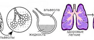
Pneumonia is an inflammation of the lower respiratory tract, involving all structures of the lung tissue, including the alveoli (cavities of the lungs involved in the respiratory process). It can have a different (but predominantly infectious) nature, and is also a complication of many diseases of the respiratory system.
The mechanism of the disease is quite simple: the pathogen, having penetrated the respiratory tract, causes inflammation in the alveoli. In the presence of a reduced bronchopulmonary barrier, transmission of infection occurs through the alveolar septa. The product of an extensive inflammatory process in pneumonia is usually exudate, the type of which corresponds to the type of disease.
As a result, the process of supplying blood vessels with oxygen, in which the alveoli participate, is disrupted, causing hypoxia. As the disease progresses, respiratory and then heart failure develops. If the course of treatment is ineffective, the patient's condition becomes severe, the disease progresses, causing further deterioration. In this case, death is possible.
Risk group
The risk group for the disease includes dogs with chronic infectious diseases, congenital disorders of the respiratory system, as well as animals with serious immunodeficiency conditions. Pneumonia often develops in weakened and emaciated dogs. Animals on duty and living on the street (hunting, guard, riding) are also at risk. Puppies and older dogs are especially susceptible.
Etiology of the disease
Depending on the etiology, pneumonia can be infectious or non-infectious.
Infectious pneumonia
Infectious pneumonia is often bacterial or viral in nature. It can develop as a primary or secondary disease against the background of influenza, bronchitis, tracheitis or tonsillitis. Depending on this, the causative agents of infectious pneumonia are:
- gram-positive microorganisms (staphylococci, streptococci, pneumococci);
- gram-negative microorganisms (enterobacteria, Escherichia coli, Proteus);
- mycoplasmas (causative agents of mycoplasmosis);
- viruses (herpes, influenza, parainfluenza, viral distemper);
- fungal infections;
- parasites (larvae of Toxocara, hookworm, filaria).
Infection of animals occurs mainly by airborne droplets, contact or hematogenous routes:
- through contact with sick animals;
- through contaminated food, dirty dishes, feces of sick animals;
- through infected blood and lymph.
Non-infectious pneumonia
Inflammation of the lungs of a non-infectious etiology in a dog is caused by the entry into the lung cavity of foreign agents of a non-infectious nature, namely:
- Aspiration pneumonia in dogs occurs after inhalation of foreign particles, foreign bodies (dust suspension, small debris), as well as when contents of the oral cavity or stomach enter the respiratory tract.
- Post-traumatic pneumonia is the body’s reaction to a violation of the integrity of the chest.
- Postoperative pneumonia is caused by complications after surgical interventions.
- Exposure to toxic substances on the respiratory system can result in toxic pneumonia.
- Allergic pneumonia. One of the symptoms of a malfunction of the immune system is inflammation of the lung tissue.
Aspiration pneumonia in cats and dogs
Aspiration pneumonia in cats and dogs
Aspiration pneumonia is a type of pneumonia that occurs in animals as a result of foreign bodies entering the respiratory tract.
Aspiration is the process of inhaling foreign substances and bodies into the respiratory tract. Aspiration leads to infection and inflammation of the lungs up to the development of necrosis of individual areas.
If a small amount of liquid food or medication enters the respiratory tract, the animal coughs and thereby removes the foreign matter from the respiratory tract. If a large number of foreign bodies enter the respiratory tract, inflammation begins to develop in the bronchi and lung tissue. If such an animal is not provided with urgent medical care in a timely manner, the animal may die from asphyxia (suffocation) or sepsis.
Aspiration pneumonia is usually divided into three types, depending on the nature of the foreign masses: chemical pneumonitis, bacterial infection of the lower respiratory tract and mechanical obstruction of the respiratory tract.
Chemical pneumonitis, or Mendelssohn's syndrome, develops when hydrochloric acid from the stomach enters the respiratory tract. Most often this occurs with megaesophagus, vomiting and when the animal is put under anesthesia on a full stomach.
A bacterial infection of the lower respiratory tract occurs as a result of infection by bacteria found in a foreign mass. It has a slower onset than chemical pneumonitis and is the most common form of aspiration pneumonia.
Mechanical obstruction of the airways is their complete or partial blockage due to the large volume of foreign masses that have entered there. To eliminate it, a thoracotomy (opening the chest cavity) can be performed.
Aspiration pneumonia in animals occurs as a result of:
- violations of the act of swallowing with pharyngitis,
- with megaesophagus,
- vomiting
- diseases of the central nervous system,
- with inept insertion of a naso-esophageal or gastric tube (when the prescribed medications can enter through the trachea into the lungs),
- when forcibly administering medications and irritating medications,
- when performing anesthesia on a full stomach.
Animals with brachycephalic syndrome, which causes narrowing of the upper respiratory tract, are predisposed to aspiration pneumonia.
Dogs suffer from aspiration pneumonia more often than cats.
Symptoms
Aspiration pneumonia is characterized by an acute course and rapid increase in symptoms:
- swallowing disorder
- cough,
- intense and rapid breathing,
- discharge from the nasal passages,
- no appetite,
- the general condition deteriorates sharply.
In severe cases, there is a progressive deterioration in the general condition, symptoms of asphyxia (suffocation), cyanosis (cyanosis) of the visible mucous membranes appear, and cardiovascular failure develops.
With chemical pneumonitis, cyanosis, shortness of breath, bronchospasm and cough with sputum suddenly occur, and the temperature rises.
Symptoms of aspiration pneumonia in cats include:
- difficulty or rapid breathing,
- increased heart rate and increased temperature,
- blueness of the mucous membranes and spasms of the respiratory tract,
- there may be a sweetish odor when breathing, which becomes more intense as the disease progresses,
- nasal discharge, sometimes reddish-brown or green in color.
Diagnostics
The diagnosis of aspiration pneumonia is made on the basis of the collected medical history (the onset of the disease immediately after medical procedures - gastric lavage, probing of the esophagus, forced administration of medications, during feeding, anesthesia, etc.).
When auscultating the lungs, moist rales are heard.
It is necessary to conduct a chest x-ray and blood tests. There is often a need to perform bronchoalveolar lavage to diagnose a bacterial infection, as well as to exclude other causes of pneumonia (fungal infection, cancer).
Treatment
Treatment for aspiration pneumonia may vary depending on the cause. It may be necessary to limit physical activity and even place the animal in a special cage.
Aspiration pneumonia always occurs urgently, so at the first moment the animal is placed in a head-down position and 2-3 minutes after swelling of the main mass of the foreign body, 3 sharp lateral compressions of the chest are performed to push out the remains. If your pet has a lung tumor or foreign body that is causing an obstruction, surgery may be necessary.
With deep aspiration, foreign masses are sucked out from the trachea and main bronchi using a probe.
Additionally, broad-spectrum antibiotics, bronchospasmolytics and glucocorticoids, bronchodilators (dilate the airways), mucolytics (thin the sputum to relieve cough) are prescribed.
Types of pneumonia according to the nature of the inflammatory process
Based on the nature of the inflammatory process, two types of pneumonia are distinguished: catarrhal and lobar.
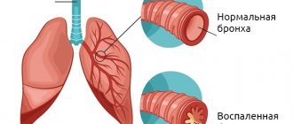
- Catarrhal pneumonia (or bronchopneumonia). The most common type of disease. The inflammatory process spreads to the tissue of the bronchi and alveoli, and is focal in nature. May develop as a complication of respiratory diseases. Focal pneumonia has less threatening symptoms and its treatment can therefore be carried out at home (but under the supervision of a doctor).
- Lobar pneumonia is acute in most dogs. It is an independent disease. It affects the entire lung or a significant part of it. It is characterized by the formation of an infiltrate in the alveoli, which can lead to swelling of the bronchial tissue. This form of pneumonia leads to respiratory problems and therefore requires hospitalization. For the most part, it is not contagious.
During the course of the disease, alveolar exudate is formed in the lumen of the bronchi and alveoli. Its composition is determined by the type of inflammatory process in the lungs:
- Purulent exudate is characteristic of complicated forms of pneumonia with the addition of pyogenic microorganisms.
- Serous exudate is a clear liquid, has a protein composition containing leukocytes and is formed in the catarrhal form of pneumonia.
- Fibrinous exudate is exuded during lobar inflammation of the lower respiratory tract. It contains fibrinogen (a special protein), which indicates increased vascular permeability.
Features of the development of pneumonia in dogs
The course of pneumonia in dogs, regardless of the forms and causes, has three stages:
- The primary stage of the disease lasts an average of five days. Pneumonia at this stage has mild symptoms. The general condition of the dog is often satisfactory.
- The secondary stage lasts no more than 10 days. At this stage, a rapid manifestation of all the symptoms of pneumonia occurs.
- The final stage. At this stage, either the dog recovers or dies as a result of irreversible changes in the lung tissue.
The course of pneumonia in dogs can be mild, moderate or severe. Inflammation in a dog can be acute or chronic.
Acute pneumonia can be caused by - traumatic damage to the respiratory tract and chest, pulmonary edema, filling of the lungs with fluid or blood, burns of the respiratory tract as a result of the dog inhaling smoke or chemical fumes, or sudden hypothermia of the dog.
Pathogenesis. Under the influence of unfavorable factors leading to inflammation of the bronchi and pulmonary lobules, their swelling, venous stagnation in the capillary network. The barrier function of the bronchial epithelium is reduced, creating conditions for the development of opportunistic microflora. Exudate accumulates in the lumen of the bronchi and alveoli, local foci of atelectasis are formed, in which purulent microflora can multiply with the formation of foci of abscessation and necrosis. Against the background of inflammation, redox processes and metabolism are disrupted, leading to disruption of trophism, blood and lymph formation and the function of the bronchi and pulmonary alveoli. In the initial stages of the disease, serous, serous-catarrhal or catarrhal inflammation occurs. As a result of the absorption of toxins and decay products from foci of inflammation into the blood and lymph, intoxication of the body is observed, gas exchange disorder due to a decrease in the respiratory surface of the lungs, and the degree of saturation of organs and tissues decreases.
Features of the course of the disease
There are several stages of development of the disease:
- The primary stage (with lobar inflammation it is called the stage of flushing or active hyperemia) lasts up to 3 days. The pathogen penetrates the lung tissue, causing inflammation. The vessels of the organ become overfilled with blood, and exudate begins to be released into the alveolar spaces.
- Secondary stage (stage of red and then gray hepatization). May last up to two weeks. The lung tissue becomes denser (taking on the consistency of the liver), as a result of which the supply of oxygen to the bloodstream is sharply disrupted. Red blood cells and then leukocytes sweat into the alveolar exudate. Clinical signs are most acute. Intoxication reaches its peak.
- The final stage. With a favorable outcome, the normal structure of the lung tissue is restored and the exudate is liquefied.
Depending on the type of pathogen, the state of the dog’s defenses and other factors, the disease can progress in different ways. There are three degrees of severity:
- Easy. No intoxication. Slight increase in temperature. The presence of a small focus of inflammation.
- Average. Moderate intoxication. The temperature may be increased slightly, but quite active formation of exudate is observed.
- Heavy. Severe intoxication. Loss of consciousness may occur. Body temperature is critically high. Active infiltration is underway. The dog is very weak and is breathing frequently. Complications may develop.
Provoking factors
Each type of pneumonia has its own risk factors that can trigger the onset of the disease.
Risk factors for infectious pneumonia
Risk factors for infectious forms of the disease are the following:
- frequent hypothermia;
- complications of primary infections: influenza, plague, tracheitis;
- chronic inflammatory diseases of the upper respiratory tract;
- weakened immunity due to insufficient production of immunoglobulin A by the body;
- poor diet, lack of vitamins and minerals;
- failure to comply with hygienic conditions for keeping animals (using dirty dishes, keeping sick and animals in the same enclosure, and so on);
- lack of timely deworming;
- eating foods with fungal infections (mold).
Risk factors for non-infectious forms
- Injuries and fractures of the chest provoke traumatic pneumonia.
- The postoperative period is often complicated by an inflammatory process in the lungs due to forced immobility for a long time.
- Aspiration of vomit into the respiratory tract during repeated vomiting can provoke aspiration pneumonia.
- Living in areas with poor ecology (with chemical production facilities nearby) is fraught with a toxic form of the disease.
- Long-term use of certain medications and chemotherapy can cause toxic pneumonia.
Causes of pneumonia in dogs
When pathogenic microorganisms (streptococci, staphylococci, mycoplasmas, chlamydia and others) enter the respiratory tract, dogs develop pneumonia.
With normal resistance of the dog’s body, the body successfully copes with them. But as soon as the dog’s immunity is weakened, these microorganisms activate their activity and penetrate the lung tissue. Weakening of the body's resistance in dogs occurs as a result of a number of predisposing factors:
- Temperature changes. Walks with dogs in winter, especially for smooth-haired breeds, should be short.
- Poor living conditions for the dog (dampness and drafts).
- Poor quality and inadequate feeding (lack of proteins, fats, carbohydrates, vitamins, macro- and microelements.).
- Contact with infected dogs.
- Injuries in the chest area.
- Weakness of the immune system (lack of immunoglobulin in the body).
- Diseases associated with metabolic disorders in the body (diabetes, uremia).
- Use of certain medications (aspirin, digoxin).
- Infectious tracheobronchitis.
- Chronic sinusitis, pharyngitis and tonsillitis.
Symptoms of the disease
The symptoms of the initial stage of the disease are very similar to the signs of any respiratory disease, so many owners do not pay attention to them. It seems to them that the dog has nothing serious. But signs such as a slight increase in temperature, a warm, dry nose, chills and fever may appear later as dangerous symptoms of lobar pneumonia:
- high fever;
- dyspnea;
- cough streaked with blood;
- clear signs of oxygen starvation, bluish coloration of the lips;
- lack of appetite, weight loss;
- hypertension, increased heart rate;
- severe intoxication;
- depression of respiratory function.
Focal pneumonia develops more often as a complication of acute respiratory infections or influenza. It is accompanied by low-grade or febrile temperature, dry or wet cough, and severe weakness. When listening, hard breathing, wheezing, and crepitus (creaking) over the source of inflammation are detected.
Diagnosis of the disease
After listening and percussing (tapping) the lung area, the doctor orders a chest x-ray. This is the most informative method for diagnosing pneumonia. The darkening and enhanced pulmonary pattern in the image will make it possible to determine the presence of a process in the lungs, its type and the extent of damage to the organ.
A biochemical blood test is no less important. The state of the blood can determine the degree of inflammation, as well as the type and cause of pneumonia. Sputum analysis helps determine the type of pathogen.
Only a professional can interpret the test results, so competent medical care in the treatment of pneumonia is necessary. In addition, the disease must be differentiated from other diseases of the upper and lower respiratory tract:
- acute tonsillitis;
- bronchitis and tracheobronchitis;
- rhinitis, sinusitis;
- lung abscess;
- tumor diseases of the lungs and tissues surrounding them.
How is the diagnosis carried out?
Diagnosis of aspiration pneumonia in dogs usually begins with a thorough physical examination by a veterinarian. He listens to the lungs with a stethoscope to look for abnormal sounds. And also prescribes:
- general analysis of urine and blood;
- radiography of the lungs in 2 projections;
- sputum bacterioscopy;
- blood chemistry.
A urine test for pneumonia will reveal protein and sometimes microhematuria. In the blood test, an increase in gamma globulins, an increase in ESR and the number of leukocytes are observed. A biochemical blood test is useful to identify concomitant diseases.
Did you know? When air enters a dog's nose, a fold of tissue located inside splits the air stream into two. One stream is for smell and the other
-
for breathing.
Additional diagnostic testing may be recommended depending on the individual patient. Thus, using bronchoscopy, a sample of material is obtained from the lungs for examination and identification of the bacterial agent.
Patient's condition with pneumonia
The treatment method is selected taking into account the patient's condition. There are three types of conditions for pneumonia:
- A stable condition may be accompanied by quite strong pulmonary symptoms, but the dog’s general well-being at this moment is close to normal: its appetite is not impaired, it exhibits normal physical activity. In this case, after visiting a veterinarian and receiving treatment recommendations, the animal can be treated on an outpatient basis.
- Unstable. In addition to pulmonary manifestations, the temperature is significantly elevated, the dog is lethargic and refuses to eat. In this case, the animal must be constantly under medical supervision in a hospital.
- Critical. Other signs of pneumonia include symptoms of acute lack of oxygen and cardiac dysfunction. The animal needs resuscitation, which consists of artificial ventilation of the lungs and maintaining the vital functions of the body.
Treatment of the disease
It is preferable to treat pneumonia in dogs in a hospital, since only there it will be complete and comprehensive.
- The main treatment for pneumonia is antibiotic therapy. They use broad-spectrum antibiotics that are effective against any type of pathogen or a complex that includes several types of drugs. These are penicillin antibiotics (Ampicillin, Gentamicin), tetracyclines (Doxycycline). They are prescribed in tablet or injection form.
- Physiotherapy. It is prescribed when acute inflammation subsides. In most cases, this is UHF heating, electrophoresis, and also a special chest massage to avoid stagnation of sputum, accumulation of alveolar exudate and pulmonary edema.
- Artificial ventilation and oxygen therapy are indispensable in severe cases.
- Infusion therapy is also possible only in a hospital setting. It is used if the animal is severely exhausted and is unable to independently support the vital functions of the body: it is in a state of extreme exhaustion or dehydration.
Treatment
First, you need to understand that pneumonia cannot be cured at home. If your pet's health worsens, do not hesitate and contact your veterinarian.
To cure pneumonia, you need to understand what caused it and the type of inflammation. An accurate diagnosis can only be made in a clinical setting, after clinical studies and on the basis of x-rays.
If it is secondary in nature (it is a complication of another disease), then first of all we treat the underlying disease.
The treatment plan is individual in each individual case. In general, it looks like this:
- Symptomatic therapy is prescribed (to alleviate the clinical manifestations).
- Broad-spectrum antibiotics.
- In some cases, medications that improve blood circulation are prescribed.
Some types of pneumonia have their own characteristics!
In case of aspiration pneumonia (when water, food or vomit enters the respiratory tract), action must be taken urgently.
These actions should be performed by a specialist, but if this is not possible. You will have to rescue the dog yourself:
- The dog must be placed in a head down position so that the foreign mass flows out on its own.
- 3 - 4 minutes after the fluid (mass) flows out, it is necessary to sharply squeeze the sides of the chest 3 times. This is necessary to clear the airways of residues.
- In severe cases, the remaining masses are sucked out using a probe. This manipulation is easier to perform in the clinic.
- Further therapy consists of prescribing antibiotics and antispasmodics, as well as drugs that improve cardiac function.
- For parasitic pneumonia, in addition to the main treatment, antiparasitic drugs are prescribed. They should be prescribed by a veterinarian after determining the type of helminth.
When treating mycotic pneumonia, additional antifungal drugs are prescribed, and it is advisable to treat the mucous membranes with Lugol's solution.
Home care and recovery
At home, every effort should be made to strengthen the body weakened by the disease:
- Continue to give antibiotics according to the regimen suggested by your doctor.
- Stop walking for a while to avoid possible hypothermia and colds, the onset of which can lead to a relapse.
- You cannot suppress the cough reflex with drugs that act on the cough center of the brain, so as not to provoke stagnation of phlegm in the lungs.
- Do regular chest massage yourself to improve sputum production in your dog.
- Bring your pet for follow-up examinations at the appointed time.
- Create a complete diet that will help restore weakened strength in a short time.
- Additionally, carry out vitamin therapy. Usually, the dog is prescribed vitamin complexes with a high content of vitamins A, C and group B.
Prevention methods
Basic preventive measures are aimed at maintaining good physical shape and immunity of the animal. It means:
- Avoid hypothermia, protect from colds and viral diseases of the upper respiratory tract.
- Vaccinate and deworm on time.
- Avoid contact with sick animals.
- Maintain a nutritious and balanced diet. The dog should receive all the vitamins and microelements necessary for its age.
- Moderate physical activity is beneficial for dogs.
If the dog receives good care and proper treatment, the prognosis of the disease is often favorable. Modern experience in the treatment of pneumonia is quite rich and allows us to cope with even a severe form of the disease.