Is it possible to cure pulmonary edema in a cat: symptoms and treatment prognosis
Cats are quite strong and hardy animals in terms of health.
But there are very dangerous diseases that a pet may not be able to cope with. Pulmonary edema in cats is a serious pathology that requires immediate attention. Due to swelling of the respiratory system, the cat begins to suffer from oxygen starvation, which can lead to irreversible consequences in the nervous system or death.
What kind of disease is this and what are its causes, we will consider in the article.
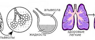
The lungs consist of two paired organs and inside have a large number of alveoli (cells). When you inhale, the alveoli fill with air, and when you exhale, they empty. All lung tissue is penetrated by small capillaries and vessels through which oxygen and nutrients are exchanged.
Pulmonary edema in cats is a serious disease in pets. The capillaries of the lungs become overfilled with blood, causing fluid to be released into the surrounding capillaries of the tissue. There are two types - cardiogenic and non-cardiogenic.
The photo shows an ultrasound of a 10-year-old cat with pulmonary edema.
Causes of a cardiac nature are called cardiogenic. This type of course occurs in heart failure. Insufficient functioning of the left ventricle provokes disruption of the pulmonary circulation, which, in turn, provokes stagnation of blood in the lungs and the release of water into the surrounding tissues.
Diseases that are provoking factors:
- cardiomyopathy;
- aortic heart disease;
- mitral heart disease;
- pulmonary embolism.
With cardiogenic factors, the lower parts begin to swell with a gradual transition to the bronchi.
In this state of affairs, the pulmonary alveoli cannot carry out normal gas exchange, as a result of which the cat experiences oxygen starvation, suffers from suffocation and dies if help is untimely. The prognosis for cardiogenic edema is unfavorable.
Pulmonary edema can be caused by electric shock.
All other causes that provoke pulmonary edema are called non-cardiogenic. The factors are:
- inhalation of hot air into the lungs;
- prolonged inhalation of toxic chemical gases;
- lobar pneumonia;
- thermal or solar overheating;
- infections of a viral or bacterial nature - pasteurellosis, plague;
- electric shock;
- brain injuries;
- the presence of septic processes;
- overdose of toxic drugs;
- renal failure;
- allergic reactions;
- asthma;
- malignant tumors.
Asthma can also cause pulmonary edema.
Diagnosis of pulmonary edema is based on anamnesis, visible symptoms, and medical history. By listening to the lungs and radiography.
Older animals with heart disease are most susceptible to the disease.
The main signs of the disease are manifested in the cat’s behavior. The pet spreads its legs wide and tilts its head, trying to inhale air. When touched, cold paws are felt. The animal may lie on its side for a long time, no longer able to rise.
Fear comes first in the gaze, the eyes become empty, and the cat’s panic is felt.
- The pet does not respond to the environment or the owner’s call.
- Pallor of the oral mucous membranes followed by cyanosis is visible.
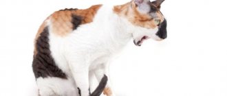
- Breathing is difficult, a hacking cough is accompanied by the release of pinkish sputum. When coughing, bubbling or gurgling sounds are heard. Foamy nasal and oral discharge may occur, with the tongue protruding.
- Rapid heartbeat followed by intermittent and weak heartbeat.
With pulmonary edema, the cat will look panicked.
It ends in paralysis of the respiratory nerves and death of the animal. The disease is very acute and has a lightning-fast course, but according to some signs it can be noticed in time and treatment can be started immediately. The onset of the disease is manifested by shortness of breath. The cat breathes more often with its stomach or open mouth. Breathing is very rapid and uneven, with periodic short coughing.
Having noticed such signs, the owner should immediately contact the clinic, otherwise delay threatens the inevitable death of the pet.
An attempt to help your pet yourself will inevitably end in the death of the latter. It's not worth even trying. You should take your cat to the clinic as soon as possible. The owner's only help may be to administer furasemide intramuscularly to remove excess fluid.
But this measure is permissible only with full confidence that the cause lies in heart failure. An important condition when going to the clinic: to prevent the animal from becoming nervous in order to avoid complications during a new attack.
Resuscitation involves the use of an oxygen cushion; in more complex cases, a tracheotomy is performed.
It is recommended to prescribe diuretics - diuretics. Defoamers and vasodilators are used. Heart medications to restore heart function. Bloodletting and novocaine blockade of sympathetic nodes are performed. After acute signs have been eliminated, the cat is placed in a cool room with good ventilation, but it is important to avoid drafts.
The hospital maintains a cool temperature with good ventilation, but without drafts
Symptomatic therapy is indicated: expectorants, antibiotics. It is important to establish an accurate diagnosis and, after eliminating the crisis, to apply specialized therapy.
conclusions
From all of the above, we can conclude: pulmonary edema is a serious illness with an unfavorable prognosis at the slightest delay, but it can be treated if pets are treated with due attention and care. A timely visit to the doctor is the key to a long life for your pet.
Heavy breathing in a cat is one of the leading symptoms of pulmonary edema, and the type of breathing changes. Normally, cats have a thoraco-abdominal type of breathing; with pulmonary edema, the type of breathing becomes abdominal, i.e. the cat breathes with its belly.
The cat wheezes when breathing. Wheezing during breathing may indicate both pulmonary edema and the presence of an inflammatory process in the trachea or oropharynx. With pulmonary edema, wheezing is gurgling, bubbling in nature, and nasal discharge is often observed.
The cat breathes with its mouth open. Normally, breathing with an open mouth is not typical for representatives of the cat family. The cat breathes poorly or heavily, suffocates and lacks oxygen; only under these circumstances does it begin to breathe with its mouth open. Sometimes cats breathe with their mouths open after periods of hyperactivity and vigorous play, but this breathing normally lasts no more than 1-2 minutes. If a cat sticks out its tongue and breathes frequently for a long time, then this is a cause for concern for owners.
Blue discoloration of visible mucous membranes and tongue in cats. Cyanosis in cats is a sign of severe respiratory failure and hypoxia (oxygen deficiency in tissues).
The cat is coughing. Coughing in cats with pulmonary edema manifests itself as an attempt by the body to free the lungs from the accumulation of fluid and mucus and is of a reflex nature. Pulmonary edema in cats is not necessarily accompanied by a cough, but if it is present, it will be a wet one, coughing up large amounts of sputum, sometimes with blood.
The cat is lethargic. Due to the lack of oxygen as a result of respiratory failure, the animal sharply loses activity, becomes lethargic, apathetic, and weakly reacts to external stimuli.
Atelectasis :: Atelectasis in a cat
With the fall of individual lobes of the lung or both lungs due to the equalization of intrapleural pressure with atmospheric pressure.
In connection with a disease of the respiratory system, atelectasis occurs when the bronchial lumen is obstructed by severely edematous mucous membrane, bronchial secretions, or aspirated vomit.
In addition, atelectasis can be caused by compression of the lung by intrapleural effusion, atmospheric air during pneumothorax, or a tumor.
The most common causes of atelectasis are obstructive forms of bronchitis, aspiration pneumonia, and pleurisy. In this case, individual lobes of the lung are more often affected. In collapsed alveoli, gas exchange does not occur, so the atelectatic organ serves only as a blood shunt.
The formation of atelectasis is the main cause of shortness of breath and cyanosis in lung disease. In areas of the lung that are excluded from breathing, secondary pneumonia develops.
The picture of the disease is mixed, characteristic of pneumonia.
Atelectatic lobes of the lung or the whole lung can be distinguished on an x-ray by the following three signs: darkening and reduction in size of the affected part of the organ; displacement of the heart and mediastinum towards the shadow; cranial displacement of the dome of the diaphragm towards darkening.
Drug therapy is limited to the treatment of pneumonia.
Non-cardiogenic pulmonary edema
As the name implies, this pathology is caused by disturbances in the functioning of the heart and is characterized by high pressure in the blood vessels. Poor pumping function of the heart (for example, due to mitral valve insufficiency, etc.) leads to accumulation and stagnation of blood in the vessels of the lungs, hence an increase in pressure and leakage of fluid from the blood capillaries into the alveoli.
This type of edema is caused by any other (non-cardiac) causes and is characterized by increased permeability of the walls of the pulmonary vessels.
The causes of this swelling may be:
- electric shock, traumatic brain injury, seizures (neurogenic edema)
- infectious and non-infectious diseases (inflammatory edema)
- gastrointestinal disorders, liver diseases, fasting, etc.
- poisoning by toxins - inhalation of carbon monoxide during a fire, snake bite, poisoning, uremia (severe intoxication as a result of kidney failure), etc. (toxic edema)
- allergic reaction, anaphylactic shock
- sepsis
- neoplasms leading to blockage of lymphatic vessels
- entry of vomit into the respiratory tract (aspiration)
We suggest you familiarize yourself with: Mussels: internal and external structure
Treatment
Most often, removal of the damaged lobe of the lung is indicated. The animal is placed in a hospital. There the patient receives the necessary infusion therapy, oxygen therapy, and painkillers. The attending physician monitors all vital signs: heart rate, breathing, blood pressure. The animal is preparing for surgery.
After the operation, the patient remains in the hospital until all physical parameters are completely stabilized. The outflow of fluid from the chest, as well as the sanitation of the chest, is carried out through installed drainages.
The prognosis for spontaneous torsion of the pulmonary lobe and timely surgical intervention is good. In the case of concomitant pathologies, it depends on the cause that caused the problem.
Diagnostics
The diagnosis of pulmonary edema can be tentatively made by anamnesis (medical history) and clinical symptoms, but the final diagnosis is made through a chest x-ray, in which the veterinarian sees a characteristic darkening of the lung tissue and a decrease in its transparency. Very often, pulmonary edema develops quite quickly and treatment must be carried out without additional diagnostics.
Treatment of pulmonary edema can be carried out exclusively by a veterinarian; self-medication or treatment “by phone” is excluded. The mortality rate for this pathology is extremely high, especially with late diagnosis. At the first sign of pulmonary edema in your pet, you should immediately take the animal to a veterinary clinic.
Treatment for pulmonary edema in cats will depend on which of the above diseases is causing it. In many ways, the prognosis depends on what kind of disease caused pulmonary edema in the cat and how promptly the treatment was carried out. If the underlying cause of pulmonary edema is successfully treated, then the prospects for a complete recovery are quite realistic.
The fastest diagnostic method is auscultation. The veterinarian listens for wheezing and gurgling in the lungs, shallow breathing. To check the functioning of the heart, it is also listened to.
An X-ray examination is required. It allows you to see the clinical picture, fluid level and “off” areas of lung tissue, blurring of the pulmonary pattern and roots of the lungs. Changes in the heart muscle are also recorded during cardiogenic shock - an increase in its size and stagnation of blood in large vessels are visible.
In general and biochemical blood tests, there will be an increase in the levels of leukocytes, nitrogen and liver enzymes (ALT and AST). To diagnose heart diseases, electrocardiography and echocardiological examination are performed.
The diagnosis of pulmonary edema is made primarily based on a chest x-ray. The picture is usually taken in two projections - frontal and lateral. With edema, the image shows darkening of the pulmonary field and congestion in large vessels. Cardiogenic edema is characterized by an increase in the cardiac shadow, and with left-sided heart failure - an increase in the left half of the heart.
Listening to the lungs with a stethoscope (auscultation) can reveal moist rales, heart murmurs and rhythm disturbances.
Pulmonary edema is a life-threatening condition. It requires immediate contact with the clinic!
Pulmonary edema in cats is a disease that can lead to the death of a pet. The structure of a cat's lungs is largely the same as a human's.
They are alveoli that are filled with air and entangled in a network of blood vessels.
Under the influence of various factors, a large amount of blood collects in the pulmonary capillaries, which contributes to a decrease in lung volume and oxygen starvation. Fluid accumulates in the alveoli and the cells become unable to breathe.
The causes that contribute to pulmonary edema in cats are divided into two types: cardiogenic and non-cardiogenic.
Cardiogenic edema occurs due to poor functioning of the left ventricle, which is a sign of heart failure. In this case, the pulmonary circulation suffers, which poorly supplies the lung tissue with oxygen and provokes fluid accumulation. The development of pulmonary edema against the background of a cardiogenic factor occurs from the lower parts of the respiratory system and ends in the bronchi.
If symptoms are not noticed in time, the animal will die.
The main heart diseases that provoke edema:
- cardiomyopathy;
- heart defects;
- cardiosclerosis;
- arterial hypertension.
It has been revealed that some cat breeds are predisposed to such diseases. Sphynx, Maine Coon, Scottish Fold, British, Bengal, Ragdoll, Abessinian and Norwegian Forest cats most often have congenital heart defects leading to pulmonary edema.
Non-cardiogenic factors include:
- overheat;
- pneumonia;
- electric shock;
- asthma;
- various types of tumor;
- brain and chest injuries;
- liver lipidosis;
- allergic reaction after anesthesia during castration or sterilization.
The first symptom of pulmonary edema in a cat is a change in its behavior. The pet is experiencing severe fear. This is visible in every movement and in the look, the animal stops responding to the situation and the calls of the owners, oxygen starvation provokes confusion in the cat and the look becomes empty.
All these manifestations indicate the need for emergency help, because the following symptoms can increase rapidly:
- the cat puts its paws forward and tilts its head down, trying to take a deep breath, but this does not happen, but only the sides swell;
- the mucous membranes of the mouth become bluish;
- in some cases, belching is observed;
- the animal tries to breathe by opening its mouth slightly, but wheezing is heard and pinkish sputum is released;
- a cough appears with gurgling or wheezing, but not necessarily;
- paws get cold;
- the cat falls to the side and can no longer get up;
- heartbeat quickens and then slows down and becomes intermittent;
- If left untreated, the cat dies from paralysis of the respiratory tract.
Signs of this disease can be noticed in advance: if the animal breathes frequently with its stomach and with its mouth slightly open, and the breathing itself is interrupted by a cough with phlegm. It is necessary to diagnose the functioning of the heart and lungs.
There is also a rapid development of the disease, which develops within a few hours. In this case, it is impossible to save the pet at home. Therefore, you need to urgently take him to the vet.
Diagnosis of edema
It is not difficult for a veterinarian to identify pulmonary edema in a cat. The doctor listens to breathing, detects wheezing and gurgling in the bronchi. An X-ray of the chest reveals the affected area with swelling, which becomes darkened.
To determine the cardiogenic factor, the animal undergoes an ultrasound of the heart, which shows an increase in muscle volume and obstructed blood movement in large vessels. An ECG detects an abnormal heart rhythm.
The final diagnosis is made after the results of a biochemical blood test. It shows an increase in white blood cell levels, as well as AST and LST.
Often the examination is combined with treatment of pulmonary edema in a cat, as symptoms develop rapidly.
Treatment and attempts to relieve symptoms at home are a dangerous waste of time and will lead the cat to inevitable death, therefore, at the first suspicion of pulmonary edema, the pet must be taken to the veterinarian.
The only thing the owner can do, provided that he is sure of the animal’s heart failure, is an injection of furosemide, which will briefly remove excess fluid from the pulmonary tract. This drug is a diuretic. This measure will allow you to gain time for transportation to the clinic.
As symptoms of swelling progress, resuscitation will be required, which involves the use of oxygen and tracheotomy.
- taking diuretic drugs that remove fluid from the body;
- glucocorticosteroids - relieve inflammation and reduce swelling;
- sedatives, prevent stress, which can provoke a new attack;
- in case of cardiac dysfunction, stabilizing medications are used;
- if swelling is caused by an allergic reaction, treatment is based on taking antihistamines.
After the exacerbation is relieved and the condition is normalized, the animal is returned to the owner with medical recommendations. It is necessary to carefully monitor the cat's condition and not provoke additional stress and stress.
Cardiogenic pulmonary edema in cats indicates a high probability of death of the animal, since stopping the acute condition does not eliminate the risk of developing a new attack.
If there is a non-cardiogenic factor in the development of this condition, the pet’s prognosis for survival is most likely, but only with timely treatment.
We suggest you read: What is the best food for cats with kidney failure?
But not only pulmonary edema, but also associated complications can pose a mortal danger:
- pneumonia;
- pneumosclerosis - fusion of the alveoli with connective tissue;
- collapse and dysfunction of the alveoli - atelectasis;
- emphysema - rupture of the lungs due to overcrowding with air.
In order to avoid such complications, the veterinarian prescribes maintenance therapy and a course of antibiotics.
The principle “the best therapy is prevention” is also relevant for the health of your pet. To avoid pulmonary edema in a cat, you need to carefully monitor the animal. At risk are sedentary animals, overweight cats, and breeds that are genetically predisposed to heart disease.
General preventive measures will help you avoid conditions that lead to heart and lung problems:
- high-quality and nutritious nutrition;
- timely vaccination and medical examination;
- insulation of electrical devices;
- eliminate the possibility of hypothermia or overheating of the animal.
The above measures will allow the cat to avoid most of the factors that contribute to the development of pulmonary edema.
The diagnosis of pulmonary edema in cats is made by typical clinical signs, heart murmurs and bubbling wheezing, which are detected during auscultation - listening to the chest with a phonendoscope.
Auscultation of the lungs
First of all, resuscitation measures are carried out aimed at relieving swelling. For this purpose it is advisable to use:
- diuretics (diuretics), which help remove excess fluid,
- foam suppressants,
- vasodilators,
- glycosides to maintain heart function: camphor, caffeine, cordiamine, adrenaline, etc.,
- bloodletting.
Then follow the generally accepted procedure:
- eliminating the cause of the disease,
- the pet is given complete rest,
- sit comfortably in a cool and well-ventilated room (but not in a draft);
- expectorants are prescribed;
- drips with calcium chloride and glucose are placed;
- antibacterial agents are used according to indications,
Occurrence and diagnosis
Flat chest usually develops within approximately the first three days of life, in some cases it appears in all kittens in a litter, in other cases only in a few or part of the litter. Less commonly, flat chest appears within the first 10 days and extremely rarely later. It is possible that cases of later development of the syndrome are associated with respiratory tract infection or pneumonia.
The main physical symptoms in mild cases include a flattened lower chest (usually a ridge-like undulation of the sides of the chest is felt along the ridge), complete flattening of the upper body in severe cases (the kitten looks like it has been squashed), difficulty breathing varying degrees, retraction of the abdomen while inhaling.
We suggest you familiarize yourself with Which cereals can be given to cats, and which ones should be avoided
The position of the chest and the movement of the abdomen resemble the state of hiccups, but the sharp spasms characteristic of hiccups here slow down or seem to get stuck: if the hiccups go away, allowing the body to return to its normal position, then this does not happen with flat chest. The syndrome may also be accompanied by digestive disturbances (see Colic, below).
Banana is used to demonstrate the shape of a flat chest
For a translation of the explanatory text to the pictures, see below. It can be difficult to determine whether a kitten is flat-chested using only a text description: in a mild case, the chest resembles the shape of a banana, with a downward curve. The rib cage of the chest is not fixed in a certain position, and flat chest can only be identified by the presence of a ridge on the sides of the rib frame.
The consequences of flat chest are delayed weight gain, breathing problems, inability to eat normally and, in a significant number of cases, death. However, due to the high percentage of kittens that survive, immediate euthanasia is not indicated and supportive care should be used to increase the likelihood of a favorable outcome (see Treatment below).
Flat chest is often misdiagnosed as pectus excavatum, which is not directly related to it, although flat chest can be accompanied by such a deformity. Although it is generally accepted that the syndrome is more common in the Burmese, it is observed in representatives of all breeds, including mongrel domestic cats, and the increased incidence of flat chest in the Burmese breed should be attributed to the availability of more data on recorded cases, and also associated with the characteristic barrel-shaped chest shape cells of a given genotype.
| Flat-chested Egyptian Mau | Rex kitten with a flat chest and a hump - a narrowing of the chest around the shoulders |
The syndrome is life-threatening in a significant number of cases (perhaps 50–60%). This is due to a lack of understanding of the reasons underlying the syndrome, untreated colic (leading to slow starvation), as well as a lack of sources of information in the veterinary literature.
Prevention issues and consequences
All diseases that may be accompanied by hyperemia (fullness of blood) of the lungs require careful monitoring in order to prevent the accumulation of fluid in the lung tissue.
The consequences of the disease are varied. There are prerequisites for damage to literally all internal organs due to oxygen starvation. Hypoxia is especially dangerous for the brain, heart muscle, liver and kidneys.
Oxygen therapy
After swelling, there is a high chance of developing other pulmonary diseases:
- atelectasis - flattening of the alveoli, as a result of which they lose the ability to fill with air,
- pneumosclerosis - the walls of the alveoli lose their elasticity, gas exchange is disrupted, lung tissue is replaced by connective tissue,
- congestive pneumonia is a secondary pathology leading to impaired blood outflow due to insufficiency of the left ventricle of the heart,
- emphysema - overflow of the bronchi with air and its penetration into the interpulmonary space.
Pulmonary hyperemia is an extremely dangerous and serious disease, in which any delay in providing first aid can cost the pet’s life.
Author of the article: Marina Nikolaevna Chuprina, veterinarian, parasitologist
Pulmonary edema is the accumulation of fluid in the intercellular space or alveoli of the lungs as a result of pathological processes. Cats, like most creatures, cannot live without oxygen. If the functioning of the lungs is impaired, its supply decreases sharply, inhibiting the functioning of the entire body and can lead to death.
How do symptoms appear?
Volvulus in an animal is not difficult to determine; the disease manifests itself in a number of acute symptoms. The pathology tends to progress quickly, the dog begins to feel spasmodic pain. The animal has impaired muscle tone in its hind legs, it is difficult for it to stand, the pet does not rise, groans, lies down and stretches its legs.
Abdominal enlargement
The main symptom of volvulus is a sharp increase in the abdominal cavity. Visually, the dog's belly begins to resemble a balloon. When palpating in case of pathology, swollen intestines and a tight abdomen can be detected. The intestine is under pressure from the enlarged organ.
Find out how to treat enterocolitis in dogs, causes and symptoms.
Vomit
The intestinal volvulus is accompanied by a strong urge to vomit with foam, and the muscles of the upper peritoneum tense. In most cases, vomit does not leave the body, since twisted organs do not allow this to happen.
Shortness of breath and irregular breathing
The animal's drooling increases, shortness of breath appears, breathing becomes difficult and hoarse. This can lead to oxygen starvation of tissues.
Stool disorders
Another sign of pathology in a pet is the absence of bowel movements. Due to the fact that intestinal patency is impaired, feces are not formed in the body.
What are the lungs?
These are 2 paired organs that are divided into several segments. Lung tissue consists of many interconnected alveoli, resembling bunches of grapes. Each cell inside contains air and is intertwined with a network of many capillaries. It is thanks to this structure that the blood is saturated with oxygen, releasing carbon dioxide into the alveoli. When fluid accumulates, a sharp decrease in the functional volume of the lungs occurs and oxygen starvation occurs.
Pulmonary edema in animals is a life-threatening condition in which accumulated fluid in the lungs presses on the alveoli and prevents the normal flow of oxygen to the body's tissues. As a result, the animal suffers greatly from hypoxia - oxygen deficiency, suffocates, suffers from shortness of breath and suffocation, and ultimately dies without a quick response.
This condition occurs not only with pulmonary diseases and infections. Edema itself is not an independent disease, it is just a symptom or rather a consequence of the development of various pathologies, each of which can provoke the occurrence of this most dangerous condition.
Just like in people, pulmonary edema in cats is a serious and very life-threatening condition, so it is only possible to save a sick animal if you go to the clinic early. Small kittens and old, obese or malnourished cats are especially susceptible to the disease. Both have weak immunity, which can poorly resist the underlying disease.
To confirm or refute a preliminary diagnosis, an x-ray is most often taken, but based on the symptoms, the veterinarian can prescribe a whole series of necessary tests and examinations.
Wen on a cat
A cat's wen is a soft, mobile formation under the skin. It is absolutely painless and when pressing on the affected area the animal does not experience any unpleasant sensations. In scientific language, wen are called lipomas. They are formed under the skin due to the growth of adipose tissue and are most often benign in nature.
A cat's wen is a reason to consult a veterinarian
Wen are absolutely painless and, even if they are small in size, do not cause any discomfort to the cat. This is why owners of sick animals most often neglect to visit a veterinarian. This is incorrect for several reasons. Firstly, the cat’s wen can grow. Secondly, the owner may mistakenly mistake a more serious neoplasm - a cyst or malignant tumor - for a seemingly harmless lump.
How to recognize a wen in a cat
You can make sure that a cat’s neoplasm is a wen only in a veterinary clinic. In addition to an external examination, the doctor will need accurate studies - x-rays and puncture biopsy - to make a correct diagnosis. An x-ray will help determine whether the tumor has metastases, and a biopsy will clarify the nature of the tumor.
Treatment of wen
It is impossible to get rid of wen by medication. They can only be removed surgically. However, veterinarians do not always consider surgical intervention appropriate. If the wen is benign in nature and small in size, most likely the doctor will suggest observing it. When the tumor grows, surgical intervention is most often recommended, since a growing wen in a cat can affect the nerve endings in the tissues, which will cause pain and discomfort.
If the lipoma is malignant, surgical removal is recommended
Source
Causes of the condition in cats
But for what reasons does pulmonary edema develop in cats? Is there any way to protect your four-legged friend? How to correctly recognize it and help a purr with such a severe pathology? There are many causes of pulmonary edema in cats. Let's look at all the possible options.
Dehydration
No matter how strange it may sound, pulmonary edema in a cat can develop both due to starvation and due to poisoning. In this case, a huge amount of water leaves through the gastrointestinal tract, and the pet becomes dehydrated.
As a result, the oncotic pressure decreases, and the liquid fraction begins to sweat out of the blood. It is this that accumulates in the lungs, causing swelling. In addition, problems with the kidneys and liver lead to a decrease in oncotic pressure.
Increased permeability of blood vessels (distress syndrome) can aggravate the condition. It develops due to poisoning with serious poisons, medications, smoke and toxic fumes, and due to previous injuries (including pulmonary injuries). Many diseases of internal organs (even pancreatitis) can lead to the vessel wall becoming less durable and capable of allowing fluid to pass through its surface into the intercellular space.
Heart failure can also cause pulmonary edema in a cat. The pet may have problems with the heart itself, or the purr is constantly tired (the heart is almost worn out). Therefore, if you already know about this illness in your pet, keep in mind that he is at risk. And for you, this is another reason to monitor him more carefully.
Other reasons
Tumors and other neoplasms can also be causes, but relatively less often than those mentioned above.
In determining the causes of pulmonary edema, there are provoking factors and direct mechanisms for the occurrence of the disease.
According to the mechanisms of occurrence there are:
- Cardiogenic. Occurs against the background of acute or chronic heart failure. The most common causes are heart defects, arterial hypertension and cardiosclerosis.
- Non-cardiogenic – quite rare in cats. Usually associated with blockage of the upper respiratory tract - swelling or paralysis of the larynx, abscesses, foreign bodies, as well as anaphylactic shock and gas poisoning.
Provoking factors include various chest injuries, concussions and electrical injuries. This also includes the entry of vomit into the respiratory tract.
Another reason for the development of the disease can be inflammatory infectious processes.
Which develop directly in the tissues of the lungs - pneumonia, tuberculosis, carnivore plague.
Neoplasms are also one of the reasons for the development of edema. As well as diseases of other organs and systems - kidneys, liver and poisons.
We suggest you read: Does sterilized dogs go into heat? Is it possible to sterilize a cat during heat and what might be the consequences?
Pulmonary edema in cats occurs due to the overflow of pulmonary capillaries with blood, accompanied by the release of the liquid part into the surrounding tissues. By its nature, the disease can be cardiogenic, that is, arising against the background of heart disease, and non-cardiogenic.
Heart reasons
Cardiogenic edema, as already noted, is the result of heart failure. As a rule, with weak functioning of the left ventricle (one of the parts of the heart), the work of the pulmonary circulation is disrupted, which leads to stagnation of blood in the lungs and subsequent leakage of fluid into the surrounding tissues.
This state can be compared to a porous sponge: up to a certain point, it swells and absorbs water without leaving a trace, but there comes a moment when there is no space left and the water flows out.
For cardiogenic causes, edema begins in the lower sections, but gradually the process moves to the bronchi.
It is worth noting that the prognosis for pulmonary edema is unfavorable, especially if it is caused by a heart defect.
Other factors
Non-cardiogenic causes of hyperemia (another name for the disease) are the following:
- inhalation of hot air;
- inhalation of potent gases with a pungent odor (for example, prolonged exposure to high concentrations of ammonia vapor will easily cause swelling);
- lobar pneumonia;
- heat or sunstroke;
- bacterial or viral infections that affect the lungs (pasteurellosis, canine distemper, etc.);
- exposure to electric current;
- brain injuries due to an unsuccessful fall or blows;
- development of the septic process;
- taking certain potent medications in dosages significantly higher than recommended;
- renal failure, when the protein concentration in the blood sharply decreases;
- allergy;
- bronchial asthma;
- malignant tumors in the lungs that interfere with normal blood supply.
It is important to understand that pulmonary edema is a serious and very severe pathology, often ending in the death of a cat.
Pulmonary edema in a cat is accompanied by coughing and difficulty breathing.
An animal suffering from heart defects, especially if it is already of advanced age, is at risk. Therefore, any self-respecting owner of a sick pet should learn to recognize the signs of impending danger. And they are:
- the cat suddenly takes a forced pose: stands with its forelimbs widely spaced and its head bowed, thereby trying to inhale as much air as possible, its sides are greatly inflated;
- paws become cold;
- after a while the animal falls exhausted on its side and never gets up again;
- the mucous membranes of the mouth turn pale or become cyanotic;
- the animal has difficulty breathing, coughs heavily, and produces pinkish sputum;
- seething, bubbling cough;
- foamy discharge may occur from the nose and mouth;
- tongue hangs out;
- the work of the heart weakens, the pulse first quickens, and then becomes weak and intermittent;
- death occurs as a result of paralysis of the respiratory center.
The behavior of the animal also changes:
- firstly, he is scared, and fear can be read in everything;
- secondly, the lack of oxygen confuses the consciousness, the gaze becomes crazy, and then empty and unseeing;
- thirdly, the cat stops reacting to its surroundings and does not recognize its owners at all.
Despite the fact that edema is an acute and rapidly developing condition, you can notice the approaching danger in advance if you carefully observe your pet and its behavior. Typically, within a few days the breathing rhythm may become disrupted:
- the animal breathes with its stomach (the abdominal stacks shake) or through the mouth;
- the number of rhythmic inhalations and exhalations per minute increases significantly (more than 40);
- the breathing itself is wheezing and labored with periodic hacking coughing.
It is clear that such symptoms do not always indicate developing edema, but the time to ring the bells has already come. It is better to play it safe and take the animal for diagnostics to a clinic, where they will order an x-ray and perform auscultation (in other words, they will listen to the lungs using a special device) and check the functioning of the heart.
Moreover, it is better to film a cough and an altered state on video - fortunately, now almost everyone has a phone with a camera. This will help the veterinarian see the progression of the disease over time, decide on the prescription of additional diagnostic tests and speed up the diagnosis.
Important! It is much easier to cure a process that is caught in a timely manner than to try to correct a neglected and altered condition, which is most often no longer compatible with life.
There is no need to console yourself with excessive hopes: in most cases, pathology is regarded as a dying condition! There is a chance to rescue an animal from the dead only if non-cardiogenic edema occurs.
Even if it was possible to stop the development of the process in the case of cardiac causes, the likelihood of a recurrence of the attack is too high.
If your cat has pulmonary edema, your cat needs immediate medical attention.
The disease requires immediate intervention from specialists; attempts to help the pet on your own will significantly aggravate the condition and hasten the approach of death.
All that the owner can do (provided that the swelling occurs due to heart failure) is to inject a solution of furosemide intramuscularly - a diuretic drug helps remove fluid from the body - and urgently go to the veterinary clinic.
Important! Using diuretics unless prescribed by your veterinarian is still not recommended.
In case of hyperemia, resuscitation cannot be avoided; further observation in a hospital is required. It is not possible to cure an animal with a telephone consultation!
When going to the hospital, it is important to try not to irritate the animal, especially at the time of the attack, so that the situation does not become even more complicated. There is no need to fuss, run, sob, cry - the cat feels all this and, naturally, gets stressed. And stress, as we know, does not lead to good things.
The resuscitation scheme usually includes the use of an oxygen cushion (chamber), in some cases tracheotomy and the administration of:
- diuretics (diuretics) to remove accumulated water in the lungs;
- defoamers;
- vasodilators;
- cardiac drugs to stabilize the heart;
- bloodletting;
- novocaine blockade of sympathetic nodes.
After the crisis has passed, the animal is placed in a cool, well-ventilated room (but ventilation does not mean a draft) and symptomatic treatment is applied: expectorants, antibiotics, etc. It is very important to determine the cause of the edema and eliminate it.
Most cases of pulmonary hyperemia end in the development of complications, which also need to be, if not prevented, then at least correctly diagnosed and treated. The most common consequences include:
- pneumonia;
- collapse of the alveoli (atelectasis);
- overgrowth of the alveoli with connective tissue (pneumosclerosis);
- emphysema - overfilling of the alveoli with air followed by their rupture.
In addition, prolonged oxygen starvation negatively affects almost all organs, especially the cells of the brain and kidneys.
For cats with heart problems, constant monitoring of the state of the cardiovascular system and heart function is necessary. It is especially important to constantly monitor animals at risk:
- obese people;
- with confirmed caryomyopathy;
- leading a “sofa” lifestyle;
- having relatives with heart problems;
- British, Scots, Macoons, Persians, Abyssinians and Sphynxes - those breeds that most often suffer from heart disease;
- drowsy and easily overtired.
Symptoms of pulmonary edema in dogs and the course of the disease
During pulmonary edema in canine representatives, slow and fast forms are distinguished.
However, their symptoms are similar:
- Inhibition of behavior. The animal is very depressed and does not respond to food.
- Rapid breathing. It is necessary to pay attention to the characteristic posture of the pet. The dog spreads its front paws and stretches its neck. In this position, she tries to straighten her airways and straighten her ribs, thereby making breathing easier for herself. In severe condition, when symptoms develop very quickly, the dog lies in a lateral position.
- Breathing changes: the animal tries to make short and jerky breathing movements. At the same time, the nostrils spread wide. Noticeably intense abdominal breathing.
- Change in behavior - the animal becomes restless.
- A dry and hoarse cough appears. It resembles a cough in people suffering from cardiac and vascular pathologies.
- The gums, tongue, and sometimes the eyelids become pale. Quite quickly a bluish tint of these mucous membranes appears. If your dog has light pigmentation, you may notice blue discoloration of the nostrils.
- Body temperature decreases.
- Unnatural discharge appears from the nostrils and mouth. Their consistency is transparent with a pink tint. The release of bloody foam is possible.
- Vesicular respiration is weakened. This can be seen using a stethoscope.
- When the chest is tapped, a dull sound is heard. This is especially noticeable in a dangerous state.
These symptoms may appear suddenly. When the current is lightning fast, there is a serious threat to the dog’s life.
Prevention
Oxygen therapy
- If your cat has a heart disease, then you should definitely carry out the treatment prescribed by your doctor and undergo regular examination by a specialist.
- Prevention of obesity and diet therapy for overweight.
- Timely treatment of infectious diseases and vaccination.
- Observation of animals at risk for heart disease - some breeds (Persian, British, Macoon, etc.), drowsy and inactive cats.
Treatment of pneumothorax in dogs
If pneumothorax occurs in a dog, it is very important to provide competent treatment. After diagnosing and confirming the pathological condition, the doctor will tell you about further actions aimed at improving and healing your pet.
If you turned to a specialist in time, without wasting time, and your dog has a small amount of air in the pleural cavity and the breathing mechanism is not impaired, then the doctor will prescribe you symptomatic therapy, give advice on caring for the animal in this condition, and prescribe the necessary medications and schedule a follow-up visit to monitor the healing process over time.
Another option, when the general condition of the animal is less than satisfactory, and there is a lot of air in the pleural cavity, respiratory function is depressed and/or heart failure is severe, round-the-clock monitoring and assistance in a hospital setting are necessary. Our Center has a comfortable inpatient department for providing care, where intensive care doctors work around the clock, providing timely care and peace. During the hospital stay, your dog will be offered long-term oxygen therapy to facilitate breathing, removal of air from the pleural cavity under the supervision of a surgeon, and, if necessary, timely administration of highly effective painkillers of various types and mechanisms of action. In some cases, drainage of the pleural cavity to remove air, surgical intervention for wounds of the chest wall, and more.
And also the opportunity to always be in touch with the doctors of the inpatient department, learn about changes in your condition and receive recommendations for further measures aimed at a full recovery.
Author of the article: veterinarian Yulia Olegovna Pavlova
Disease prognosis
It depends on the size of the tumor, the presence of metastases and damage to regional lymph nodes:
- When the tumor is single , located among healthy tissues, and does not have metastases, the prognosis is cautious. This is due to the fact that adenocarcinoma and carcinoma often recur. The average life expectancy is about a year.
- With multiple tumors , as well as damage to regional lymph nodes, the prognosis is unfavorable. If the neoplasm has penetrated into neighboring structures and given distant metastases, then the prognosis is also unfavorable. The average life expectancy in these cases does not exceed 4 months.
- The prognosis worsens when the tumor is located on the periphery of the lung , since in this position it is difficult to determine the boundaries of the tumor, and it easily metastasizes here.
- With single solitary, discrete tumors that do not have metastases, after successful surgery, life expectancy increases greatly, can be several years or more.
What you need to know about a dog in shock
Pulmonary edema can contribute to the development of shock in a dog. In this case, there is a sharp stop or a weakening of the most important functions of the body almost to zero. As a rule, due to pulmonary edema, a torpid type of shock develops.
Its signs are as follows:
- lack of reflexes to any external stimuli;
- shallow and very weak breathing;
- immobility;
- weak pulse, sometimes thread-like;
- constriction of the pupils;
- blue discoloration of the mucous membranes of the body.