Diseases of the respiratory system are often accompanied by complications. One of the most dangerous is pulmonary edema, which causes an acute lack of oxygen in dogs. Without timely help, the animal may die from suffocation! It is very important not to lose sight of the first symptoms of illness and to be able to provide first aid if necessary.
What is pulmonary edema in dogs?
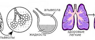
All mammals are characterized by pulmonary respiration based on gas exchange. When you inhale, oxygen enters the body, which is necessary to maintain its vital functions. With exhalation, the process is reversed: the breakdown product, carbon dioxide, formed as a result of metabolism, comes out.
All the described actions occur thanks to the work of the alveoli - small vesicles located at the very end of the bronchi. These numerous cells are surrounded by blood vessels and capillaries. Failure in this entire system occurs due to a number of diseases that disrupt the circulation of fluid inside the chest.
Characteristics of the phenomenon and its danger
Stagnation of intervascular fluid and excess blood inside the vessels impairs respiratory function, depriving the blood flow of the required amount of oxygen. In such cases, the body removes excess mucus and water along with a wet cough. But with extensive swelling, sputum separation is ineffective, so without the help of a veterinarian or owner, the pet may die.
Blood accumulating in the vessels increases internal pressure, increasing vascular permeability. Fluid in a dog's lungs seeps through the holes that appear and gradually fills the space outside the cells, including the alveoli.
A similar situation occurs when the pressure is too low due to insufficient protein. This element is part of the plasma, so its lack causes a difference between the pressure inside the vessels and the space between the cells. Trying to compensate for the difference, the body increases permeability, pushing part of the plasma out of the vessels.
Fluid accumulating in the alveoli reduces the space available for gas exchange. The level of oxygen in the blood decreases, and the amount of carbon dioxide, on the contrary, increases.
The heart and brain, which consume the maximum amount of oxygen, receive the greatest impact. The pulse rapidly decreases to a threadlike state, causing loss of reflexes. Breathing becomes difficult and gradually fades away, reducing the chances of saving the animal.
Stages
Edema develops in 2 stages. At the first stage (interstitial), the fluid fills the connective tissues, and at the second (alveolar) it moves into the alveoli. Sometimes the transition between these stages takes only a few minutes, so the pet must be given first aid without waiting for the veterinarian.
Causes of pulmonary edema in dogs
The development of the condition is caused by the presence of major heart problems. Disturbance of the pulmonary circulation is a provoking factor for the occurrence of edematous processes in all adjacent organs. Previous ailments include:
- Various congenital heart defects, especially those associated with ventricular dysfunction.
- Arterial hypertension of symmetrical or asymmetrical type. Increased pressure forces fluid into the lungs.
- Improper functioning of the heart valves, including lack of synchronization.
- Blockage of any of the pulmonary arteries, completely or partially.
- Hypertrophy (enlargement) of the heart muscle. The fibers are stretched, the heart cannot pump a normal volume, and stagnation occurs.
- The aorta does not have enough volume for the storage function.
Other common causes include the following:
- Pneumonia and severe bronchitis.
- Severe bruises to the chest that occur in collisions with cars or after being hit by people.
- Most often, doctors attribute pulmonary edema to the presence of cancer. This is almost 90% true in the old age of the pet.
- Closed-type traumatic brain injury with temporary or permanent brain dysfunction.
- Snake bites and massive wasp attacks can lead to pulmonary edema due to an allergic reaction.
- Blood poisoning caused by non-healing wounds, especially on the pads of the feet.
- Severe shock in small breeds can cause edema in the lungs. This can be caused, for example, by fear from an explosion or gunshot.
- Allergy to excessive amounts of medication or anesthesia.
- Chronic diseases of the excretory system, in which fluid is not removed from the body.
- Poisoning with pesticides, especially poisons intended for pest control.
- With extensive burns to the skin, salt imbalance and general disruption of the circulation of fluids in the body may occur.
- The presence of general exhaustion in dogs on an incorrect diet.
- Excessive exposure to direct sunlight, heat stroke and loss of consciousness.
- Presence of infection in the blood.
- This is one of the complications after chest surgery.
- Blood transfusion under the wrong conditions causes pulmonary edema.
- Long-term problems with the gallbladder, pancreatitis and biliary dyskinesia, the presence of stones.
- Epilepsy attack with loss of consciousness.
- Presence of anaphylactic shock. Therefore, it is important to give injections only in the clinic.
There is a misconception that sterilization or castration can lead to pulmonary edema. Typically, pulmonary edema in dogs occurs due to complications from anesthesia. This is possible after any surgical intervention.
Classification by causes
Depending on the cause, pulmonary edema in dogs can be cardiogenic or non-cardiogenic. In the first case, the complication is provoked by diseases of the cardiovascular system, in the second - by all others not related to the activity of the heart.
Cardiogenic
Cardiogenic pulmonary edema in dogs occurs when capillary blood flow in the pulmonary circulation slows down. This is what causes this type of pulmonary edema in dogs:
- sudden changes in internal pressure;
- cardiosclerosis;
- heart failure;
- dysfunction of the cardiac aorta and valve;
- congenital and acquired heart defects;
- pulmonary artery blockage;
- thickening of the ventricular wall (hypertrophic cardiomyopathy).
All of these pathologies lead to a reduction in the volume of arterial blood enriched with oxygen. The left half of the heart does not receive the required volume, and the right half suffers from excess. The resulting pressure difference provokes tissue swelling.
Non-cardiogenic
The main causes of the non-cardiogenic type are protein deficiency and intoxication. Lack of one of the most important nutrients occurs due to a poor diet, kidney disease and cirrhosis of the liver. There are many more reasons for intoxication. These include:
- poisoning by poisons (chemicals, bites, medications, household chemicals, plants);
- oncology;
- infections;
- overheating and burns;
- pneumonia and bronchitis;
- sepsis;
- chronic diseases of the urinary system that disrupt the natural outflow of fluid;
- pancreatitis;
- electric shock.
Also, thinning of the capillaries can be caused by chest and head injuries, excessive exercise, allergies, stress, asphyxia and epilepsy. The risk group includes overly emotional pets of small breeds and animals with chronic diseases.
Postoperative
The postoperative appearance should be considered separately. It occurs after surgery due to a negative reaction to anesthesia, which makes it possible to classify it as a non-cardiogenic type.
Despite this, deterioration of the condition is observed only in animals with chronic heart diseases, that is, the main causes of the cardiogenic type. That is why, before performing surgery, the surgeon must disclose to the owner all possible risks.
Causes of edema
Pulmonary edema in dogs can be an independent disease, but it can also develop against the background of some other pathologies.
The causes of cardiogenic edema are most often diseases of the cardiovascular system:
- various heart defects;
- hypertonic disease;
- rheumatism;
- ischemia;
- formation of a blood clot in the pulmonary artery.
Non-cardiogenic edema can be triggered by various external influences and diseases not related to heart failure:
- head injury;
- neoplasms, inflammation or rupture of blood vessels in the brain;
- pneumonia or complex form of bronchitis in dogs;
- inhalation of gases or chemicals;
- suffocation due to compression of the respiratory tract or due to a foreign object entering it;
- stressful state;
- electric shock;
- bite of a poisonous snake or insect;
- heatstroke;
- severe allergic reaction.
As we can see, pulmonary edema in a dog has a wide variety of causes and can occur, for example, after piroplasmosis, which the dog became infected with through a tick bite while walking outside. Therefore, you should carefully monitor your pet on walks and at home so that it does not overheat, do not go into dangerous places, give it all the necessary vaccinations on time and use protective equipment against the bites of dangerous insects.
Forms of the disease
Depending on the speed of development and severity of symptoms, the pathology can be acute or chronic. Most often, there is a rapid deterioration of the condition, which requires immediate response and prompt assistance.
Acute
It manifests itself in the form of a severe cough and shortness of breath that does not stop even during rest. Develops against the background of hypertension, heart defects and myocarditis. The most alarming sign of this form is the appearance of pink foam at the mouth.
Chronic
Breathing problems only occur with increased activity. Despite this, changes within the body are reflected in weight. It decreases, and the dog’s condition is gradually complicated by other symptoms. This form is typical for animals with slowly progressing diseases of the urinary system.
Symptoms of the condition
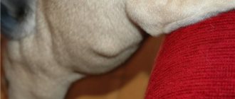
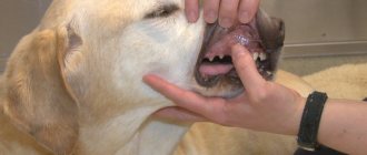
The main sign of pulmonary edema in dogs is difficulty breathing. A sick animal breathes quickly and shallowly, without closing its mouth and spreading its front paws wide. His nostrils flare actively. Due to developing pathology, usual activity is lost.
As the situation worsens, the described symptoms are supplemented by coughing and wheezing. During attacks, pinkish foam appears from the mouth. The mucous membranes of the oral cavity turn blue or pale. Your pet should be taken to a veterinarian as soon as possible to prevent suffocation.
What is pulmonary edema?
The main organ responsible for breathing is the lungs, consisting of two parts, which are divided into small “lobes” - segments. The segments, in turn, are formed from pulmonary vesicles or alveoli, surrounded on all sides by the finest vessels - capillaries.
Pulmonary edema in dogs occurs due to an excessive amount of blood and plasma in the capillaries and vessels; as a result, blood pressure “squeezes” exudate out of them, filling the alveoli and penetrating into the interstitial space. The process by which fluid accumulates in the lungs occurs gradually, from bottom to top, which makes it possible to detect the deterioration of the dog’s health and provide him with timely help.
Diagnostics in a veterinary clinic
To make a diagnosis, it is necessary to listen to the chest and check the function of the heart. The veterinarian determines the presence of extraneous noises, respiratory rate and heart rhythm. A four-legged patient must undergo a chest x-ray to identify pathological changes.
The exact cause of fluid stagnation will be determined with the help of additional studies: ultrasound, pleural puncture, analysis of blood clotting and its gas composition. The latter method is very informative for pulmonary artery thrombosis.
Diagnostics
The final diagnosis is established after studying the history and symptoms, examining the animal and analyzing laboratory tests. The life of the pet depends on the speed of diagnosis.
- Chest X-ray in several projections.
- Stethoscopy (listening to the lungs).
- Blood chemistry.
- Tapping the chest.
- Analysis of gas composition and blood clotting.
- Ultrasound of the heart.
- Pleural puncture (puncture).
- Blood pressure measurement.
First aid
After alarming symptoms occur, it is necessary to take your pet to the veterinarian as soon as possible. Until this moment, the owner can only alleviate the animal’s condition by slowing down the development of pathology until medical assistance is provided.
What to do first
What to do if your dog develops pulmonary edema:
- Place the dog on its side and limit its mobility. Activity is accompanied by an impressive consumption of oxygen, which is already in short supply.
- Try to replenish your air supply by opening all the windows in the room or car.
- Promptly remove any discharge that comes from your nose or mouth.
Try to calm your pet down with gentle words and strokes. Excessive excitement will only worsen his condition.
Elimination of shock
Shock is eliminated by artificial ventilation, or tracheostomy, which involves inserting a special tube into the trachea. Oxygen may also be given intravenously. All these procedures increase the pulmonary surface, normalizing the functioning of the pulmonary circulation. The alveoli are again saturated with oxygen, so for complete recovery all that remains is to eliminate the cause of the pathology that has arisen.
Prohibited actions
Pets are very good at picking up on their owners' emotions, so try not to panic and act judiciously. Do not try to make your pet feel better with diuretics unless advised by your veterinarian. With severe dehydration, they can aggravate the existing situation, accelerating the increase in symptoms.
If the attack occurred at night, do not wait for the morning to come. Many veterinary clinics operate around the clock and accept patients in serious condition without an appointment.
Causes of the disease
There are two types of pulmonary edema in dogs:
- Hydrostatic edema accompanies diseases of the cardiovascular system.
- Membranous edema is caused by exposure to toxins.
Hydrostatic edema can have two development mechanisms:
- The large volume of blood in the vessels causes its pressure to increase significantly. The permeability of vascular walls increases. As a result, the liquid portion of the blood enters the interstitial (extracellular) space and then fills the alveoli.
- Low oncotic pressure of the blood (pressure of the protein component of the blood - plasma), which occurs due to insufficient protein content, creates a large difference in the pressure of the fluids located in the vessels and in the intercellular space. Physical laws require that this difference be equalized. Therefore, part of the fluid passes through the walls of blood vessels, filling the interstitial spaces.
Edema of the membranous type is based on damage to the walls (membranes) of blood vessels as a result of exposure to external toxic substances or autotoxins. As a result, the fluid enters the intercellular space through the damaged vessel walls.
Cardiogenic and non-cardiogenic edema
In dogs, this disease can have various causes. It depends on them what type of disease occurs. There are two of them: cardiogenic and non-cardiogenic.
Cardiogenic pulmonary edema occurs much more frequently in dogs. It can be classified as hydrostatic type. Provoking factors are:
- heart failure (congenital or acquired);
- hypertension;
- blockage of a pulmonary artery by a thrombus.
The non-cardiogenic type of edema can be either hydrostatic or membranous.
The hydrostatic type develops if the dog has pathologies in which blood protein is reduced, namely:
- cirrhosis of the liver;
- kidney diseases;
- diet poor in proteins.
A common cause of hydrostatic edema is the uncontrolled use of diuretics (Furosemide).
Any pathology and injury accompanied by severe intoxication of the body can lead to the development of membranous edema:
- bites of snakes and poisonous insects;
- sepsis;
- infectious diseases;
- allergic and autoallergic reactions;
- electric shock;
- heat or sunstroke.
The cause may also be trauma and mechanical damage to the chest, leading to pleurisy or pneumothorax.
Treatment of pulmonary edema in dogs
After stabilization with oxygen therapy and drainage of excess fluid, the main treatment will be aimed at eliminating the underlying cause and maintaining the functioning of the affected organs. For this purpose, the four-legged patient may be prescribed the following medications:
- antibiotics effective against pneumonia and other infectious diseases;
- diuretics used in the treatment of cardiogenic pathology;
- vasodilators, dilating blood vessels;
- bronchodilators that eliminate spasms;
- glucocorticosteroids that relieve inflammation and allergic reactions;
- sedatives that reduce stress levels;
- respiratory stimulants that normalize heart function.
For the first few days, the animal remains in the hospital as it receives infusion therapy. The injected solutions thin the blood, facilitating the removal of excess fluid.
Treating Fluid in the Lungs in Dogs
Treatment will depend on the cause of the fluid, but in either case, the first step is to stabilize your dog. Oxygen therapy may be started along with antibiotics to prevent pneumonia.
Cardiogenic pulmonary edema
In this case, oxygen, rest and diuretics (to speed up fluid removal) will be used. Vasodilators (to dilate blood cells) are also needed. Your veterinarian will carefully monitor your pet's blood pressure, heart rate, and breathing rate during hospitalization. Repeated x-rays may be ordered by your veterinarian to monitor lung fluid levels. Heart disease is a chronic problem, so it is possible that swelling may return.
Non-cardiogenic pulmonary edema
Control of the causative factor is an important part of the treatment protocol in this case. Depending on the cause and severity, your dog may improve quickly with oxygen therapy. Antibiotics, intravenous fluids and colloids, diuretics, and anti-inflammatory drugs will be administered as needed, depending on the cause of the swelling. Again, check blood pressure, respiratory rate, body temperature and oxygen saturation regularly during treatment.
In both cases, your dog will experience minimal stress during the hospitalization.
Complications and prognosis for future life
The main complication that occurs with an acute lack of oxygen is heart failure. Also, against the background of pathology inside the chest, fibrosis of the pulmonary tissue, emphysema, atelectasis (collapse of the pulmonary lobe), pneumonia and sepsis can develop.
An animal left without timely assistance risks dying from cardiogenic shock and obstruction of the respiratory tract. For a favorable prognosis, not only the speed of decision-making is important, but also the cause that caused respiratory failure.
Is it possible to cure the disease completely?
The cardiogenic type is more difficult to treat and takes longer to treat than the non-cardiogenic type. The diseases that caused it often recur, so patients with this diagnosis are given a very cautious prognosis. They have to be monitored by a veterinarian for the rest of their lives to prevent exacerbations of existing heart pathologies.
Intoxication is dangerous due to the rapid development of complications, but is highly treatable with timely treatment. The chances of recovery from diseases of internal organs and injuries depend on their neglect.
How long will the pet live?
With successful relief of shock and intensive care, the pathology does not affect life expectancy. An unfavorable prognosis is typical only for acute pneumonia, which can lead to death within 2 months.
Reasons for appearance
By their nature, the causes of this dangerous condition are divided into cardiogenic and non-cardiogenic. The first group includes those associated with pathologies of the cardiovascular system. The second included everyone else.
Cardiogenic
Some heart diseases in dogs cause blood flow in the capillaries to slow down. This causes the pressure in the pulmonary circulation to slow down, and fluid from the blood vessels is retained and accumulates in the lungs.
Cardiogenic causes:
- Congenital heart defect.
- Hypertension.
- Heart valve dysfunction.
- Cardiosclerosis.
- Blockage of the pulmonary artery.
- Heart failure.
- Hypertrophic cardiomyopathy.
- Cardiac aortic dysfunction.
Non-cardiogenic
This group of reasons has nothing to do with the functioning of the cardiovascular system. Excess fluid in the lungs accumulates due to thinning of the capillaries, which occurs against the background of general disorders in the dog’s body.
Non-cardiogenic causes:
- Pneumonia.
- Chest injuries.
- Oncological diseases.
- Traumatic brain injuries.
- Snake bite.
- Sepsis.
- Severe stress.
- Electric shock.
- Asphyxia (suffocation).
- Allergy to medications and anesthesia.
- Chronic kidney diseases.
- Severe poisoning with pesticides.
- Burns.
- Anorexia.
- Heatstroke.
- Bacterial blood infections.
- Postoperative complication after surgery.
- Reaction to blood transfusion.
- Pancreatitis.
- Epileptic stroke.
- Anaphylactic shock.
To avoid the development of complications incompatible with life, immediately show the dog to a doctor. Immediate emergency assistance several times increases the dog’s chances of a complete recovery.
Pulmonary edema after spay/neuter surgery
A pathological condition can develop in an animal after sterilization or castration surgery. This is a reaction to anesthesia. However, this condition is possible only in animals that have chronic heart disease. Be sure to consult with a veterinary cardiologist before spaying your pet. Do not put your animal at risk of death!