Spread of endocardiosis in dogs
Endocardiosis of atrioventricular valves in dogs
is the most common cardiac pathology in dogs of toy and small breeds of the older age group. It should be noted that Cavalier King Charles spaniels, terriers, dachshunds, poodles, Pekingese, miniature pinschers, bulldogs, as well as other representatives of small and dwarf dog breeds are most predisposed to mitral regurgitation (Figure 1).
Figure 1 – Breed and sex predisposition of dogs to endocardiosis of atrioventricular heart valves
A correlation has been established for the occurrence of mitral valve endocardiosis in dogs
with other diseases characterized by connective tissue dysplasia: tracheal collapse, intervertebral disc prolapse, rupture of the anterior cruciate ligament, etc.
For canine endocardiosis
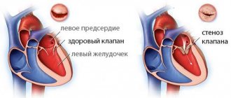
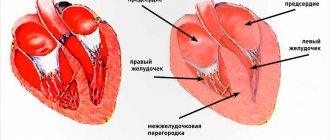
A degenerative process in the chords and leaflets of the bicuspid and tricuspid valves is characteristic. As this pathology progresses, it is characterized by the formation of insufficiency of the bicuspid and tricuspid valves located between the left and right atrium and ventricle. Loose closure of the cusps of the atrioventricular heart valves during ventricular systole causes the appearance of mitral and/or tricuspid regurgitation, the so-called backflow of the next portion of blood into the left and/or right atrium.
The reasons for the above pathological process have not yet been fully elucidated, but a typical change in the structure of connective tissue is typical. Endocardiosis
usually begins to debut in dogs over 5 years of age (Figure 2).
Figure 2 – Age-related susceptibility of endocardiosis in dogs of toy breeds (dependence of incidence on age)
However, it should be noted that the frequency of occurrence of this pathology clearly correlates with the age of sick animals. In dogs aged 5-7 years, the frequency of diagnosing endocardiosis of the mitral and tricuspid valve is on average about 10%, in dogs aged 9-11 years - about 20%, in dogs aged 13-15 years - about 30%, over 17 - more than 55%. According to statistical data regarding gender predisposition (Figure 1), it has been established that in males myxomatous degeneration of the heart valves
occurs significantly more often (1.5-1.6 times at p≤0.01). Genetic determination regarding endocardiosis in animals is obvious and does not require proof.
Mitral valve endocardiosis in dogs: treatment
Unfortunately, it is impossible to completely cure mitral valve endocardiosis in dogs.
impossible, the most important thing is to maintain the dog’s health at a sufficient level.
In this case, therapy consists of reducing stagnation in the systemic and pulmonary circulation, improving heart function, and minimizing stress on the heart muscle. For this purpose, drugs such as ACE inhibitors, diuretics, cardiotonics, agents that reduce platelet aggregation, corticosteroids as prescribed by a doctor, and metabolic drugs are used.
The medications are taken constantly, as the disease is chronic and gradually progresses. It is also necessary to ensure that signs of intoxication do not increase when administering drugs. Then the medications need to be discontinued and, under the supervision of a doctor, other medications that are safer for this particular dog should be selected.
Etiology of endocardiosis in dogs
Etiology of endocardiosis of atrioventricular valves in dogs
is not known exactly, but the hereditary genesis of this pathology is quite obvious. The genetic determination of myxomatous degeneration in dogs can be indirectly confirmed by the high incidence of this pathology in certain breeds. Cases of familial form of endocardiosis of atrioventricular heart valves in dogs have been repeatedly described, when an identical pathology is diagnosed in blood relatives (probands). It was previously established that dogs of dwarf and small breeds, middle-aged or elderly, and more often males (and more severely) are most often affected. The penetrance of familial forms of endocardiosis increases with the age of affected dogs. It is obvious that there are dormant genes that encode the development of lesions, deformations, and proliferation of connective tissue in the heart valves. It is likely that the genes encoding endocardiosis of the atrioventricular heart valves in dogs can be activated under the influence of inflammatory, stress or other factors that are still poorly understood.
dogs are highly affected
Cavalier King Charles Spaniel breed.
This breed has been proven to have polygenic inheritance involving age and sex alleles. This thesis is confirmed by clinical data, according to which myxomatous degeneration of heart valves
in males is more malignant than in females, although the severity of the heart murmur is the same in them. Some large dog breeds also suffer from endocardiosis, such as German Shepherds. However, large breeds of dogs do not typically develop heart failure.
Predisposition:
The highest predisposition and early onset of the disease was observed in dogs of the Cavalier King Charles Spaniel breed. Polygenic inheritance with the influence of sex and age is assumed. Also at risk are representatives of the following breeds: toy and miniature poodle, miniature schnauzer, chihuahua, Pomeranian, fox terrier, cocker spaniel, Pekingese, Boston terrier, miniature pinscher, whippet. Among larger breeds, endocardiosis can occur in Dalmatians, German Shepherds, and Ridgebacks.
Pathogenesis of myxomatous degeneration of heart valves in dogs
The pathogenesis of heart valve endocardiosis in animals is complex and poorly understood. The presence of multiple factors in the development of pathology has been established, which include metabolic disorders of connective tissue, degeneration of collagen fibers, endothelial function and sharply increased physical stress on the heart valve leaflets. It should be noted that endocardiosis in dogs develops very slowly; clinical symptoms of the disease appear already with significant deformation of the valves, cardiac remodeling and the development of chronic heart failure.
In the early stages of the disease, small nodules appear at the ends of the valve leaflets. Subsequently, these nodules become larger and merge to form large plaques. Next, shortening, thickening and deformation of the atrioventricular valves occurs. Histological studies have established signs of myxomatous valve degeneration that are similar to pathological changes in human mitral valve prolapse. Degeneration of the collagen fibers of the valve leaflets occurs with the accumulation of acidic mucopolysaccharides in them, which causes nodular degeneration, deformation and insufficiency of the affected valve. Similar pathological processes also occur in tendon strings. When the latter are lengthened, a parachute-like protrusion of the mitral valve leaflet into the lumen of the cavity of the left atrium is possible. This pathological condition in dogs is called mitral valve prolapse. For some breeds of dogs, prolapse of the atrioventricular heart valves is an important part of the pathogenesis. As a concomitant pathology with myxomatous degeneration of heart valves in dogs, endomyocardial fibrosis, intramural coronary arteriosclerosis, intramural myocardial microinfarctions and local myocardial fibrosis can develop.
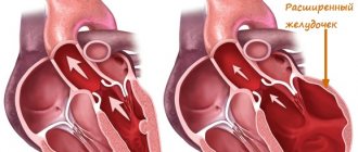
The affected (degenerated) valve loses its ability to close tightly. Therefore, at the next stage of the disease, insufficiency of the bicuspid or tricuspid valve is formed (mitral or tricuspid regurgitation, respectively). Further stages of endocardiosis in dogs are associated with the development of chronic heart failure. The body has powerful compensatory mechanisms, thanks to which hemodynamics in sick dogs remain at a sufficient level for a long time. The most important mechanism for compensating for pathology in the early stages is tonogenic dilation of the left and right atria, and subsequently of the left and right ventricles. Thanks to this, it becomes possible to implement the Frank-Starlig mechanism, which is based on the property of the myocardium to increase the force of contraction with greater diastolic stretching of the cardiomyocyte. In the early stages of the disease, the contractility of the left ventricular myocardium increases markedly. This process is also influenced by the activation of the sympathetic nervous and adrenergic systems.
Additional compensatory mechanisms of the body during cardiac decompensation are represented by a weakening of the tone of the parasympathetic nervous system, activation of the renin-angiotensin-aldosterone system, an increase in the concentration of natriuretic peptide, and concentric hypertrophy of the left ventricle.
The implementation of the above compensatory mechanisms allows the majority of sick dogs to remain in a stable and active state for a long time. With significant dilatation of the left atrium, a dry cough appears. Subsequently, dyspnea, tachypnea and symptoms of heart failure develop - intolerance to physical activity. In the future, pulmonary edema may develop. With mitral regurgitation, sick dogs often develop pulmonary arterial hypertension, and subsequently develop congestion in the systemic circulation.
In the later stages of the disease, significant remodeling of the myocardium of all chambers of the heart occurs and systolic dysfunction and severe biventricular heart failure with cardiogenic cachexia, a systemic inflammatory reaction, and activation of “lethal” cytokines in the blood serum are formed. Congestion in the lungs and intestines causes the activation of opportunistic bacteria living on the mucous membranes, which can cause hypostatic pneumonia, congestive catarrhal bronchitis and congestive gastroenterocolitis. Congestion of internal organs (liver, kidneys, endocrine glands) causes the development of multiple organ failure. The death of animals occurs in most cases (approximately 85%) due to the progression of chronic heart failure or sudden cardiac death syndrome develops with a frequency of about 15%. The latter develops as a result of the development of complications - rupture of the atria with cardiac tamponade, separation of the chordae tendineae with fulminant pulmonary edema, thromboembolism, atrial or ventricular fibrillation.
What is mitral valve endocardiosis?
Mitral valve endocardiosis, also called myxomatous degeneration of the heart valves , can affect the mitral valve, which separates the left atrium and left ventricle, and the tricuspid valve, on the right side of the heart. Most often - 60% of cases of damage to the mitral valve, 33% - degeneration of both valves, and 6% - only the tricuspid valve.
Endocardiosis of the mitral valve in dogs is an organic disease; during its course, degenerative changes occur in the structure of the connective tissue that makes up the chords and leaflets of this bicuspid valve.
.
First, small nodules appear, which enlarge and connect with each other, eventually forming growths and plaques. The tendinous strings or chords of the valve also become thicker and coarser. The valve itself thickens, becomes deformed, and its flaps no longer close tightly. A pathology of the mitral valve occurs; it does not cope well with its function, passing a stream of blood from the ventricle into the atrium (mitral regurgitation). heart failure develops with all the ensuing consequences.
This disease is one of the most common in dogs; according to statistics, it is diagnosed in 60% of visits to the veterinarian due to cardiovascular ailments.
It must be said that mitral valve endocardiosis in dogs progresses very slowly
, since the body uses powerful compensatory mechanisms to ensure efficient functioning of the heart and blood vessels.
It is believed that the disease debuts no earlier than the age of five, but usually the owners consult a doctor when the disease is already causing visible changes in the health of the four-legged animal. The older the dog, the more often endocardiosis is found in it.
Dogs of toy and medium-sized breeds are susceptible to this disease: dachshunds, toy poodles, Pomeranians, Chihuahuas, Cavalier King Charles spaniels. Large breeds include German Shepherds.
Mitral valve endocardiosis in dogs: symptoms
A disease such as mitral valve endocardiosis in dogs develops over years, but at first it does not make itself known. Only during preventive or routine examinations, for example before vaccination, can a doctor hear a heart murmur
.
1Cough is one of the main signs of this disease.
Initially it occurs after exercise and feeding. Subsequently, the frequency of coughing increases. A cough occurs because the enlarged left atrium puts pressure on the bronchus, from which reflex signals go to the brain, which provokes a cough. 2An enlarged abdomen and ascites are a consequence of heart failure, stagnation of blood in the systemic circulation, especially in the hepatic veins. The liquid component of the blood penetrates through the vessels into the abdominal cavity and accumulates there, which can be seen in the dog’s swollen belly. 4 Increased water consumption - the dog drinks more, but there is no need to limit it in liquids. The dog's appetite usually does not suffer and remains good despite the illness.
Complications of mitral valve endocardiosis in dogs
With further progression of chronic heart failure
congestion in the lungs, intestines, and internal organs increases, which weakens their functioning.
Under conditions of blood stagnation, bacteria inhabiting the mucous membranes are activated, thereby provoking the development of inflammatory processes in the lungs, bronchitis, and gastroenterocolitis. Pulmonary arterial hypertension is common in affected dogs. Death occurs either due to
an increase in chronic heart failure or due to cardiac death syndrome (thromboembolism, atrial rupture, pulmonary edema).
Mitral valve endocardiosis in dogs: diagnosis
When diagnosing mitral valve endocardiosis in dogs, the doctor will listen to the dog’s heart and lungs, identify typical heart murmurs, and prescribe blood tests: general and biochemical. An X-ray examination is mandatory, in which you can see an enlarged shadow from the left atrium and ventricle, upward displacement of the bronchus due to pressure of the left atrium, pulmonary edema, congestion in the pulmonary veins, and an enlarged liver. Echocardiography can effectively identify a number of pathological changes in the heart.
Clinical signs and symptoms of endocardiosis in dogs
Endocardiosis of atrioventricular heart valves
in dogs it can be subclinical for a long time. The first symptom that is usually accidentally recorded in patients is a systolic murmur in the projection of the mitral and/or tricuspid valve and a weakening of the first heart sound, that is, an auscultatory picture of bicuspid or tricuspid valve insufficiency develops. Subsequently, a paroxysmal nonproductive cough occurs. Further symptoms of myxomatous degeneration of heart valves in dogs are associated with the development of chronic heart failure syndrome, namely: decreased tolerance to physical activity, congestion in the lungs, dyspnea, orthopnea, tachypnea, pulmonary edema, variable rales in the lungs, dullness of the pulmonary field upon percussion. Cough, decreased endurance for physical activity, and tachypnea are the most common complaints of owners of sick animals. Tachypnea more than 30 times per minute at rest or during sleep in dogs with endocardiosis is formed when blood stagnation develops in the lungs. This symptom is an independent predictor of the development of acute pulmonary edema and should be carefully taken into account by owners of sick dogs. Coughing attacks are very variable, most often manifesting at night or early in the morning, as well as with increased physical and psycho-emotional activity. Alveolar pulmonary edema causes the development of respiratory distress syndrome and wet cough. Occasionally, foamy, bloody fluid may be discharged from the nasal passages, but more often than not large amounts of such fluid will leak from the animal's lungs only after death. The course of mitral valve endocardiosis in dogs is characterized by periods of exacerbation and remission. Symptoms of collapse or fainting (syncope) may occur due to severe coughing, fatal arrhythmias, or rupture of the left atrium or appendage. Symptoms of tricuspid valve insufficiency are usually hidden by more severe symptoms and clinical signs of mitral valve insufficiency. When the right atrioventricular valve is damaged, right ventricular heart failure develops, the main symptoms of which are ascites with peripheral edema of the hind limbs, hydrothorax with edema of the forelimbs, signs of congestive colitis (hyporexia, tenesmus, defecation disturbance).
The most typical symptom of myxomatous mitral valve degeneration is a holosystolic heart murmur, localized at the left apex of the heart (fourth to sixth intercostal space). Noise tends to radiate in all directions. Mild mitral valve insufficiency may not manifest itself as a murmur, but may be manifested by a short protosystolic click. Psycho-emotional arousal, as well as physical activity, can increase the intensity of the heart murmur. In dogs with endocardiosis of the atrioventricular heart valves, the degree of heart murmur in most cases significantly correlates with the stage of the disease and the functional class of heart failure. In some patients with endocardiosis of the atrioventricular valves of the heart of dogs, auscultation reveals a gallop rhythm (sound S3). A typical symptom of tricuspid valve insufficiency is a holosystolic murmur, which is best heard on the right at the apex of the heart. This noise is well carried to the jugular veins and is associated with their pulsation, which makes it possible to differentiate it from the radiating noise of mitral regurgitation.
Auscultation of the lungs reveals hard, accentuated, vesicular breathing and crepitant rales at the end of inspiration, which are best heard in the caudoventral parts of the lungs. They are harbingers of pulmonary edema. Fulminant pulmonary edema can cause the development of bilateral rales of different sizes and the absence of vesicular respiration in the posterior and lower parts of the lungs. Pulmonary edema is also accompanied by orthopnea - the inability to lie on its side, a position in which the dog takes a forced sitting position with the forelimbs widely spaced.
Also for canine heart failure caused by endocardiosis
, other symptoms less significant for diagnosis may occur:
- small wave pulse
- caudal displacement of the cardiac impulse and the posterior percussion border of the heart
- tachycardia
- jugular veins overflowing with blood
- positive venous pulse
- systolic trembling of the cardiac region upon palpation
- hepatomegaly
- splenomegaly
- ascites
- hepatojugular reflex (increased severity of jugular vein overflow when pressing on the liver)
Forecast:
In the SVEP study of Cavalier King Charles Spaniel dogs that had a heart murmur in the absence of heart enlargement, the median time to symptoms of heart failure was significantly longer than 3 years. An article by Borgarelli et al (Borgarelli M et al, 2008) showed that in a more mixed group of dogs with mitral regurgitation that did not suffer from clinical manifestations of heart disease, less than 50% of such animals died from complications of this disease during the follow-up period.
X-ray diagnosis of endocardiosis of atrioventricular valves in dogs
X-ray examination of the chest for endocardiosis of atrioventricular valves in dogs
makes it possible to identify:
- enlarged shadow of the left atrium and left ventricle
- dorsal displacement (“lifting”) of the bronchus by the dilated left atrium, which causes compression of the left main bronchus and the development of cough due to irritation of the receptor apparatus of the respiratory tract
- When performing fluoroscopy, it is possible to detect dynamic collapse of the main bronchus during a coughing attack (sometimes during breathing at rest)
- enlargement of the shadow of the right atrium and ventricle
- interstitial pulmonary edema or congestion in the pulmonary veins (in the hilar, dorsocaudal, bilaterally symmetrical zone)
- dilatation of the caudal vena cava
- hepatomegaly
- visualization of the border between the lobes of the lungs
- sometimes pleural and/or abdominal effusion
Stages of process development
For the first time, all stages of the process were summarized, systematized and studied by veterinarian Whitney, and this was back in 1974. There are four in total:
- At the first stage, the left atrium and ventricle do not change in any way, but small “nodules” appear on the mitral valve itself, that is, places of degenerative tissue changes.
- The second stage is characterized by the fusion of damage, and they also begin to involve the valve chords.
- When the disease reaches stage 3 , there are numerous growths in the form of plaques on the valve itself, the chords are thickened and noticeably “coarser” than normal. The thickness of the MC itself also increases significantly, and flexibility decreases. The basal portion of the valve is thickened and there may be areas of calcification (mineralization) and bleeding.
- 4th degree process is characterized by the fact that the valve tissue undergoes rapid degradation, the shape of the latter is greatly distorted, and the edges are curled. In especially severe cases, the chords tear or completely lose elasticity, which allows the valve to dangle when the ventricle contracts, like an open window. At this stage, the valve may resemble an open parachute in the sky.
Electrocardiographic diagnosis of endocardiosis in dogs
For endocardiosis of the atrioventricular valve in dogs, electrocardiographic examination is not very informative
. Electrocardiograms (ECG) of sick dogs can reveal nonspecific signs:
- Increased electrical activity of the left atrium and left ventricle
- Less commonly, increased electrical activity of the right atrium and right ventricle
- The presence of hypoxia of hypertrophied myocardium, which is manifested by ST segment depression and T wave inversion
- supraventricular extrasystole
- sinus tachycardia
- ventricular extrasystoles
- persistent or paroxysmal supraventricular tachycardia
- atrial fibrillation
Echocardiographic diagnosis of heart valve endocardiosis in dogs
Using echocardiography in dogs with endocardiosis
a number of pathological changes are revealed:
- the leaflets (especially the septal ones) of the mitral and/or tricuspid valves are deformed, thickened, shortened, with nodes at their end parts
- increase in the anteroposterior size of the left atrium and the ratio of the diameter of the left atrium to the size of the aortic root
- an increase in ESR in the initial stage of the disease; subsequently, ESR and the mitral-septal separation index also increase (EPSS indicator, which correlates with the severity of systolic dysfunction)
- hyperkinesis of the free wall of the left ventricle and interventricular septum, with their normal size or slight hypertrophy
- at the initial stage there is an increase in the shortening fraction and ejection fraction of the left ventricle, at the later stage there is a decrease in them
- systolic prolapse with parachute of the leaflet into the atrium cavity
- sometimes it is possible to detect a rupture of the chordae tendineae
- regurgitant jet with turbulence using color Doppler mapping
Distribution of mitral (bicuspid) valve dysplasia in dogs
Congenital mitral valve disease (mitral valve dysplasia)
is a congenital heart disease in dogs; the disease is less common in cats. Dogs of the following breeds are predisposed to mitral valve dysplasia: bull terriers, German shepherds, Great Danes, etc. Due to the development of mitral valve dysplasia, mitral insufficiency and systolic regurgitation of blood into the left atrium develops. Any component of the bicuspid valve complex (valve leaflets, chordae tendineae, papillary muscles) can be affected, but multiple components of the bicuspid valve are most commonly affected.
Treatment of dogs with endocardiosis of atrioventricular heart valves
Therapy for dogs with endocardiosis of the mitral and tricuspid heart valves lies in the correction of heart failure syndrome: elimination of symptoms (dyspnea, cough, ascites, etc.), increasing survival and quality of life, preventing cardiogenic cachexia, multiple organ disorders. The approach to the treatment of dogs with endocardiosis is differentiated and depends on the stage of the disease and the functional class (FC) of the heart failure syndrome (Table 3).
Table 3 — Differentiated approach to the treatment of dogs with endocardiosis of atrioventricular heart valves
| Stage and functional class of CHF | I FC | II FC | III FC | IV FC |
| A | ACEI? | ACE inhibitor pimobendan? thiazide diuretic? | — | — |
| B | ACE inhibitor pimobendan? | ACE inhibitor pimobendan? amlodipine? thiazide diuretic? | ACE inhibitor pimobendan furosemide amlodipine? thiazide diuretic? oxygenation? antibiotic therapy? | ACE inhibitor pimobendan torsemide spironolactone thiazide diuretic oxygenation digoxin? amlodipine? antibiotic therapy? laparocentesis? |
| C | ACE inhibitor pimobendan spironolactone thiazide diuretic? | ACE inhibitor pimobendan spironolactone amlodipine? thiazide diuretic? | ACE inhibitor pimobendan spironolactone thiazide diuretic digoxin? amlodipine? furosemide? sildenafil? antibiotic therapy? oxygenation | ACE inhibitor pimobendan digoxin torsemide spironolactone thiazide diuretic amlodipine? sildenafil? antibiotic therapy laparocentesis? oxygenation |
| D | — | — | ACE inhibitor pimobendan furosemide thiazide diuretic spironolactone antibiotic therapy oxygenation laparocentesis oxygenation sildenafil? digoxin? amlodipine? albumin infusion? antitussives? bronchodilators? Metabolics? | ACE inhibitor pimobendan torsemide thiazide diuretic spironolactone antibiotic therapy laparocentesis albumin infusion (reinfusion of ascitic fluid) oxygenation digoxin? amlodipine? sildenafil? dobutamine? antitussives? bronchodilators? symptomatic therapy? |
The treatment regimen for dogs with myxomatous degeneration of atrioventricular heart valves indicated in Table 3 is approximate and can be significantly supplemented or adjusted by a veterinary doctor depending on the specific clinical case, the presence of concomitant pathology or complications.
The most important aspect of the treatment of dogs with degenerative disease of the mitral and tricuspid valves is the blockade of neurohumoral processes in the body. The basic drugs for the correction of CHF syndrome in dogs with endocardiosis are ACE inhibitors: enalapril, benazepril, ramipril (the initial stage of circulatory failure is the minimum dose, disease progression is the maximum). When using ACE inhibitors, it is necessary to monitor the level of azotemia and creatinine in the blood serum.
Pimobendan is also considered the “gold standard” of therapy, which should be prescribed at the earliest stages of the disease. This drug is used as an inodilator agent, in the early stages at a dose of 0.2 mg/kg 2 times a day, for CHF refractory to therapy - 0.3 mg every 8 hours.
To correct minor congestion in the pulmonary and/or systemic circulation, we recommend using a thiazide diuretic (hypothiazide) or small doses of furosemide. In case of severe stagnation, use a combination of a loop diuretic (furosemide or torsemide) with hypothiazide and veroshpiron (high dose). If refractoriness develops with long-term use of diuretics, adding a carbonic anhydrase inhibitor for 3 days every 10-14 days may be useful. In the earlier stages of the disease, low doses of spironolactone can be used to block the body's neurohumoral reactions. Doses and frequency of use of diuretics depend on each specific clinical case. It is undesirable to use a sharp increase or decrease in diuretics.
Amlodipine is used for endocardiosis of AV heart valves in dogs to reduce blood pressure and dilate blood vessels. When amlodipine is combined with pimobendan and/or dobutamine, their pharmacological effects may be partially neutralized.
Digoxin may be useful in treating dogs with mitral and/or tricuspid heart valve endocardiosis. In the early, compensated stages of the disease, low (adaptogenic) doses of digoxin can be used; in case of systolic dysfunction of the left ventricular myocardium, standard therapeutic doses can be used. The cumulative properties of the drug should be taken into account. With the development of the tachysystolic form of atrial fibrillation, digoxin is the drug of choice. It is prescribed to correct heart rate, most often in combination with diltiazem or a beta blocker.
Sildenafil is prescribed to sick dogs for high pulmonary hypertension. It should be added that in dogs with endocardiosis of the mitral and tricuspid heart valves, with stagnation of blood in the pulmonary circulation, congestive bronchitis or hypostatic pneumonia often develops, while no increase in body temperature is detected in sick animals. Therefore, it is recommended to conduct a general clinical blood test. For leukocytosis, elevated ESR and/or neutrophyllosis, it is recommended to use antibiotic therapy. Antitussives and sedatives can be used as symptomatic therapy. If respiratory failure occurs, sick dogs are prescribed oxygen therapy. For pulmonary edema - use of high doses of furosemide or torasemide intravenously, oxygen inhalation, use of nitrates; after stabilization of the condition - standard therapy.
In severe stages of heart failure with ascites, laparocentesis is necessary. With severe ascites, oral forms of drugs practically do not work, so intravenous or intramuscular diuretics may be required. With the development of stages of heart failure refractory to therapy, intravenous administration of albumin may be useful, which can reduce ascites and partially eliminate the phenomenon of refractory to diuretics.
In the later stage of the disease, the use of glucocorticosteroids (prednisone or dexamethasone) to slow cachexia and stimulate appetite may be useful. However, glucocorticoid hormones have the ability to retain fluid and sodium in the body, so they can increase congestion. Among the drugs that have bronchodilator properties, aminophylline can be prescribed. Among the means of auxiliary therapy for endocardiosis of the mitral and tricuspid heart valves in dogs, hepatoprotectors, cardioprotectors, metabolic agents, vitamins, nootropics, antioxidants can also be used. These drugs do not affect patient survival, but may be useful for slowing cachexia, stimulating appetite, reducing the severity of cytolysis syndrome, improving metabolism and general condition. Important in the treatment of dogs with myxomatous degeneration of the mitral and tricuspid valves is nutritional support - diet therapy with diets with a limited content of kitchen salt, complete in amino acid and mineral composition and a sufficient concentration of omega-unsaturated fatty acids.
Thus, endocardiosis of atrioventricular heart valves in dogs
is a fairly common pathology with a poorly understood etiology and complex pathogenesis, requiring veterinary doctors to have certain skills in diagnosing the disease and treating animals. see DCM.
Literature
- Illarionova V.K. Morphological and functional parameters of the heart of dogs in normal conditions and with atrioventricular valve insufficiency. Author's abstract. dis... cand. vet. Sciences: spec. 03.00.13 – “Physiology”; 16.00.01 – “Diagnostics and therapy of animals” – M. – 2006. – 21 p.
- Quantitative echocardiographic [corrected] evaluation of mitral endocardiosis in dogs using ratio indices // J. Vet. Intern. Med. – 2005. – Vol. 19, N. 4. – P. 542–552.
- Immunohistochemical characterization of the extracellular matrix in normal mitral valves and in chronic valve disease (endocardiosis) in dogs / [H. Aupperle, I. Marz, J. Thielebein et al.] // Res. Vet. Sci. – 2009. – Vol. 87, N. 2. – P. 277–283.
- Donnely KB Cardiac valvular pathology: comparative pathology and animal models of acquired cardiac valvular diseases / KB Donnely // Toxicol. Pathol. – 2008. – Vol. 36, N. 2. – P. 204–217.
- Moon HS The cardiac sodium-calcium exchanger gene (NCX-1) is a potential canine cardiac biomarker of chronic mitral valvular isufficiency / [HS Moon, E. Choi, C. Hyun] // J. Vet. Intern. Med. – 2008. – Vol. 22, N. 6. – P. 1360–1365.
- Boswood A. Valvular heart disease in the dog / A. Boswood // Veterinary Focus. – 2008. – Vol. 18, N. 3. – P. 25–30.
- Chronic mitral valvular disease in dogs: assessment of functional clinical stage and echocardiographic measurement of the mitral valve / [RAL Muzzi, LAL Muzzi, RB Araujo, DA Lazaro] // Arq. Bras. Med. Vet. Zootec. – 2009. – Vol. 61, N. 2. – P. 337–344.