Liver diseases in dogs
Liver diseases have recently been increasingly reported in companion animals of various species, breeds and ages. Liver lesions account for a significant percentage of non-communicable diseases in animals, including cats and dogs. The occurrence of liver disease in cats and dogs is most often caused by poor-quality and unbalanced nutrition, and the use of drugs that have a hepatotoxic effect.
Secondary hepatitis and hepatosis can develop in animals with infectious, invasive and non-contagious diseases, these include autoimmune pathologies, endocrinological disorders - diabetes mellitus, Cushing's disease, neoplasms of the liver and other organs, etc. This is the largest gland, which is indirectly or directly involved in all metabolic processes occurring in the body.
Liver diseases in dogs and cats are divided into inflammatory and non-inflammatory. The cause of liver disease in cats and dogs can be viral and bacterial infections, helminth and protozoan infestations.
In addition, intoxication and poisoning often lead to inflammatory processes in the liver. Diseases may be in the nature of neoplastic processes of liver neoplasm, autoimmune pathologies, endocrinological disorders.
Inflammatory diseases are often accompanied by tissue degeneration. A sharp increase in them in the blood may indicate an increase in membrane permeability, necrosis and lysis of cells in the affected liver tissues during acute and chronic inflammation, or the presence of an oncological process that destroys liver tissue. For diagnostic purposes, total, indirect and direct bilirubin are determined in blood serum. The level of bilirubin can increase with acute and chronic hepatitis, hepatosis, liver cirrhosis, leptospirosis and blood parasitic diseases, as well as with obstruction of the bile ducts of the liver, cholelithiasis, liver tumors.
Of course, the diagnosis is made by examining a complete list of biochemical parameters in combination. Clinical blood test. Hematological studies can detect only nonspecific changes that occur in liver diseases: microcytic, normocytic, normochromic, non-regenerative anemia. Leukocytosis and neutrophilia can often be detected in acute bacterial hepatitis and liver tumors.
When examining urine, it is possible to detect a decrease in urine density, hematuria, bilirubinuria, and crystalluria. When examining stool, the presence of helminths and parasites that can affect the liver is excluded, as well as consistency, color, smell, biochemical indicators, including pigments and enzymes, which may indicate impaired liver function.
Only on the basis of such a comprehensive diagnosis is it possible to most accurately diagnose an animal with liver pathology and prescribe its subsequent treatment. There are also combined diseases, and therefore treatment is always selected individually for each animal. Acute and subacute processes in the liver should be treated comprehensively and over a long period of time so that the process does not progress to the stage of chronic inflammation.
In the presence of chronic inflammation, regular dynamic monitoring of the animal’s condition should be carried out, blood tests, and liver ultrasound should be performed in order to timely diagnose periods of relapse and remission of inflammation. You can always take all of the above tests in our clinic.
Experienced doctors and laboratory assistants will individually select the necessary test schedule for your animal. Liver diseases in cats and dogs Liver diseases and hepatopathy can have a variety of symptoms, since the liver is the most important intermediate metabolic organ. It is necessary to clarify that the criteria for systematization and rational classification of liver diseases may be different.
This is due to the fact that the same causative etiological factor is viruses, poisons, or others. Classification I. Primary liver diseases. Acute processes. Secondary liver diseases. Liver damage due to neoplasms and tumor-like diseases. Liver damage during pregnancy. Liver damage due to endocrine diseases. Liver damage due to circulatory disorders. Storage diseases: fatty liver, hemochromatosis, glycogenosis.
Diseases of the liver and nervous system, hepatolenticular degeneration. Hepatitis is a parenchymal inflammation of the liver, accompanied by granular degeneration, fatty infiltration and destruction of liver cells. It can occur either in an acute form for several days or in a chronic form for several months.
As a rule, it develops as a consequence of gastroenteritis, poisoning, long-term drug therapy, infectious leptospirosis or parasitic diseases.
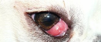
Characteristic: jaundice, mucous membranes and conjunctiva colored yellow, pruritis, discoloration of stool, dark urine, loss of appetite, gastroenteritis, bradycardia and arrhythmia, enlargement or reduction of the liver, pain on palpation of the liver. Nonspecific symptoms: depression, fever, weakness, exhaustion, diarrhea, vomiting, dark urine, black feces, epileptic seizures. Liver failure manifests itself in both acute and chronic forms.
The functioning of the liver sharply deteriorates, which leads to severe self-poisoning of the body and the development of hepatic coma. It is observed in all cats, both old and young, but, as a rule, develops against a background of obesity due to excessive accumulation of fatty acids in the liver. One of the auxiliary factors contributing to lipidosis may be stress. In all cases, there is an increase in the level of lipolysis and, as a consequence, an increase in the level of fatty acids that must be processed by the liver, which functions as a regulator of fatty acid metabolism.
Fatty acids in the liver are either oxidized to extract energy or form a lipoprotein complex and transported to other tissues. Any inhibition of fatty acid oxidation or lipoprotein synthesis promotes the accumulation of fatty acids by the liver. First aid: force feeding, drugs that stimulate and increase appetite.
The disease occurs in hypertrophic and atrophic types. There are primary and secondary cirrhosis. Connective tissue grows along the periphery of the hepatic lobules, putting pressure on Kupfev cells and capillaries.
Atrophying and dying hepatocytes are replaced by connective tissue proliferation. Increasing stagnation of blood in the portal vein system leads to ascites. Obstructive jaundice occurs. Atrophic cirrhosis occurs more often than hypertrophic cirrhosis. In hypertrophic cirrhosis, toxicosis leads to proliferation of the stroma and atrophy of the parenchyma. The liver slowly increases in volume. Portal hypertension, edema, and ascites occur. Abdominal dropsy Ascites A chronic secondary disease associated with difficulty in resorption of peritoneal fluid into the blood and lymph circulation system and the accumulation of this fluid in the abdominal cavity.
The reason for the accumulation of a large volume of transudate in the abdominal cavity is its weak outflow. Outflow difficulties may depend on the state of the portal hepatic circulation, insufficiency of the heart, kidneys, and hydremia. All liver diseases, during which its volume changes, the tension of the capsule, and the function is sharply upset, for example, cirrhosis, cancer, echinococcosis, can lead to stagnation of blood in the portal vein system and a decrease in the absorption of fluid from the abdominal cavity.
Similar consequences are caused by congestion in the systemic circulation that occurs due to diseases of the heart, lungs, and improper metabolism. Abdominal dropsy is often a local manifestation of edematous disease. Upon external examination, symmetrical bilateral protrusion of the lower and lateral parts of the abdominal wall, difficulty breathing, fatigue, emaciation, and swelling of the lower parts of the body are noticeable.
The animal is reluctant to change position, lie down or sit. Their body temperature is normal, the mucous membranes are pale. The appearance of jaundice is an unfavorable sign. When palpating the abdominal walls, fluid fluctuations are felt. Bowel sounds are often weakened. Dullness is determined by percussion, and with a test puncture of the abdominal wall, a clear straw-yellow liquid with a small protein content flows out. The course of ascites is chronic, severe, the disease lasts for months and even years.
When jaundice and edema appear, the outcome of the disease is unfavorable. Depending on the etiological factors, their strength and duration of exposure, fatty degeneration - fatty hepatosis, amyloid degeneration - liver amyloidosis and other types of degeneration may predominate. Fatty hepatosis, fatty degeneration, liver steatosis is a disease characterized by the accumulation of triglycerides in hepatocytes and disruption of the basic functions of the liver.
There are acute fatty hepatosis, toxic liver degeneration, and chronic fatty hepatosis, which occurs much more often than the first. Fatty hepatosis is registered as a primary, and more often as a secondary concomitant disease. The causes of primary hepatosis include feeding poor quality, spoiled feed. Toxins of pathogenic fungi, protein rotting products, and rancid fats are especially dangerous for the liver.
Hepatosis occurs when feeding animals with low-quality fish meal, meat and bone meal, feed yeast, rancid fats, spoiled meat, fish, etc. The possibility of liver damage from nitrates, nitrites, pesticides and other mineral fertilizers, which are contained in food products in increased quantities, cannot be ruled out. As a concomitant disease, hepatosis develops in obesity, diabetes, poisoning and many other diseases, which are based on metabolic disorders and the functions of endocrine organs.
Liver dystrophy is often a consequence of infectious and invasive diseases, chronic diseases of the gastrointestinal tract, kidneys, uterus, heart and other organs. Acute fatty hepatosis develops quickly, its clinical manifestation is characterized by signs of general intoxication and jaundice. Appetite is absent or reduced. The liver is often enlarged, soft, and slightly painful. Dogs experience anorexia, numbness, loss of strength; vomiting, diarrhea, general muscle weakness, sometimes cramps, often a scaly or nodular skin rash.
In acute hepatosis, animals can die in a very short time or after 1-2 weeks. With chronic hepatosis, the symptoms are mild. Depression, general weakness, and loss of appetite are observed. The liver is moderately enlarged, with a smooth surface, painful on palpation and percussion.
Yellowness of the mucous membranes and skin does not appear or is very slight. Body temperature is normal. In the blood in acute and chronic hepatosis, a decrease in glucose content, an increase in bilirubin, and cholesterol are noted.
In the case of concomitant hepatosis, characteristic signs of the underlying disease are noted. Acute fatty hepatosis is accompanied by severe liver failure and often leads to the death of the animal.
Symptoms and treatment of liver failure in dogs
It is not for nothing that the liver is called the biochemical laboratory of the body. Of its many functions, detoxification is the most important. Its violation leads to gradual poisoning of the animal’s body with metabolic products (ammonia, fatty acids), and this is fraught with the development of coma and death.
Liver pathologies in dogs are relatively rare, since its functional reserves are quite large. Many diseases have a long hidden course. While the animal feels well, its body is already poisoned by metabolic products. This is dangerous, since late diagnosis may lead to untimely provision of veterinary care, and it will not be possible to save the animal.
Loss of the ability to eliminate toxins occurs due to severe damage to the parenchyma (liver tissue). The complex of diseases that accompany these disorders is collectively called liver failure. The pathology occurs with severe intoxication, metabolic disorders, neurological signs, jaundice, edema, and dyspeptic syndrome. In this article we will look at what are the causes of liver failure in dogs, what are the main symptoms and treatment of this disease.
Cholangitis in dogs: symptoms, treatment
Their problem is that these ailments remain undetected for a long time due to the specific characteristics of the organ. This is the name for inflammation of the bile ducts in the liver. Divided into three types:. Neutrophilic cholangiohepatitis is characterized by the infiltration of large numbers of neutrophils into the portal areas of the liver and directly into the bile ducts. The disease occurs when pathogenic microflora penetrates from the small intestine. E. coli, various types of staphylococci, streptococci, clostridia, pathogenic fungi and salmonella - all of them can easily lead to this outcome. Predisposition factors include: congenital or acquired pathologies of the biliary system, various anomalies in the development of the gallbladder, adhesions resulting from an infectious disease, obstruction of the bile duct, parasitic infestations. The lymphocytic type of the disease develops in a similar way. Diseases of all types described above appear rather blurred.
The liver is an important organ responsible for the breakdown of nutrients and the synthesis of many molecules such as albumin, blood clotting factors, cholesterol, glucose and many others. The liver has a phenomenal ability to regenerate.
WATCH THE VIDEO ON THE TOPIC: Canine distemper is the most dangerous disease, but it begins with ordinary and harmless symptoms - don’t miss the moment
Causes and types
Cholangitis occurs due to obstruction of the bile ducts and associated infection. The patency of the ducts can be difficult due to the formation of stones, scarring, removal of the gallbladder, cysts, various parasites, etc.
Infection usually occurs in two ways:
- ascending route (from the intestine);
- descending path (through blood and lymph).
- There are acute and chronic cholangitis.
In this case, acute cholangitis occurs:
- catarrhal (with the occurrence of swelling of the mucous membrane of the bile ducts);
- purulent (pus forms and spreads to neighboring organs);
- diphtheritic (necrosis occurs during inflammation);
- necrotic (occurs due to the entry of enzymes into the pancreas, destroying the mucous membrane);
- The chronic form can be latent, recurrent, septic, abscessing, sclerosing.
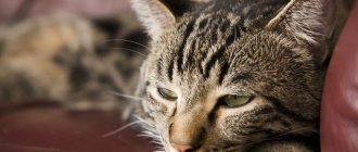
Cholangiohepatitis in cats
The wide prevalence of cholangiohepatitis in cats is associated with the peculiarities of their anatomy: the pancreatic duct and the gallbladder ducts connect before emptying into the duodenum. Therefore, inflammation of the small intestine or pancreatitis, inflammation of the pancreas also leads to inflammation of the bile ducts, cholangitis. The acute form occurs more often in young cats. It begins with a sudden refusal to feed and lethargy. Vomiting appears, body temperature often rises, and the abdominal area is painful. With acute hepatitis, dehydration quickly occurs.
Catalog.
Diagnosis of diseases of the hepatobiliary system should be based on medical history, the results of a clinical examination and laboratory tests, and the results of instrumental research methods.
Dog, Labrador, not neutered male, 3.5 years old. Reason for treatment: abdominal enlargement, loss of appetite, lethargy. History of piroplasmosis - 5 months before the onset of these symptoms. Vaccination with a complex vaccine 8 months before the onset of these symptoms. VIDEO ON THE TOPIC: Allergy symptoms in dogs.