How would you react if you woke up in the morning and your dog had a cherry eye? For a responsible pet owner, this will obviously cause a bit of shock followed by a lot of worry. If you are completely unaware of this condition, it is best to call your veterinarian to find out what it is. The first thing you will hear will be described as "cherry eye" and you will also be told not to panic as this is not a life-threatening condition.
However, a red blob sticking out of your dog's eye is definitely confusing, and as a pet owner, you should make an effort to understand what causes cherry eye in a dog and what steps you can take to deal with it.
So what is cherry eye?
Medically called “third eyelid adenoma,” it is a condition in which the lacrimal gland located on the third eyelid of a dog’s eye bulges. It appears as a red blob from the inner corner of the eye near the dog's nose and seems quite painful, although this does not explain why your dog may not complain about it.
© shutterstock
A closer examination of a dog's eye would reveal that, unlike the human eye, it consists of three eyelids. Among them, the upper and lower eyelid are always visible, while the third eyelid remains hidden from view and serves the dual purpose of protecting the eye from dirt particles and keeping it moist. This third eyelid is also home to the tear gland, and when it pops out from the corner of the eye as a red blob, the condition is called cherry eye .
Although this disease in itself may not be life-threatening, it deserves a certain degree of attention because there have been cases where it has become infectious. If you notice that your dog is constantly scratching his eye or there is thick or watery discharge in the area, it is time to visit the veterinarian. Another characteristic of cherry eye is that it is never limited to one eye—sooner or later the other eye is bound to burst.
Cherry eye syndrome in spaniels
Dog eye diseases
https://www.irish-terriers.lv/kinologiya/2009-06-26-09-14-09.html
Structure of the eye
There is no doubt that the eye is an extremely complex organ, the structure of which is difficult to understand even by those who do not consider themselves new to the world of cynology. Here you cannot do without detailed illustrations and manuals, so a collapsible eye the size of a small ball helped those present to clearly see the desired part of the organ of vision throughout the entire seminar. I will not go into these details - for those who do not know the topic, a small article will not replace the chapters of an intelligent textbook. I will briefly recall only the main points. The organ of vision is located in the eye orbit - a bony cavity formed by the bones of the skull. The eye is protected by eyelids: the upper eyelid, where the eyelashes grow, and the lower eyelid. In the inner corner of the eye there is the so-called third eyelid, which is usually visualized in dogs with a raw constitution.
The eye has three membranes: the outer one, which is divided into the anterior transparent part, the “sight glass” of the eye - the cornea and the opaque protein shell - the sclera; the middle one, consisting of the iris, in which the pigment cells that determine the color of the eye are scattered, and two more parts that are not visible without special equipment - the ciliary body and the choroid; and, finally, the inner one - the retina or retina. The nerve layer of the retina contains photoreceptors - rods and cones, which provide light and color perception. The light-refracting media of the eyeball are the lens and the vitreous body. In the center of the iris there is a round hole that is not related to the structure of the eye - the pupil. The mucous membrane located under the eyelid and lining the eyeball with the cornea is called the conjunctiva.
What causes cherry eye in dogs?
Despite extensive research, the exact cause of cherry eye has yet to be determined and remains one of the unsolved mysteries of the canine world. A number of conclusions have been drawn as to why this occurs, and the reasons range from physiological to genetic in nature.
A generally accepted fact about cherry eye is that it occurs when the connective tissue that attaches the tear gland to the eye weakens. When this happens, the lacrimal gland loses its anchor and moves out of its place, thus protruding above the white part of the eye like a cherry. However, what weakens connective tissue still eludes experts, and this is where speculation arises.
The occurrence of cherry eye is also associated with genetic factors, which lead experts to believe that some dog breeds are more vulnerable to the condition than others. Most flat-faced, wide-mouthed breeds such as beagles, bulldogs, pugs, poodles, etc. have been found to have weak eye tissues and hence are considered more prone to cherry eye. But this distinction is not sacrosanct, and this condition can affect any dog, regardless of its breed.
Cherry eye in dogs - a game of terms and definitions
There are a number of different definitions around the pathology, which in its appearance reflects the appearance of a red and often painful visible formation in the inner corner of the dog’s eye, some of which are erroneous.
A condition in which a dog’s third eyelid becomes inflamed, resulting in loss of shape and elasticity of the organ, is most correctly called third eyelid prolapse, or prolapse. The pathological process is based on inflammation of Gardner's lacrimal glands, followed by blockage of their ducts. Lacrimal secretion accumulates in the loose and very tender parenchyma of the third eyelid, it swells and forces the organ to move into the external environment.
The term third eyelid adenoma in dogs, the treatment of which will be discussed below, is often used to refer to prolapse of the nictitating membrane, but does not accurately reflect the pathogenesis of the disease. The fact is that “adenoma” in medicine and veterinary medicine is usually called a benign cancer. Although prolapse of the third eyelid resembles an outwardly developed neoplasm, there are no pathological processes on the basis of which the process could be classified as cancerous. In fact, this is a banal inflammation of the tissues of the third eyelid, leading to an increase in organ cells (hypertrophy). Hyperplasia - an increase in the number of cells in the pathological focus, is not observed.
As for the definition of “cherry eye in dogs,” this term is exclusively “folk” and should not be indicated in official documents, for example, in the “diagnosis” column. Let us repeat once again - the official name of the disease is third eyelid prolapse in dogs, or prolapse.
Common Cherry Eye Symptoms
Besides the characteristic symptom of protrusion of the round and red lacrimal gland, other common symptoms associated with cherry eye include:
- Squinting of the eye, especially if the dog is in pain;
- Dry eyes due to swelling and bulging of the lacrimal gland;
- In some cases, cherry eye may be accompanied by swelling in the surrounding areas, but this does not always happen;
- A discharge—watery or thick—from the affected eye, indicating infection of the lacrimal gland as a result of exposure;
- Visual impairment.
While at first it's just a cherry on top of your dog's eye, the situation can quickly get worse if ignored. A dry eye can cause a dog to scratch it in an attempt to relieve irritation, and this can lead to more serious problems such as bleeding and injury. Instead of waiting for the condition to worsen, you should take your dog to the vet immediately and explore possible treatment options.
© shutterstock
Symptoms of third eyelid adenoma in dogs
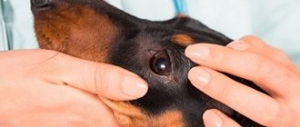
Adenoma of the third eyelid is difficult to confuse with any other disease. With this disease, the gland of the third eyelid enlarges and falls out from the inside of the eye. This formation looks like a red or bright pink mass, which is why people call this disease “cherry eye.” The gland can reach up to 15 mm in diameter, and may periodically disappear and reappear. If a dog has already developed a similar formation in one eye, it is possible that the second eye will soon be affected. Moreover, a mucous clear discharge may also be observed in the dog's eye. Often adenoma occurs together with conjunctivitis.
Cherry eye treatment
Cherry eye itself should not be a concern for pet owners, but there are a number of secondary problems that it can create that should be considered. The longer cherry eye remains in place, the higher the chance of more serious complications occurring, and this is the reason why treatment should be sought as early as possible.
Problems solved by timely treatment:
- Returning the lacrimal gland to its rightful place and restoring the third eyelid in the process;
- Minimize the likelihood of discharge from the affected eye;
- Protect your eyes from injury;
- Prevent irritation and dry eyes, which cause discomfort for the dog;
- Allows the lacrimal gland to resume its function of maintaining eye moisture.
Treatment options for ridding your dog of cherry eye have evolved over the years and range from using gentle topical products to invasive procedures such as surgery. There was a time in the past when the only treatment was to remove the tear gland, causing the eye to become permanently dry. This method was then abandoned because it not only removed moisture from the eye, but also made the organ susceptible to infection, especially variants of conjunctivitis. As a result, the dog had to be taken to the vet many more times to be treated for dry eye and infection.
Gradually, more treatment options have emerged, which are discussed as follows:
- Current products . They need to be applied to the surrounding area, where their purpose is to reduce swelling and inflammation. At the same time, they are also designed to protect the eye from secondary infections that may occur if the lacrimal gland is exposed. However, topical medications are only intended to provide temporary relief and do not cure cherry eye. They simply serve as short-term pain relief until a more specific treatment plan can be made.
- Operation . Instead of removing the tear gland and subjecting the dog to a lifetime of desiccation, experts recommend surgical repositioning, which returns the eyelid to its original position through a small procedure. There are several methods that can be used to achieve this result, and the choice of method depends on the ease of implementation and the comfort of the dog, as well as its owner.
A dog owner should be aware that choosing a surgical procedure is by no means a guarantee that cherry eye will not recur in either eye in the future. Since there are no preventative measures, you will need to treat your dog every time this occurs.
Cherry eye
A few days ago I got an eight week old Chihuahua puppy. On the first day we noticed that his eye was a little watery, but we didn’t think anything of it. We took him to the vet the next day because something red was coming out of his eye! The vet gave us ointment and pushed the red thing back, but it popped out again two days later, just as the vet expected. She suggested piercing the cherry eye or removing it. Could you talk a little more about these two treatment options for cherry eye? I've heard that piercing doesn't always work and is expensive. Removal may lead to other eye problems, including permanent blurring of the eye. What would you suggest? Thanks for the help! Amber Holmes
Dear Amber. Cherry eye or third eyelid gland prolapse is very common in small dog breeds. Prolapse of the gland itself does not cause discomfort or damage the eyes, so treatment can mainly be classified as cosmetic procedures. Most people choose to treat cherry eye as it looks quite unattractive. If the gland does not return to its normal position with steroid ointment, then the only treatment option is surgery. Most of the eye tears are produced by this gland, so its removal leads to dryness of the eye and blurred vision. If this happens, you can control eye hydration with medications, but it is still better to prevent the disease. The best method of surgery is a technique in which the gland is inserted into the mucous membrane of the eye. The only risk with this method is rubbing the cornea with a small part of the suture (which can be easily corrected by removing the interfering suture); re-popping of the cherry eye is very rare. The technique of attaching the gland to the eye socket can be a botched operation, so I don't often recommend it. At this time, due to potential problems in the future, I advise removing the gland if its attachment has failed and it is actually interfering with the eye. In the last 16 years of my practice, I have removed the gland only once and have had wonderful results using the gland insertion technique. About Dr. Sheri Weaver
Dr. Sheri Weaver
Dr. Weaver graduated with honors from the University of Georgia Veterinary Medicine. She founded a place of excellence, an animal hospital that teaches children how to care for animals, and devotes time and resources to participating in rescue organizations.
Cherry Eye.
Don't lose us, subscribe to the VKontakte page vk.com/26dogs.
Forecast
Regarding the surgical procedure, the prognosis is very positive, considering that after the operation the eye returned to its normal state. While removal of the gland entails the use of eye drops on your dog for the rest of his life, the repositioning will ensure that the lacrimal gland resumes its function of lubricating the eye as it once did.
After surgery, you must ensure that your dog is properly rested to recover, and he will need to wear an Elizabethan collar throughout the recovery phase to protect him from scratches and friction.
Once the cherry eye has been treated, now is the time for you as a pet owner to start monitoring the other eye to ensure the same problem does not occur. Sometimes pet owners choose to have surgery on both eyes at the same time as a preventative measure, but this is the exception rather than the rule.
Despite the surgery, there is a 20% chance that the gland will slip out and stick to your dog's face again. When this happens, everything returns to normal for you, as well as for your beloved dog.
Author of the article : Olivia Williams.
Eye diseases in dogs and their treatment
Any disease of the organs of vision occurs with symptoms characteristic of the pathology. The task of the animal owner is to promptly pay attention to the signs that appear and begin treatment in order to prevent the disease from becoming severe.
Barley
Barley on the eye is a painful pathology that occurs as a result of pathogenic bacteria entering the eyelash follicle or the pores of the sebaceous glands of the eye. A round white lump appears on the animal's eyelid.
A dog's stye on the eye can be located both on the outside and on the inside of the eyelid. Symptoms of the inflammatory process are swelling of the organ of vision and a feeling of soreness in the eye. Microorganisms that get into the follicle or sebaceous pore multiply very quickly, leading to an increase in the size of the white tubercle and its further opening.
Anti-inflammatory and antimicrobial ointments (Hydrocortisone, Erythromycin or Tetracycline) are used as therapy for barley. It is also necessary to use eye drops for dogs containing an antibiotic (for example, Levomycetin). Both eyes need to be treated at once.
Leukoma
The light cloudy film covering the pupil of the eye is absolutely painless, but causes discomfort to the animal.
An eyesore in a dog can appear as a result of an infectious or fungal disease. For example, a disease such as lichen sometimes causes a cloudy pupil. Leukoma causes your pet to constantly squint in bright light and hide in dark corners.
The eye may water, clear discharge appears, and sometimes it becomes purulent. The cornea of the organ acquires a yellow tint and a rough surface. With a sore eye, the dog sees absolutely nothing.
Treatment of leukoma involves the use of antimicrobial agents (drops, ointments), as well as the use of drops that relieve inflammation from the eyes (Tobrex, Taufon) and vitamin preparations for tissue regeneration. Very rarely, veterinarians succeed in completely getting rid of the cataract, but it is quite possible to improve vision.