Types of salivary glands and their functions in the body
But before we describe the essence of the disease, we need to list the types of salivary glands and their functions in the body:
- Small glands. They are located in the subcutaneous tissue near the lips and cheeks, as well as on the palate, tongue, floor of the mouth (there are especially many of them under the tongue).
- Large glands. They are located in the subcutaneous tissue near the ears, eyes, as well as under the tongue and in the lower jaw.
But much more important is the functional classification of the salivary glands:
- Serous. The saliva they secrete contains a lot of protein.
- Mucous. As is easy to understand, in their case the secreted secretion is enriched with mucus.
- Glands of mixed type.
The function of all glands is very important. They secrete saliva, which moistens the food and begins the initial process of fermentation. If there is little saliva, the animal will develop gastritis very quickly, and it will certainly suffer from recurrent diarrhea and/or constipation.
Finally, we note that inflammation of these organs is called “sialoadenitis” (more precisely, the main, basic type of the disease).
Home Remedies for Mumps in Dogs
Some remedies that can provide some relief to our dog's condition are applying cooled wipes to the area with or without substances with anti-inflammatory properties such as aloe vera or chamomile. Another remedy that can provide some relief from pain and inflammation due to its anti-inflammatory properties is a paste of fresh ginger root placed directly on the inflamed area.
While these remedies can be an excellent addition to veterinary treatment, we insist that it is very important to see a professional to diagnose and treat the disease.
Author of the article : Laura Garcia Ortiz . I am a veterinary graduate who graduated in 2021 and have been living with a wonderful 6 year old European cat ever since. I later completed my master's degree in feline medicine as I have always loved animals.
Bibliography
- V. Ausina, S. Moreno. (2005). SEIMC Treatise on Infectious Diseases and Clinical Microbiology. Panamericana Medical Publishing.
- Mumps in dogs. Available at: https://perritoshc.mx/index.php/k2-extra-field-groups/perritos-y-mascotas/item/5589-paperas-en-perros
- Autonomous University of Barcelona. Diseases of the salivary glands. Available at: https://ddd.uab.cat/pub/llibres/1914-1930/60248/patterespanidom_a1914-1930t2f1r1x2.pdf.
Reasons for the development of inflammatory processes
Here are the main reasons for the development of inflammatory processes:
- Injuries to the mouth or head area. This happens when eating bones, as well as in fights. A separate type of injury is iatrogenic injuries to the salivary glands, which a dog can receive during certain medical or diagnostic veterinary procedures.
- Dogs on walks often pick up and chew all sorts of garbage. The ingress of foreign bodies from such “goodies” is another reason for the development of inflammatory processes in the salivary glands.
- Many bacterial, viral, and other diseases of the oral cavity (infection can easily rise through the salivary ducts to the glands themselves).
- Fungal pathologies. They are often the cause of chronic inflammatory processes.
- The formation of stones (calculi) in the salivary gland itself.
- Autoimmune diseases , when the body begins to attack and destroy itself.
- Tumors of a malignant nature.
- Idiopathic inflammation , the cause of which remains unknown.
Interesting! Veterinary statistics made it possible to identify breed predisposition. Most often affected are German shepherds, many varieties of poodles, some terriers, as well as boxers and dachshunds.
Types of inflammation of the salivary glands
In general, the types of inflammation of the salivary glands are quite diverse, but in practice there are only two types. They are the most common and typical, and therefore we will talk about them.
Sialadenitis
As we wrote above, sialadenitis is a classic type of inflammation of the salivary glands. Most often it differs in the nonspecific nature of its occurrence, i.e. appears against the background of injuries, blood parasites and systemic infectious diseases. Often accompanied by severe swelling and suppuration.
The disease is quite dangerous, since if left untreated, the pathological process almost always leads to organ necrosis.
Mumps
This pathology is much less common (compared to the previous version). In humans, by the way, mumps is called “mumps.” The disease is of viral origin, the pathogens come from the paramyxovirus family. As a rule, this pathology affects not only the salivary glands, but also other glandular organs (testes, mammary glands, endocrine glands). It occurs quite easily; in many cases, the presence of the disease is indicated only by increased salivation.
Mumps (mumps)
The incubation period lasts from 11 to 23 days (usually 15-19 days). In some patients, 1-2 days before the development of the typical picture of the disease, prodromal phenomena , manifested by chilling, headache, pain in muscles and joints, dry mouth, and unpleasant sensations in the area of the parotid salivary glands. More often, the disease begins acutely with chills and an increase in body temperature from low-grade to high; fever persists for no more than 1 week. However, there are often cases of the disease occurring with normal body temperature. Fever is accompanied by headache, general weakness, malaise, and insomnia. The main manifestation of mumps is inflammation of the parotid, and possibly also the submandibular and sublingual salivary glands. In the projection of these glands, a swelling appears, painful on palpation (more in the center), having a dough-like consistency. With a pronounced increase in the parotid salivary gland, the patient's face takes on a pear-shaped shape, and the earlobe on the affected side rises. The skin in the area of swelling is stretched, shiny, difficult to fold, and its color is usually unchanged. More often the process is bilateral, involving the parotid gland on the opposite side after 1-2 days, but unilateral lesions are also possible. The patient is bothered by a feeling of tension and pain in the parotid region, especially at night; When the tumor compresses the Eustachian tube, noise and pain in the ears may occur. When pressing behind the earlobe, severe pain appears (Filatov's symptom). This symptom is the most important and early sign of mumps. The mucous membrane around the opening of the Stenon's duct is hyperemic and edematous (Mursu's symptom); Hyperemia of the pharynx is often noted. In some cases, the patient cannot chew food due to pain, and in even more severe cases, functional trismus of the masticatory muscles develops. Decreased salivation, dry mouth, and decreased hearing are possible. The pain continues for 3-4 days, sometimes radiating to the ear or neck, and gradually subsides by the end of the week. Around this time or a few days later, swelling in the projection of the salivary glands disappears. With mumps, regional lymphadenopathy is usually not noted.
In adults, the prodromal period is observed more often and is characterized by more pronounced clinical manifestations. In addition to general toxicity, catarrhal and dyspeptic symptoms are possible during this period. The acute phase of the disease is usually more severe. Much more often than in children, lesions (possibly isolated) of the submandibular and sublingual salivary glands are observed. With submaxillitis, the salivary gland has a doughy consistency and is slightly painful, elongated along the lower jaw, which is recognized when the head is tilted back and to the side. Swelling of the subcutaneous tissue around the gland sometimes spreads to the neck. Sublinguitis is manifested by swelling in the chin area of the same nature, pain under the tongue, especially when protruding it, local hyperemia and swelling of the mucous membrane. Swelling in the projection of the salivary glands in adults persists longer (2 weeks or more).
Mumps can occur in various clinical forms, which is especially important when diagnosing this disease. There is no generally accepted classification of clinical forms of mumps. A number of authors (S.D. Nosov, N.I. Nisevich, etc.) proposed classifications of the disease, but they had significant drawbacks and did not find wide practical application. The classification of V.N. Remorov was more successful.
Classification of clinical forms of mumps . A. Manifest forms: 1. Uncomplicated: damage to only the salivary glands, one or more. 2. Complicated: damage to the salivary glands and other organs (meningitis, meningoencephalitis, pancreatitis, orchitis, mastitis, myocarditis, arthritis, nephritis). According to the severity of the course: - mild (including erased and atypical); - medium-heavy; - heavy. B. Inapparent form of infection. B. Residual phenomena of mumps: - testicular atrophy; - infertility; - diabetes; - deafness; - dysfunction of the central nervous system.
In the classification of manifest forms of mumps, two criteria are used: the presence or absence of complications and the severity of the disease. Next, the possibility of an inapparent (asymptomatic) course of infection is indicated and, for the first time, residual phenomena are identified in the classification, which persist for a long time (usually lifelong) after the elimination of the mumps virus from the patient’s body. The need for this section is determined by the severity of the consequences of mumps (infertility, deafness, etc.), which practitioners often forget about.
Uncomplicated forms include those cases of the disease when only the salivary glands (one or more) are affected. In complicated forms, damage to the salivary glands is also an obligatory component of the clinical picture, but in addition, damage to other organs develops, most often the glands (genital, pancreas, mammary, etc.), as well as the nervous system (meningitis, encephalitis, Guillain-Barré syndrome) , myocardium, joints, kidneys.
Criteria for the severity of mumps are related to the severity of fever, signs of intoxication, as well as the presence or absence of complications. Uncomplicated mumps is usually mild, less often it is of moderate severity, and in severe forms there are always complications (often multiple).
Mild forms of mumps include diseases that occur with low-grade body temperature, with the absence or mild signs of intoxication, without complications.
Moderate forms of mumps are characterized by febrile temperature (38-39.9°C), prolonged fever and severe symptoms of general intoxication (chills, headache, arthralgia and myalgia), significant enlargement of the salivary glands, more often - bilateral parotitis, and the presence of complications.
Severe forms of mumps are characterized by high body temperature (40°C and above), a prolonged increase (up to 2 weeks or more), pronounced signs of general intoxication: asthenization, severe weakness, tachycardia, decreased blood pressure, sleep disturbance, anorexia, etc. Mumps is almost always bilateral, complications are usually multiple. Toxicosis and fever occur in the form of waves, each new wave is associated with the appearance of another complication. Sometimes a severe course is not observed from the first days of the disease.
Complications of mumps . With mumps, complications most often manifest themselves in damage to the glandular organs and the central nervous system. In children with diseases, one of the common complications is serous meningitis. The incidence of this complication exceeds 10%. Mumps meningitis accounts for about 80% of all serous meningitis in children. Meningitis develops 3 times more often in men than in women. As a rule, symptoms of damage to the nervous system appear after inflammation of the salivary glands, but simultaneous damage to the salivary glands and nervous system is also possible (in 25-30%). In 10% of patients, meningitis develops before inflammation of the salivary glands, and in some patients with mumps, meningeal symptoms are not accompanied by pronounced changes in the salivary glands (probably, by the time meningitis develops, mild changes in the salivary glands have already passed). Meningitis begins acutely, often violently (usually on the 4th-7th day of illness): chills appear, body temperature rises again (up to 39°C and above), severe headache, vomiting, and soon severe meningeal syndrome (stiff neck) develops , Kernig's, Brudzinski's symptoms). The cerebrospinal fluid is transparent, flows out under pressure, the protein content increases to 2.5 g/l, cytosis up to 1000 in 1 μl, the content of chlorides and sugar is usually not changed, sometimes a fibrin film may fall out. Symptoms of meningitis and fever disappear after 10-12 days, sanitation of the cerebrospinal fluid occurs slowly (up to 1.5-2 months).
Some patients, in addition to meningeal symptoms, develop signs of encephalitis (meningoencephalitis) or encephalomyelitis. Patients experience impaired consciousness, lethargy, drowsiness, uneven tendon and periosteal reflexes, paresis of the facial nerve, sluggish pupillary reflexes, pyramidal signs, hemiparesis.
Orchitis is more often observed in adults. Their frequency depends on the severity of the disease (in moderate and severe forms of orchitis occurs in approximately half of the patients). Signs of orchitis are observed on the 5-7th day from the onset of the disease and are characterized by a new wave of fever (up to 39-40 ° C), the appearance of severe pain in the scrotum and testicle, sometimes radiating to the lower abdomen. The testicle enlarges, reaching the size of a goose egg. Fever lasts 3-7 days, testicular enlargement lasts 5-8 days. Then the pain goes away, and the testicle gradually decreases in size. Later (after 1-2 months), signs of testicular atrophy may appear, which are observed in 50% of patients who have had orchitis (if corticosteroids were not prescribed at the beginning of the development of the complication). In case of mumps orchitis, pulmonary infarction was observed as a rare complication as a consequence of thrombosis of the veins of the prostate and pelvic organs. An even more rare, but extremely unpleasant complication of mumps orchitis is priapism (prolonged painful erection of the penis with filling of the cavernous bodies with blood, not associated with sexual arousal).
Acute pancreatitis develops on the 4-7th day of illness. Sharp pain in the epigastric region, nausea, repeated vomiting, fever appear; upon examination, some patients experience abdominal muscle tension and symptoms of peritoneal irritation. An increase in urine amylase activity is characteristic, which persists for up to a month, while other symptoms of pancreatitis are observed within 7-10 days.
Damage to the hearing organ sometimes leads to complete deafness. The first sign is the appearance of noise and ringing in the ears. Labyrinthitis is indicated by dizziness, vomiting, and lack of coordination of movements. Typically, deafness is one-sided (on the side of the salivary gland affected). During the period of convalescence, hearing is not restored.
Arthritis develops in approximately 0.5% of patients, more often in adults, and in men more often than in women. They are observed for the first time 1-2 weeks after damage to the salivary glands, although they may appear before the glands change. Large joints (wrist, elbow, shoulder, knee and ankle) are most often affected. The joints become swollen, painful, and serous effusion may appear in them. The duration of arthritis is usually 1-2 weeks; in some patients, symptoms of arthritis persist for up to 1-3 months.
It has now been established that the mumps virus in pregnant women can cause damage to the fetus. In particular, children experience a peculiar change in the heart - the so-called primary myocardial fibroelastosis.
Other complications (prostatitis, oophoritis, mastitis, thyroiditis, bartonylitis, nephritis, myocarditis, thrombocytopenic purpura) are rare.
Acute and chronic forms of inflammation
In addition to the previous classification, the disease can be divided according to the nature of the inflammatory process. There are acute and chronic forms of inflammation, differing in the course and manifestation of clinical signs:
- The main distinguishing feature of the acute form is its rapid, sudden development. In this case, the general condition of the dog suffers, the animal may refuse to eat and even drink; upon visual examination, one may notice swelling, purulent exudate, etc.
- The chronic form proceeds secretly. During periods of relapse, you can see signs characteristic of the acute type, but in a more moderate and smoothed version. During remission, the animal appears completely healthy. This course is typical for incompletely cured fungal or bacterial infections.
Symptoms
Clinical manifestations are characteristic and easily recognized:
- profuse drooling;
- the dog has difficulty swallowing;
- the pet refuses to eat;
- a swelling forms above the inflamed gland;
- the dog moves away when touched;
- the inflamed surface is hot;
- if suppuration develops, the dog suffers from pain and cannot sleep;
- the mouth is constantly slightly open, there is an unpleasant smell from it;
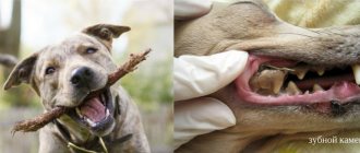
- tooth enamel darkens;
- plaque appears and tartar forms;
- gingivitis develops, which leads to tooth loss;
- the dog loses weight, the coat loses its metallic shine.
Symptoms and first signs of inflammation
The following symptoms and first signs of inflammation are distinguished:
- A sharp increase in salivation. The dog drools constantly and so intensely that all the fur in the mouth and even the chest becomes wet. In long-haired pets, it can turn into unpleasant-looking tangles and icicles. In these same cases, the dog often swallows, and involuntary swallowing movements may remain after recovery.
- In more severe cases, a small amount of blood may be released from the mouth.
- There are problems with swallowing food.
- Frequent swallowing movements contribute to irritation of the vomiting center, causing the pet to vomit from time to time or have involuntary vomiting spasms.
- While eating, the dog may moan and whine. But not out of pleasure, but because it hurts him to eat.
- The animal strongly resists attempts to open its mouth slightly, as this also gives the dog unpleasant sensations.
- The dog completely or partially loses his appetite.
- Due to lack of interest in food, progressive exhaustion develops.
- Development of fever of a constant or intermittent type.
- Swellings filled with watery fluid or ulcers appear under the skin in the head area and in the oral cavity itself.
- Neurological symptoms. This, in particular, includes protrusion of the eyeball and increased lacrimation.
Methods for treating inflammation of the salivary glands in dogs
In veterinary medicine, there are the following methods for treating inflammation of the salivary glands in dogs:
- Medication.
- Surgical.
First, let's look at the first type (as the most common and not very effective):
- The pet is prescribed broad-spectrum antibiotics.
- So that the dog can eat normally without suffering from pain, he is given painkillers and sedatives.
- The inflammatory process is stopped using anti-inflammatory corticosteroids.
Accordingly, surgical treatment involves a more radical approach to therapy:
- The salivary ducts are “pierced”, restoring their patency and capacity.
- When there is advanced purulent inflammation and/or necrosis, the affected gland is completely excised.
- If the veterinary surgeon is experienced and highly qualified, it is possible to restore damaged gland ducts.
- If there is a risk of further spread of the infection, the damaged gland(s) are excised and its ducts are ligated (ligated).
- In case of cystic lesions of the glands, the cysts are opened and the damaged tissue is excised. Sometimes the gland can be saved (if the gland is small, there is no need to mess with it - removal of the organ is preferable).
Important! If the disease is of a tumor nature, it is preferable to combine surgical and drug treatment methods. First, the tumor is excised and then “finished off” with chemotherapy.
Symptoms of mumps in dogs
The first localization of the mumps virus is the parotid glands, causing their painful swelling with an increase in area, giving them the characteristic appearance of mumps. The affected dog exhibits the following clinical signs:
- Inflammation of the parotid glands is more or less apparent;
- Redness and/or pus in the gland;
- Hardening of the glands due to an increase in connective tissue;
- Heat;
- Pain;
- Anorexia;
- Decay;
- Lethargy;
- Weight loss.
Depending on the severity of the process, inflammation of the submandibular glands can be prolonged and even affect the facial nerve, causing facial paralysis. If you notice any symptoms of mumps in your dog, you should definitely contact your veterinarian.
List of drugs and antibiotics
Here is a short list of drugs and antibiotics used to treat inflammation of the salivary glands:
- Antibiotics from the cephalosporin group (Ceftriaxone, Cefazolin).
- Itraconazole, miconazole, terbinafine, and antibiotics from the griseofulvin group are prescribed for the treatment of fungal inflammation.
- Dexamethasone is a standard representative of corticosteroids.
- Analgin can be used for pain relief, and barbiturates can be used as sedatives (only as prescribed by a veterinarian and only those that are approved for veterinary use).
Dog feeding diet
There is no need to somehow balance the diet of a dog with this disease, the only problem is the consistency of the food:
- For the first five days, the dog will have to be fed cereals (buckwheat, boiled rice), boiled to a creamy consistency (sort of risotto).
- In order for the dog to eat porridge, meat broth or baby meat puree is added to it.
- You can also use high-quality dry food as a feed, after soaking it in the same meat or even vegetable broth.
In general, there are no other feeding features. If the dog’s condition is stable and one or two small glands are affected, you can feed the pet exactly the same as before the illness.
Caring for an animal at home
Before visiting a specialized institution, the pet should be placed in a warm and dry room, without drafts. After a diagnosis has been made and treatment has been prescribed, it is important for the owner to follow the specialist’s recommendations. The sick pet is given complete rest. Due to the fact that eating is difficult, concentrated feeds are completely excluded from the diet. Feed the dog warm food with a semi-liquid consistency. It is useful to give a weakened animal meat broth.
Physiotherapy will help speed up your pet’s recovery. On the recommendation of a veterinary specialist, the owner can independently apply alcohol compresses and heat the inflamed area using a Minin lamp.
When an abscess forms, the attending physician also prescribes anti-inflammatory ointments, for example, Ichthyol, Vishnevsky. The owner’s task is to regularly apply ointment to the affected area.
For information on the use of Minin's blue lamp, watch this video: