How does a dog become infected?
Females are viviparous and produce larvae that accumulate. They can be detected in dirofilaria immitis in the evening, and in dirofilaria repens at night, at which time mosquito activity is highest.
The larvae, which enter the mosquito with blood, go into its Malpighian vessels, where they mature to become invasive. After this, they move to the head and gather around the mouthparts.
After a mosquito bite, heartworms end up in the blood, where they then find their localization.
Etiology
The causative agent is the roundworm Dirofilaria immitis. A sexually mature individual is localized in the right ventricle of the animal’s heart. As their numbers increase, parasites occupy the space of the atrium and pulmonary artery.
Dirofilaria viviparous worm. Microfilariae larvae are released by sexually mature individuals directly into the bloodstream. The larva is microscopic, the adult reaches 25-30 mm. They migrate with the bloodstream, being carried into various organs, and can be transmitted intrauterinely.
Microfilariae with a drop of blood are swallowed by mosquitoes during bites. The larva remains in the insect's body for 15-17 days and is transferred to the lower jaw. By biting another dog, the intermediate host, a mosquito, infects it. At the same time, the tendency of infection of short-haired dogs is several orders of magnitude higher than that of long-haired dogs. Once in the subcutaneous tissue, connective and adipose tissue, it sheds repeatedly over 2.5-3 months and then penetrates the bloodstream. Having reached the cardiac cavities with the blood flow, it stops there and after 3-4 months reaches the sexually mature stage. The entire development cycle in the body of a permanent host takes place in 7-8 months. One individual can secrete up to 30 thousand microfilariae per day. The duration of sexually mature forms is up to 3 years.
Dirofilariasis in dogs symptoms
The first - subclinical form has nonspecific symptoms. There is a fickle appetite. From the nervous system it manifests itself as convulsions, collapses and cuts.
The animal gets tired quickly.
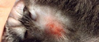
The second form is cutaneous. The skin of the extremities, back and head is affected. At the first stage - hair loss, the second stage - the skin turns red, pustules with pus appear, at the final stage - ulcerations.
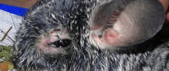
A typical symptom is resistance to antibacterial and anti-inflammatory drugs. The third form of the disease is pseudotumor.
It is expressed by tumor-like growths on the skin surface of the thighs, back, metatarsals and mammary glands. Ulcers appear in which serous contents cover the surface of the skin.
The fourth is cardiopulmonary. It is characterized by renal failure, anemia of the mucous membranes, prostate cyst, enlarged spleen, liver and ascites.
The dog becomes weak and short of breath. Electrocardiography shows hypertrophy of the ventricle and right atrium.
Symptoms
Initially, the pathology manifests itself with uncharacteristic symptoms. Fatigue, mild depression, inability to perform heavy physical activity, shortness of breath. Further, a gradual decrease in body weight and the manifestation of cough after physical activity, cyanosis of the mucous membranes are visible.
On auscultation, heart murmurs are clearly audible.
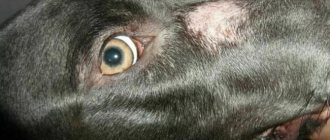
Being in the subcutaneous tissue, the parasite causes itching. As a rule, such places are parts of the body that are not covered with hair, the interdigital space on the limbs. The skin takes on a red-burgundy hue. The dog licks and chews the itchy part, which causes sores and dermatitis.
In cats, dirofilariasis is manifested by attacks of vomiting and breathing difficulties, as well as atypical manifestations.
The prognosis of the disease is favorable with timely, qualified treatment.
Diagnosis of dirofilariasis in dogs
Treatment of dogs infected with d.repens ends successfully almost every time. For the treatment of animals whose body was attacked by d.immitis, with moderate severity, the prediction of a favorable result is low, and in severe forms, it is doubtful.
Before starting treatment with macrofilaricidal therapy, it is necessary to make a prognosis for the animal’s life. It may happen that death occurs much sooner during treatment than without treatment.
Because certain drugs can provoke a strong accumulation of microfilariae in the vascular bed, and vascular embolism occurs (strokes, necrosis, paresis).
The most important thing in concluding an intravital diagnosis is the detection of microdirofilaria in the blood. Most of them are found in dogs affected by d. immitis, can be detected in the evening.
In case of defeat d. repens - at night. To diagnose an animal during life, scientists have proposed many methods. For example, the Knott method is often used in practice.
It consists in the fact that in order to find out whether a dog is sick, they take a test for dirofilariasis from its venous blood and dilute it with formalin and mix it.
The remaining sediment is combined with methylene blue, and within 5 minutes the sediment should become colored, after which it can be examined for the presence of microfilariae.
In many countries around the world, kits of reagents are produced for the detection of heartworm antigens and serological diagnostics in an enzyme-linked immunosorbent test.
After the death of the animal, diagnosis is made through a pathological autopsy and identification of helminths and their localization.
How do you know if an animal has heartworm disease?
Diagnosis is carried out comprehensively: starting with collecting anamnesis, examination, clarifying any symptoms, taking blood for biochemical and general analysis - i.e. usual routine manipulations at appointments, but the most important contribution to the diagnosis comes from the laboratory, cardiac ultrasound and x-ray.
Suspicion may already be caused by the result of a general blood test, the form of which may show signs of anemia and a decrease in the number of platelets, as with babesiosis; On the leukocyte formula of this form, neutrophilia and pronounced eosinophilia are possible. Next, a blood smear is made for microscopy and microfilariae are detected (or not); The Knott test is done for the same purpose. Enzyme-linked immunosorbent and immunochromatographic tests are carried out to detect the D.immitis antigen - a negative result for the antigen in the presence of microfilariae in the blood may indicate that the animal has a cutaneous form of the disease. Antigen tests are repeated after 6-7 months. The most accurate method of research with species differentiation is the polymerase chain reaction (PCR), it allows you to detect the DNA of D.immitis and D.repens. Ultrasound of the heart (echocardiography) and x-rays are special research methods. On ultrasound it is possible to see the enlargement of the right side of the heart and adult specific D.immitis. On an x-ray you can see the expansion of the pulmonary artery and its branches, infiltrates and enlargement of the heart. Echocardiography and X-ray examination make it possible to differentiate the cardiopulmonary form of the disease from the cutaneous form. Diagnosis of the skin form of the disease, in addition to the above studies, also includes an external examination of the animal’s body: searching for nodules, alopecia, papules, and inflammation.
Treatment of dirofilariasis
Often owners who have learned about the diagnosis ask to euthanize the dog, although in many cases this should not be done. Although the treatment is very expensive, it can be done.
To destroy mature heartworms, medications containing arsenic or ivomec are usually used.
Treatment of dirofilariasis mainly involves the use of the following deworming drugs: brovanol-plus, levamisole, macrolide drugs, thiacetarsamide. Heartworm disease in dogs can now be treated with the effective but very expensive melarsomine.
Sometimes, due to the individual characteristics of the body, some dogs may experience toxicosis or other side effects.
For a speedy recovery, medications are prescribed that activate the reticuloendothelial system and the functioning of the red bone marrow.
In order to avoid heartworm disease, you need to know some precautions. The first priority should be to prevent dogs from coming into contact with blood-sucking insects.
During the warm season, animals are most vulnerable, so you should definitely buy an insectoacaricidal collar for your dog; it repels up to 98% of mosquitoes and prevents infection for up to six months.
Periodically, you need to do tests for the presence of helminths in the animal’s body and take preventive medications.
Treatment
Ivermectin is used for treatment. The drug has low toxicity for warm-blooded animals, and in the parasite it blocks the transmission of nerve impulses, which leads to paralysis and death. The instructions for the medication do not regulate its use in dogs, but practice shows its high effectiveness. The drug is administered once, subcutaneously, at a dose of 1 ml per 30 kg of live weight.
Mature parasites that die in the cardiac cavity undergo rapid lysis and are carried out of the heart through the bloodstream.
Ivermectin is the main active principle of the drug Dironet AVZ. Using tablets (1 per 10 kg of body weight) or suspension (1 ml per 10 kg of body weight), you can carry out prevention, starting from the moment mosquitoes fly, once a month. As soon as the flight of insects has stopped, the drug is stopped.
You can use the drugs “Advocate”, “Advantix” (Fizer).
During the period of therapeutic measures, animals are not subjected to physical activity and are provided with complete, easily digestible, balanced feeding.
Treatment and prevention
Approaches to the treatment of dirofilariasis in animals and humans vary greatly. When treating animals, preference is given to chemotherapy using melarsomin hydrochloride and doxycycline. Surgical treatment in animals is used extremely rarely.
When treating human dirofilariasis, preference is given to surgical treatment. In the cutaneous form, a skin incision is made above the node, followed by its enucleation along with the parasite. In the pulmonary form, accompanied by severe symptoms, thoracoscopy is performed with atypical resection of a section of the lung containing an inflammatory focus with a parasite in the center.
Clinical picture of the disease
As a rule, heartworm disease in cats occurs between the ages of 3 and 6 years. Pets and feral cats have the same chance of becoming infected. The clinical signs of this disease are characterized by nonspecificity.
As a rule, damage to the respiratory system is detected. Often, with dirofilariasis in cats, a cough is found that is similar to the cough of asthma. In addition, apathy, loss of appetite, vomiting, and neurological problems are noted. In some cases, sudden death occurs. It is worth noting that in this disease, damage to the cardiopulmonary system is rarely combined with neurological symptoms.
Laboratory diagnostics
Laboratory diagnosis of helminthiasis comes down to identifying microfilariae circulating in the blood and antigens (proteins) of the body of an adult worm.
- 1Examination of blood samples under a microscope.
- 2PCR (determination of parasite DNA in the blood/tissues of the body).
- 3ELISA (enzyme-linked immunosorbent assay), immunochromatographic analysis - available tests with high sensitivity and specificity, are used to detect Dirofilaria antigens circulating in the blood.
- 4Serological studies to determine antibodies against Dirofilaria.
- 5Biopsy of affected organs, microscopy, immunohistochemical examination of the obtained material.
If there are complaints from the lungs (fever, chest pain, cough), the patient may be prescribed a chest x-ray. In the pulmonary form, changes in the lung parenchyma can be detected (foci of shading with smooth contours, spherical, oval, homogeneous in structure), expansion of the pulmonary artery contour, pleurisy with pleural effusion. (5) For differential diagnosis with other lung diseases, the patient may be prescribed a computed tomography scan of the chest.
Diagnostics
To detect dirofilariasis in dogs, laboratory and hardware diagnostic methods are used. The veterinarian may prescribe the following for your dog:
- a blood test to determine the presence of microfilariae (parasite larvae);
- echocardiography, which in this disease reveals organic changes in the heart and/or signs of heart failure;
- immunological blood tests, which are carried out to exclude other diseases with similar symptoms.
A popular method for diagnosing dirofilariasis is a blood test for the presence of pathogens. It is based on the reaction of specific components of the test preparation to a protein secreted by the body of the female parasite. The reliability of such a rapid analysis is high; in 60-80% of cases it turns out to be positive, even if there is only one adult female worm in the dog’s body.
Human dirofilariasis
The likelihood of dirofilariasis developing in humans exists wherever this helminthiasis is recorded in dogs. The medical literature mentions 1,782 cases of human dirofilariasis, of which 1,410 are the subcutaneous/ocular form, 372 are the pulmonary form.
In Russia, 622 cases of the subcutaneous form and 3 cases of the ocular form of the disease have been registered. In the European Union, 586 cases of the subcutaneous form and 33 cases of the pulmonary form of the disease have been registered.
There are 50 cases reported in South America, most of them in Brazil. There have been 116 human cases of infection in the United States.
2.1. Pulmonary form
The pulmonary form of dirofilariasis in humans is characterized by the formation of nodules in the lung tissue (an inflammatory reaction of the body in response to the presence of the parasite).
Figure 3 - Pulmonary dirofilariasis (Human Pulmonary Dirofilariasis), source Medscape
When the parasite larvae reach the small branches of the pulmonary artery, they cause blockage of the lumen of the artery and lead to the development of inflammation around it. As a rule, during examination, single nodes in the lung tissue are recorded in the patient. In rare cases, multifocal damage to the pulmonary arteries is possible with the development of multiple pulmonary nodules.
Spherical or oval shadows with clear edges and uniform density are recorded on the radiograph. More often the lesions are located along the periphery of the lung. The pulmonary form of dirofilariasis is extremely rarely accompanied by any symptoms and is detected during a routine examination (fluorography of the chest organs). In rare cases, the disease may be accompanied by a cough, sometimes mixed with blood or purulent sputum, chest pain, general weakness, and muscle pain. (8)
2.2. Subcutaneous and ocular form
The subcutaneous form of dirofilariasis develops as a result of infection with D. repens, which leads to the formation of subcutaneous nodules (a mature worm surrounded by an inflammatory infiltrate and reactive proliferation of connective tissue).
In the formation of the inflammatory infiltrate, the body’s immune response to foreign proteins of the parasite plays an important role: immune cells produce cytokines, active substances that stimulate the influx of neutrophils and lymphocytes from the blood to the site of the parasite.
In turn, neutrophils produce growth factors that stimulate the growth of dense connective tissue around the parasite. Over time, under the influence of inflammatory cells, the parasite dies and only a nodule of inflammatory cells remains with a dead, calcified parasite in the center, surrounded by a dense fibrous capsule.
Subcutaneous nodules have a hard, elastic consistency and can increase in size over several weeks or months. Erythema (redness) develops on the skin in the area of subcutaneous nodules.
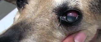
The incidence of detection of the ocular form of dirofilariasis has increased over the past decade. The ocular form is characterized by damage to the orbital zone, eyelids, conjunctiva and vitreous body.
As a rule, the ocular form is accompanied by the following symptoms: conjunctivitis, vision impairment, even loss, the appearance of “floating spots”, “spots” when looking at light objects. Complications are possible in the form of retinal detachment, glaucoma, opacification of the vitreous body and lens.
With orbital localization of the parasite, inflammation and swelling of the eyelids, ptosis (drooping of the upper eyelid), and discomfort in the eye area develop. (9)