Patella luxation - what is it?
To understand the nature of a dangerous disease, it is necessary to understand some concepts of veterinary terminology:
- “patella” translated from Latin means kneecap. It protects the joints of the femur and tibia from spontaneous displacement to the side, and the tendon from friction;
- Luxation is a shift of the kneecap. It is customary to distinguish between 2 types of pathology:
- lateral, with this type of defect the movement is carried out to the outer side of the thigh. If there is pathology on both hind limbs, you can visually notice an X-shaped curvature of the paws;
- The medial type of pathology is characterized by a shift of the patella to the inside of the paw, which causes a shift of the tibia and, as a result, osteochondritis.
Patella test
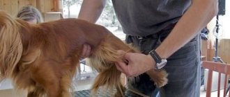
Patella luxation in dogs can occur as a consequence of injury or be a genetically inherited pathology. And, if the first option is force majeure, then the development of the second can be predicted. Important: animal owners who plan to breed the breed, in order to avoid the birth of sick animals, must conduct a special test for patella in breeding dogs.
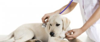
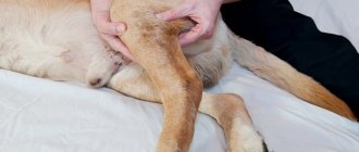
The expert test is performed by palpation. The purpose of the assessment is to ensure that the patella is stable. This diagnostic procedure must be performed by a veterinarian who has undergone special training and is authorized to perform this procedure.
The test can be done in our clinic. The result of the expert assessment is recorded in a special certificate. This is done in order to guarantee future owners of the offspring that there is no genetic mutation.
How does the disease manifest?
Small breed dogs, such as Spitz, toy terriers, Chihuahuas, and pugs, are most often susceptible to this dislocation. If not treated correctly, a dislocation can provoke severe degenerative changes in the pelvic bones. There are several degrees of the disease:
- First degree. With such an injury, the kneecap may well return to its usual position on its own;
- Second degree. When bent, the kneecap takes on an unnatural position;
- Third degree. The dislocation is very noticeable to the naked eye when the dog moves;
- Fourth degree. The cup is always in an unnatural position, the dog practically does not walk and experiences severe pain.
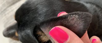
The symptoms are quite clear. It is clear that the joint is not in its normal position. The animal is painful to any touch or palpation, the area of the kneecap itself is hot and enlarged.
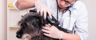
For diagnosis, visual inspection, palpation, ultrasound and x-ray examination are used.
Causes
Pathological movement of the patella in dogs can be due to a number of reasons:
- abnormal attachment of the patellar ligaments;
- injury during play, road accident, fall;
- deformations of bone tissue and joints as a result of any illness or old age;
- hip dysplasia;
- torsion of the lower leg;
- bending of the lower part of the hip bone or its congenital subluxation;
- displacement of the crest of the tibia.
Lameness in this disease can be intermittent, i.e. the animal limps, then stops, then the limping returns. The animal may not experience any pain.
Diagnosis of patella defect
Diagnosis of patella in dogs consists of identifying pathology and assessing the degree of luxation. A veterinarian can give an opinion after performing the following diagnostic procedures:
- carrying out visual observation of the animal’s gait;
- determining the degree of dislocation using special tests;
- X-ray examination of the limb in direct and lateral projection using radiographic equipment.
The listed diagnostic methods make it possible to differentiate this pathology from other diseases with similar symptoms.
Dogs may occasionally have joint problems. Among these types of diseases, luxation of the kneecap, which is called patella, stands out unpleasantly. The disease provokes significant problems in the activity of the animal’s musculoskeletal system, sometimes reaching the complete inability to move in severe cases of the disease.
Veterinary medicine divides patella dislocation into four degrees, based on the complexity of the disease. When a dog is diagnosed with the first degree, the kneecap comes out of its own groove during the animal's rest period, without putting stress on the sore paw. So the dog makes its first movements after waking up, limping on one of its hind legs, resting on three limbs. After a short period of time, the joint takes its usual position, and the dog walks as if everything is fine with it. In the second degree, most of the time the patella is located in the wrong position, the hip bone is turned at best by 30 degrees, there is a medial shift of the knee cup, the femoral joint moves off the axis to the outside. Such a disruption in the functioning of the hind paw causes the dog to not stand on the affected paw, and when the cup can be adjusted, a specific click is observed, which causes friction of the patella on the knee joint, which in the future can cause the final wear of the bone.
The third stage is characterized by regular displacement of the cup. In this case, the hip bone is turned out only 30 - 50 degrees, the hock joint leaves the vertical trajectory and is located to the right or left in relation to the knee joint. The groove containing the kneecap narrows slightly, becoming flat. The dog begins to limp, avoiding resting on the sore paw. At the fourth stage of dislocation of the patella, the entire hip bone becomes rotated in the internal direction and moves 50-90 degrees from the usual vertical state, the patella is located from the inside of the joint, the groove of the cup is covered with bone tissue, losing its own functions. At this degree of dislocation, the dog cannot lean on the affected paw at all.
Diagnosis of the disease
The dog owner is able to recognize the first signs of patella dislocation by looking at the asymmetrical state of the hind legs; often in this case, the pet, immediately after becoming upright, jumps only on three legs, after a few steps restoring the deformed joint. By feeling the knee at this time, you can feel its swelling and the release of the knee cup from its groove. Establishing a more detailed diagnosis requires macroscopic and microscopic analysis of the affected joints, as well as x-ray examination of the direct and lateral surfaces.
Patella treatment
Patella dislocation in dogs can be treated using conservative and surgical methods. When the disease is in the first or second degree, you can limit yourself only to general therapy. The most effective in this situation may be anti-inflammatory, chondroprotective agents, which help prevent future wear and tear of the knee joint. At the same time, it should be noted that the analgesic ingredients contained in these medications can show a false picture of the recovery of an animal that does not limp for a certain time. At the same time, the patella will continue to wear out all this time, so after using such products you should definitely consult a veterinarian. Some hope for relief can be brought by a diet that reduces the feeding of carbohydrates, which provoke inflammatory processes in the pet.
Stages three and four of patella in dogs require surgical intervention. There are two options for operations to restore the knee joint - trochleoplasty and trochlear chondroplasty. At the same time, surgery should be considered as a last resort, since post-operative life is very difficult for the dog - it needs to stay in a cage for several weeks or even months, avoiding any physical activity that could affect the recurrence of the disease.
0
Classification of patella dislocations
The classification of patellar displacement is determined by four degrees:
- I - the kneecap spontaneously shifts from time to time, and then returns to its original state. Most often, such a defect is caused by ligament weakness. The animal may limp slightly;
- II – the patella often dislocates during flexion and returns to its original position when the knee joint is extended. The dog tries not to stand on the sore limb, the toes are turned inward;
- III - the kneecap is constantly displaced, can be reduced, but immediately returns to its abnormal state;
- IV - the patella is always in a displaced position, attempts to straighten it do not yield results. All degrees of pathology are accompanied by lameness when walking. Therefore, if you experience symptoms of a displaced patella, you should not delay visiting the clinic. Only a surgeon will be able to make the correct diagnosis and prescribe therapy depending on the severity of a particular clinical case.
Classification of patella luxation in dogs
First of all, it is worth noting that dislocation of the patella, displacement of the patella, is possible in two directions:
- inside;
- out.
When the kneecap slips inward, it is called medial displacement, and outward, it is called lateral displacement. In addition to the directions of displacement, there is a classification based on the severity of the dislocation, characteristic disorders and symptoms. In total, this classification has 4 degrees:
- Intermittent dislocation of the kneecap.
- Frequent dislocation.
- Stable dislocation with possibility of reduction.
- Persistent dislocation with impossibility of reduction.
The first degree of patellar dislocation is characterized by the appearance of intermittent claudication. As a rule, lameness occurs after sleep or prolonged lying. The patella, which has shifted in a relaxed joint, does not immediately take its natural position, causing the dog to limp or move on three legs for a short time. This condition passes quickly enough, the patella returns to its anatomical boundaries, and manifestations of lameness disappear.
The second degree is expressed in constantly recurring, long-term displacement of the kneecap. Lameness in this case is almost permanent. Periodically, the dog, sparing the limb, does not step on it. This severity level is considered moderate. The animal can be helped by manual reduction of the patella, performed by turning the tibia outward. This reduction is usually performed under anesthesia.
This degree of dislocation allows the dog to move more or less normally, but prolonged displacement of the kneecap leads to its abrasion. The friction of the patella on the bone produces a crunching sound that occurs when trying to return the limb to its normal position. Sometimes a crunching sound is heard when walking.
Third degree LP. It is characterized by a constant anatomically incorrect location of the patella, a strong change in the position of the dog’s limbs, and a rotation of the hip bone. This degree of dislocation can still be corrected manually.
The fourth, most severe degree is characterized by severe eversion of the hip bone, its significant deviation from the vertical plane. Help with such a dislocation of the kneecap in a dog can only be provided through surgical intervention.
https://youtu.be/https://www.youtube.com/watch?v=y6SpCDuP1QY
_
Treatment
Treatment of patella luxation in dogs does not require emergency surgery. However, it is not recommended to postpone your visit to the clinic. The longer a four-legged pet limps, the lower the likelihood of complete rehabilitation after surgery.
Drug therapy provides only short-term beneficial effects. The main treatment method is surgical correction of the patellar defect. There are several types of surgical correction. In our clinic, surgeons use a combinatorial version of therapy. This is the optimal method that allows not only to eliminate the defect, but also to prevent relapse.
It is worth noting that modern veterinary surgery recognizes kneecap replacement as one of the most effective treatment methods. During the operation, the animal is implanted with implants from KYON (Switzerland). The surgeons of our veterinary center underwent theoretical and practical training from the authors of this technique in a Swiss clinic.
Need for surgery
Summarizing all of the above, when diagnosing patella displacement in dogs, we recommend surgery. The sooner it is done, the greater the chance of completely eliminating the defect. This guarantees an increase in the animal’s quality of life and the prevention of disability. The optimal age for surgical intervention in such a clinical situation is up to 1 year.
Our clinic is equipped with advanced diagnostic and surgical equipment. Our specialists have extensive experience in performing operations to eliminate patella luxation, and therefore guarantee a favorable outcome of surgical treatment if you visit the clinic in a timely manner.
The meaning of patella in dogs
The patella (patella, also known as the kneecap) is a small but extremely important bone. It is involved in the formation and functioning of the knee joint. Anatomically located on the front of the joint inside the ligament. Since the main distribution of weight falls on the hind limbs, nature has strengthened the knee joint with a kind of block - a small, movable bone, the patella.
In addition to functioning as a block, the patella prevents abrasion of the bones of the knee joint by directly sliding the patellar ligament along them. The normal location of the kneecap is in the grooves of the bones that form the joint. For the knee joint, and the entire limb as a whole, to function properly, the kneecap must occupy an anatomically correct position. With pathology, injury, or weakening of the ligamentous apparatus, the patella is capable of shifting and changing position relative to its natural one.
A change in the normal position of the patella in dogs, relative to the normal one, is called luxation, from the Latin luxatio - dislocation, displacement.
This pathology has been recorded in dogs of all ages - from puppies to older animals.
In large dogs, luxation of the patella usually occurs as a result of injury, twisting of the hind limbs, an unsuccessful jump, or a blow to the knee area. In small, decorative dogs, this disease is often hereditary. Animals that show LP (non-traumatic) are excluded from breeding. There is a breed predisposition of dogs to this pathology. Among the breeds with the most frequently reported cases of LP are:
- Pomeranian Spitz;
- Cocker Spaniel;
- Yorkshire Terrier;
- chow-chow;
- Shar Pei;
- Japanese chin.