One of the most common dermatological diseases is microsporia in dogs, popularly referred to as “ringworm”. The disease is considered highly contagious; not only pets, but also people can catch the disease from an affected dog.
Most often, very young and elderly family members with weak immunity become infected from a pet. Dermatological pathology takes a very long time and is difficult to treat; in puppies, an unpleasant illness without the timely administration of specific medications can result in death. If you notice the first signs of infection on your pet’s body, it is recommended to contact a veterinary clinic as soon as possible.
What is microsporia in dogs
To get a complete picture of skin pathology, it is necessary to understand the question of what microsporia is in dogs. Microsporia is a dermatological disease caused by a microscopic pathogenic fungus of the genus Microsporum. Most often, dogs, cats, rabbits, chinchillas and humans are affected by a fungus of the species M. Gypseum. Pathogenic microsporia form a huge number of spores that retain their vital activity in the external environment for quite a long time. Thanks to this feature, the pathology is widespread throughout the world.
Dogs become infected through contact with a sick animal, person or care items.
The source of the pathogen can be skin flakes or hair with spores. In the natural environment, pathogenic fungi live on mice, rats and stray animals. Even a lap dog can catch the disease from humans or other pets. The pathology occurs at any time of the year and is observed in all breeds of dogs.
When microsporia enter the body of a pet, they penetrate into the inner layers of the skin, destroying hair follicles. The proliferation of a pathogenic fungus in the affected area is accompanied by the production of toxins that cause local damage and the appearance of hairless, round areas with a clear boundary.
In the absence of treatment, multiple inflammatory foci form, a secondary infection occurs, and pyoderma and sepsis develop.
Etiology
The causative agent of the disease is a microscopic fungus of the genus Microsporum. In horses - M. eguinum, in other animals the pathogen can be M. lanosum, M. canis, M. pelineum, M. gypseum, M. nanum.
Under a microscope, microsporiums can be seen inside the hair, and the spores form a sheath on the root of the hair. The fungus is cultivated on Sabouraud's medium, wort agar at 26-28 ᵒC. Growth is read 6-7 days after sowing. It looks like a folded gray-yellow coating with airy, fluffy gray-white mycelium. Microscopy of a pure culture reveals single, pear-shaped, spindle-shaped, oval microconidia.
In the external environment, the pathogen exhibits high resistance. In hair and scrapings, the fungus retains its pathogenicity for up to 5 years, and in pus for up to 8 months. Resistant to freezing. When boiled, it dies within a few minutes; when heated to 80ᵒC, it dies after 2 hours; when treated with dry steam, it is inactivated after 30 minutes. Solutions of formaldehyde are inactivated in 15 minutes, caustic alkali 5% in 20 minutes.
Young animals are most susceptible to the pathogen. The source is sick and recovered animals, as well as contaminated environmental items. Sick stray animals are especially dangerous; rodents also carry this disease.
In large cities, outbreaks appear in the autumn, cats get sick most often.
The pathogen settles on the epidermis, disrupts tissue trophism and causes an inflammatory process with a corresponding clinical picture.
Differences between microsporia and trichophytosis
Ringworm is a collective name for several dermatological diseases; most often in dogs this term refers to microsporia and trichophytosis. Pathologies have specific treatment, so it is extremely important to correctly determine the type of disease.
Differences between microsporia and trichophytosis:
- Both pathologies are caused by pathogenic fungi. The causative agent of microsporia belongs to the genus Micrisporum, and trichophytosis - Trichophyton. It is possible to identify species differences between fungi only when they are grown on nutrient media.
- With ringworm, clearly defined alopecias are formed with short, broken hair, as if it had been cut. With microsporia, the hair shafts are longer than with trichophytosis.
- Microsporia in dogs is diagnosed in 90% of cases, trichophytosis accounts for the remaining 10%.
- To identify the type of pathogen, examination using a Wood's lamp is used . When exposed to luminescent irradiation, microspores glow with emerald light. Trichophytosis cannot be detected by this diagnostic method.
Symptoms of the disease
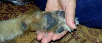
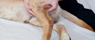
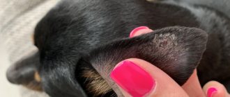
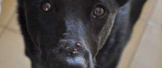
Microsporia in dogs can manifest itself in quite a variety of ways. Basically, the lesions are localized at the base of the tail, on the limbs, head near the ears and are round spots of irregular shape. The fungus can even affect a dog's toes.
Skin affected by the fungus begins to redden and thicken . The coat suddenly loses its healthy appearance, and its hairs seem to stick together. Severe itching occurs, the dog begins to scratch the sore spot, and as a result, the disease spreads to other parts of the body.
Microsporia often occurs in animals that have:
- Hormonal imbalance.
- Improper metabolism.
- Lack of vitamins A and C.
Microsporia can occur in different forms:
- Superficial.
- Deep.
- Erased.
- Hidden.
The latter form most often occurs in dogs that are over a year old. All forms are found in younger animals. allergic reactions may . Treatment in this case is carried out with antihistamines.
At the very beginning of the development of the disease, the skin is not yet inflamed and has a normal appearance. As microsporia progresses, crusty patches appear and begin to peel off.
The superficial form of ringworm is the most common and is characterized by hair loss with the formation of bald patches. Untimely treatment provokes the addition of a secondary infection .
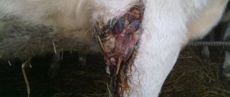
The deep form has pronounced symptoms. The skin becomes crusty, small and large spots form. Small ones often merge into one large lesion, but this form is very rare.
Symptoms and first signs of the disease
The disease can have several forms of manifestation, the severity of which is determined by the age of the animal and the state of immunity. Depending on the clinical signs, it is customary to distinguish between superficial, deep, erased and hidden forms of skin pathology. Erased and hidden varieties are characteristic of dogs older than one year, superficial and deep microsporia occurs in young puppies, elderly and weakened dogs.
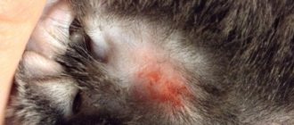
After microsporia penetrate the animal’s body for a long period of time, the skin has a normal, healthy appearance. At the end of the incubation period, the first signs of the superficial form appear: hairless, round spots with a clear gray border are formed . The skin in the affected lesions becomes dry, red and rough, the hair may be briefly broken off, sometimes sticky hairs or only black awns remain in the lesion. Alopecia can form on the scalp, abdomen, anal area and between the fingers.
A distinctive feature of microsporia is the attachment of pathogenic fungi to the base of the coat.
Hair from the affected area breaks off easily; upon careful examination, white dandruff, which is microsporia, can be found at the tip. On reddened, inflamed skin, scales first appear, then ulcers, ulcers and erosions form.
Symptoms and clinical signs of microsporia
The first clinical signs appear 3-5 weeks after pathogenic microorganisms enter the pet’s body.
At the initial stage of the disease, the skin has a normal appearance, there is no inflammation, so it is impossible to recognize infection. As it progresses, flaky, red, inflamed, rounded areas appear on the skin.
Typical localization of lesions for microsporia: tail and base of the tail, paws (especially the front ones), muzzle and ears, as well as fingers and claws. The coat loses its healthy appearance and seems to stick together. The dog experiences severe itching and constantly scratches the affected areas, spreading the infection throughout the body. Hair loss occurs, round bald areas covered with a crust are formed. Their edges are clearly defined.
If left untreated, small lesions merge into large ones, covering a large surface of the body.
Microsporia in dogs has the following forms:
- Erased or atypical. Determined only by laboratory methods. The lesions look like warts rather than circles. Summer seasonality is pronounced.
- Hidden. Characteristic of adults. It is asymptomatic, but with an inflammatory process and itching, which causes the pet to scratch the skin. Hair loss occurs; Allergy is also possible.
- Superficial. External symptoms are clearly expressed: round spots of a pinkish color with clear boundaries. The lesions are covered with white scales. The infection spreads quickly and causes hair loss.
- Deep. A purulent inflammatory process begins in the hair follicles. The skin around the hairs is covered with dense crusts and a gray-white coating.
The sooner the pathology is detected and treatment is started, the faster the pet will recover. A late visit to the doctor is fraught with an increase in the number of lesions and the addition of a secondary infection.
Incubation period
A pet of any age can become infected with microsporia, but most often dermatomycosis affects young animals under one year old, older individuals and dogs with a weakened immune system.
The incubation period from the moment of infection to the appearance of the first symptoms of the disease is 45-47 days. A healthy dog with a strong immune system can be a carrier of microsporia without clinical signs of pathology.
In addition to age and immune status, contributing factors for the development of the disease are:
- poor quality dog feeding;
- violation of conditions of detention;
- vaccination;
- weaning;
- change of diet;
- hypothermia;
- viral, bacterial and parasitic diseases;
- non-communicable diseases;
- lack of vitamins and microelements;
- allergy;
- metabolic and hormonal regulation disorders.
Symptoms
The incubation period is from 20 to 40 days. The disease can occur latently, in an erased form, superficially, and affecting the deep epidermis and dermis.
Hidden and superficial forms appear in kittens. In this case, the skin of the muzzle and paws, sometimes the tail, is covered with hairless spots with white-gray scales. Hair is occasionally visible on the surface of the spots, but it is broken off.
In dogs, the muzzle, back, and occasionally paws are affected. The extent of the site can be significant.
In fur-bearing animals, the fungus is mainly localized around the eyes, at the base of the ears, and sometimes on the limbs. Often the form is papules and pustules, which burst to form light yellow crusts.
Horses suffer from the superficial form. The head, withers, neck, back, and sometimes limbs are affected. The diameter of the hairless areas is 3-4 cm.
In deep processes, all animals experience inflammatory processes with redness and the formation of exudate, which allows the accompanying microflora to multiply and provoke purulent processes.
In pigs, the fungus appears as red spots with a limited area. The stubble falls out, the area of inflammation becomes covered with brown crusts.
The fungus does not cause itching, but the secondary microflora causes inflammatory reactions, which provoke scratching.
Tests and diagnostic methods
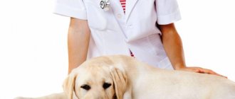
A veterinarian should make the diagnosis and prescribe appropriate treatment. Several diagnostic methods are used to identify drusen of a pathogenic fungus.
Clinical examination
It is possible to assume the presence of microsporia in a sick animal during a clinical examination only if there is a pronounced superficial form of the disease, characterized by the formation of round lesions. The deep form in the presence of complications is very difficult to visually diagnose. It is almost impossible to determine the erased and latent forms of microsporia only through a clinical examination.
Luminescent diagnostic method
To identify microsporian mycelium, a Wood's lamp is used as a source of ultraviolet radiation. To ensure a high-quality examination, it is not recommended to apply ointments or solutions to the affected areas, which can cause an incorrect result.
To carry out the fluorescent diagnostic method, the sick animal is placed in a dark room and the beam of a Wood's lamp is directed at the affected area. When exposed to ultraviolet radiation, the mycelium of pathogenic fungi begins to glow green.
Microscopic examination
Using a microscopic diagnostic method, it is possible to detect fungal mycelia and changes in the skin and coat characteristic of dermatomycosis. The type of specific pathogen cannot be determined using this research method. To analyze and identify a pathogenic fungus, a microscopic examination takes a skin scraping from the affected area and a few hairs from the animal’s body.
Culture diagnostic method
Microscopic and luminescent methods are used for rapid diagnosis of microsporia, but the most reliable result can only be obtained by inoculating the pathogen on nutrient media. Despite the energy consumption and duration of the cultural method, it makes it possible to accurately determine the type of pathogenic fungus, which is necessary for a veterinary specialist to prescribe competent treatment for the disease.
Diagnostics
Despite the very typical clinical signs of microsporia detected during the initial examination of the dog, the diagnosis is made after a series of examinations. This will make it possible to differentiate the pathology from allergic dermatitis, scabies, hypovitaminosis A, and especially from trichophytosis and scab, which give similar symptoms but are caused by other types of fungi.
To determine the nature of the disease, a veterinary laboratory conducts a microscopic examination of hair and skin scrapings taken from the affected area. The type of fungus is determined by sowing the test material into a nutrient medium, identifying the pathogen and its variety. An effective method for diagnosing microsporia is the fluorescent method of examination with a Wood's lamp. With this method, you don't even need to take scrapings: the dog is placed in a dark room and a thin beam of UV rays is directed at it, in which the spores of the Microsporum fungus glow green.
Treatment of microsporia in dogs
The duration of treatment for microsporia in dogs directly depends on the severity of the disease, the severity of the disease and the health status of the pet. In healthy animals with a strong immune system, the disease may occur without clinical symptoms, or minor skin lesions may resolve without the intervention of a veterinarian.
For the treatment of puppies, elderly or weakened animals with microsporia, aggressive drugs are used over a long course. The prognosis for the disease depends on the severity of the pathology and the timeliness of the dog owner’s visit to the veterinary clinic.
Treatment regimen
Treatment of the disease is aimed at destroying the causative agent of the disease and secondary pathogenic microflora, restoring the skin, stimulating the immune system and increasing the overall resistance of the body.
Treatment of microsporia in dogs: methods and medications
Today, there are many effective and high-quality drugs that veterinarians use to combat pathogenic fungi. In addition, over the past years, specialists have developed several standard treatment regimens.
I would like to note that in most cases, veterinarians do not encourage treatment of microsporia at home, since this often leads to infection of all people and animals living in the house.
Approximate treatment plan for lichen
Please note that the treatment regimen given below is just an approximate one, and it must be adjusted based on the realities of a particular case:
- The animal is always prepared before treatment. The fur around the lesions is trimmed and even shaved. If the dog is small, it is very advisable to cut it completely. This will greatly facilitate the treatment procedures and make them more effective.
- If the disease occurs in a superficial or erased form, and the skin lesions are small and few in number, only local treatment is allowed. Salicylic ointment, Yam ointment, as well as regular alcohol tincture of iodine have proven themselves to be effective. Note that before applying ointments, it is advisable to treat areas of the skin with water with the addition of a couple of drops of the same iodine.
- To remove scabs and scales from the surface of the skin, it is advisable to wash a sick pet with shampoos with a keratolytic effect once every three days. At the same time, you can use shampoos with an antifungal effect, and regular Nizoral is good for this.
- In the first week, the dog must be bathed in solutions of antifungal drugs.
- Other measures depend on the condition of the particular animal. Thus, it is recommended to prescribe immunostimulants and antimicrobial drugs in cases where pathogenic fungal microflora, etc., are added to a fungal infection.
- A high-quality and balanced diet is a must. We recommend that you consult with your veterinarian and select a suitable holistic veterinary food.
Basic medications for the treatment of microsporia
The following drugs can be used directly to destroy pathogenic fungi:
- Ivamerol. This is a very effective (but expensive) medicine, available in the form of a solution. It is diluted in boiled water in a ratio of 20 ml of medicine per liter of water, after which it is used to bathe a sick dog. If the dog is small, it must be seated in the solution up to its neck and kept in it for at least five minutes.
- Irunin and all other intraconazole derivatives (Clotrimazole, Miconazole). They are usually used in a dose of 10 mg per kilogram of the dog’s live weight. These drugs are given for ten days. The remedy is powerful and can often cure mild cases without the use of other drugs, but if it is given for too long, the dog may develop problems with the liver and kidneys.
- Antibiotics from the griseofulvin group help well. They are prescribed at a dose of 18 mg per kilogram of live weight.
Gamavit (stimulates specific resistance), as well as multivitamin complexes, are often prescribed as auxiliary therapy. To prevent bacterial skin lesions, broad-spectrum antibiotics from the cephalosporin group are prescribed.
Folk remedies for the treatment of microsporia
You can use folk remedies only in cases where the disease occurs in the mildest forms, skin lesions are few and their total area is small. In other cases, there is no need to waste time on “self-medication”.
At home you can use the following:
- Birch tar. Apply in its pure form to the affected areas of the skin no more than twice a day for a week. If visible inflammation, swelling, itching and signs of allergy develop, treatment should be stopped immediately.
- A mixture of tar and laundry soap in the form of a slurry. This mixture is also used in the form of applications to affected areas of the skin. They are applied three times a day, exposure time is from 15 to 20 minutes, after the procedure the mixture is washed off with cool water. Overexposure may result in chemical burns to the skin.
- It is also recommended to bathe a sick pet in infusions of chamomile, calendula and oak bark (1:1:1). This will help prevent the development of severe inflammatory reactions on the skin.
Caring for a sick dog
Microsporia is a very insidious disease; for a complete cure, in addition to correct diagnosis and prescription of specific medications, proper care for the sick dog is essential, consisting of simple actions:
- isolation of the four-legged patient from all pets and family members;
- trimming long hair to avoid the spread of fungal spores and better penetration of medications;
- it is necessary to burn all toys, bedding and care items, and regularly disinfect new bedding;
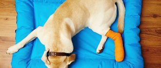
- To prevent scratching and the spread of infection throughout the body, it is recommended to trim the claws short and put a special protective collar on your pet;
- food should be easily digestible and balanced; low-fat soups, cereals, boiled pureed vegetables and fermented milk products are best suited for feeding a dog during the period of treatment and recovery;
- if you refuse to drink and feed, you must forcefully drink water and feed your little friend pureed food from a syringe;
- The best medicine for a weakened pet is the attention of the owner, so it is necessary to encourage the pet during all procedures with gentle, comforting words.
Important! All manipulations with a sick animal must be done with sterile gloves and hands must be thoroughly washed with soap and running water after any procedure. During the period of treatment and recovery, it is advisable to prevent contact between small and elderly family members with a sick dog.
Vaccination and vaccines
For the prevention and treatment of microsporia in dogs, vaccines against dermatomycosis are widely used: Vakderm, Mikolam, Mikoderm, Polivak, Mikkanis. Thanks to vaccination, a persistent immune response occurs, so it is recommended to vaccinate pets at a very tender age.
The use of vaccines against dermatomycosis in dogs is considered effective only if they are used in combination with fungicidal external and internal drugs.
Immunobiological preparations are administered intramuscularly at intervals of 10-21 days until the symptoms of the disease completely disappear and the pathogen is absent in animal tests.
Danger to humans
Unfortunately, a sick pet poses a real danger to humans. Infection of people occurs directly through contact with the fur of a pet or through care items, dishes and toys. Children, the elderly and weakened people are at risk; most often, the peak incidence is observed in the autumn-winter period when immunity deteriorates.
In humans, pathogenic fungi cause damage to hair follicles, skin and nails. Ringworm develops according to the same scenario as in animals. Healthy people do not become infected with an unpleasant disease, but in children the pathology can begin with the formation of one rounded lesion and develop into a generalized form with a wide area of damage. Antifungal creams and ointments are used to treat the disease.
Prevention of microsporia
In the case of ringworm, prevention is better than cure. Thus, you can protect not only your four-legged friend, but also all household members and other pets from severe pathology.
Prevention of microsporia consists of the following measures:
- avoiding contact with stray dogs and cats
- vaccination with the drugs “Vakderm”, “Mikolam”, “Mikoderm”, “Polivak”, “Mikkanis”;
- regular washing of the animal’s limbs after each walk;
- quality feeding and proper care;
- use of vitamins and mineral supplements;
- timely treatment of infectious and non-infectious pathologies.
Disinfection of living quarters
Disinfection of living quarters is one of the main components of therapeutic measures for microsporia in dogs. The pathogen can remain active for a long time outside the animal’s body, due to which re-infection of a pet or family members can occur at any time.
It is advisable to throw away all toys, care items and bedding. New bedding must be changed daily by subjecting replacement underwear to boiling, washing at high temperatures and double-sided ironing.
During treatment, the animal must be placed in a separate room, excluding the possibility of contact with other pets and family members.
Upholstered furniture and carpets must be thoroughly vacuumed and treated with chlorhexedine steam; the room must be regularly wet cleaned with formaldehyde, chlorhexedine or chlorine. Clothes must be washed with any chlorine-containing substance.
Treatment of microsporia
To make a correct diagnosis, laboratory tests are performed using two methods .
- The first method is to take broken hairs from the damaged area of skin and scrape off the scales.
- The second method allows you to distinguish ringworm from scab. The dog is taken into a dark room and irradiated with a mercury-quartz lamp. If it is microsporia, then under the influence of the drug the hairs affected by the spores will stand out in emerald color against a dark background.
Interesting: Paraanal glands in dogs: cleansing and causes of inflammation
Treatment for ringworm takes a long time and is very difficult. The dog must be kept in a separate room and constantly cleaned so that family members do not become infected.
Every day, the animal should be treated with antifungal drugs, lubricating the affected areas of the skin with a binary solution of iodine and 10% salicylic alcohol. Iodine monochloride also helps. During the first three days, the sore spot is soaked in a 3–5% solution without removing the crust. After this, the affected area is washed with soapy water and cleaned. Subsequently, the skin is lubricated with a 10% solution.
Your veterinarian may prescribe antibiotics. 0.25% trichocetin helps very well. It is applied in the form of a suspension to the dog’s sore skin once every 6-8 days. Along with it, another antibiotic should be given orally - griseofulvin. Several courses are carried out for 20 days, with a break of 10 days. It is recommended to administer microderm or vakderm intramuscularly.
Preparations such as zoomicol, vedinol, tsipam or black walnut ointments are very effective. It is better to treat puppies with homeopathic remedies (traumeel, engystol). They are used until complete recovery.
Even if the dog has recovered completely, if the room is not cleaned sufficiently, it can get sick again . Therefore, the entire apartment should be treated with a solution of 2% formaldehyde and 1% sodium hydroxide. In addition, the animal must be under the supervision of a veterinarian for another 45 days, avoiding contact with sources of infection.