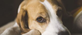
Types of otitis
Depending on the type and form of the inflammatory process, the nature of the ear discharge and the course of the disease, there are five main classifications of the disease.
1)By origin
There are three types of otitis media:
- Primary. It occurs as a result of injury, water getting into the ear, hypothermia, or due to congenital abnormalities. Often leads to ear infections.
- Secondary. Appears against the background of bacterial and viral infections, diseases of the upper respiratory tract.
- Idiopathic variety. Despite the presence of symptoms characteristic of the disease, diagnosis does not allow one to determine the exact causes that provoked the inflammatory processes.
2) By localization
Anatomically, the ear consists of the auricle, the tympanic cavity and the membranous labyrinth. Depending on the location of the appearance of the inflammatory process, the following are distinguished:
- Outer. Inflammation of the outer ear and ear canal to the eardrum.
- Average. When inflammation occurs in the middle ear (tympanic cavity).
- Interior. The rarest type of otitis is when the inner ear (membranous labyrinth) becomes inflamed.
The most common otitis externa in dogs is otitis externa, which is the least dangerous and can be easily treated. If the infection penetrates beyond the eardrum, inflammation may spread to the brain and nervous system.
3) According to the form of the disease
According to the severity of the disease, acute and chronic otitis media are distinguished:
- The acute form has more severe symptoms. It is easier to treat. But if no measures are taken in time, the acute form can develop into chronic.
- The symptoms of the chronic course of the disease are not so pronounced, but can worsen with relapses. Chronic otitis media is difficult to treat. Therapy is aimed at preventing the occurrence of new attacks of the disease and suppressing acute symptoms.
4) By type of discharge
Based on the nature of the discharge, exudative and purulent otitis in dogs are distinguished:
- In the first case, a viscous yellow-green liquid accumulates in the middle ear.
- In the second, there is abundant separation of earwax.
As a rule, at the very beginning of the disease, the inflammatory process affects one ear, and soon spreads to the second.
5) According to the causative agent of inflammation
Depending on the pathogen, veterinarians identify 5 main sources of otitis media in dogs:
Parasitic
The most common cause of inflammation is ear mites Otodectos cynotis. Another cause of a parasitic type of otitis media in dogs is flea dermatitis.
Allergic
It occurs against the background of allergic reactions of the animal’s body to food components or external factors (household chemicals, hygiene products, medications, reagents, etc.). In addition to inflammation of the ears, allergies are often accompanied by other manifestations: skin rashes, hair loss on other parts of the pet’s body.
Traumatic
Occurs as a result of injury.
Verrucous
Characterized by the appearance of multiple warts. Which block the ear canal and prevent the natural release of wax. As a result, inflammation occurs.
Infectious
The infectious form includes fungal, bacterial and viral otitis media in dogs. Characterized by increased body temperature and copious purulent discharge.
Fungal otitis appears due to decreased immunity or prolonged use of antibiotics.
Bacterial occurs as a result of viral infections or injuries.
Otitis externa in dogs
Svetlana Belova, Estonian University of Life Sciences Photos of the author are used in the article
Introduction
Otitis externa (otitis externa) is an inflammation of the external auditory canal (EA), which extends from the auricle to the eardrum (ET) and consists of skin lining cartilaginous tissue.
As in other parts of the body, the skin of the NSP has an epidermis and a dermis containing adjacent structures: hair follicles, sebaceous and sweat glands. The sweat glands in the NSP are called ceruminal or sulfur glands.
Earwax (cerumen) consists of a mixture of secretions from the sulfur and sebaceous glands and dead cells of the stratum corneum of the epidermis. Accumulated wax is removed from the lumen through the constant movement of cells in the upper layers of the epidermis from the ear canal towards the exit from the ear canal. This important mechanism for clearing NSP is called epithelial migration.
A patient with otitis externa is usually a patient with a dermatological problem.
Etiology
Otitis externa (OE) is not a diagnosis, it is just a clinical symptom that can be caused by many reasons. A poor understanding of the true cause—or causes—of OI, and, as a consequence, an incorrect approach to this clinical sign, often leads to failures in treatment and disappointment for both the animal’s owners and the doctor. To better understand all the factors that may play a role during OI, they have been divided into four main categories.
Predisposing factors increase the risk of OI, but alone are not able to cause inflammation. Examples: stenosis and/or excess hair in the ear canal, large heavy floppy ears, too much earwax secretion, increased humidity in the ear ear, too frequent and aggressive cleaning of the healthy ear with cotton swabs, obstruction of the ear ear with polyps or tumors.
Primary causes directly cause inflammation in the NSP. The most common primary causes are ear mites and allergic skin reactions. Less often in practice we encounter OI caused by keratinization disorders or autoimmune diseases.
Secondary causes are infections (bacteria and/or yeast) that complicate existing OI. They will not cause otitis in a healthy ear. It is a very common mistake to consider infection as the primary cause of OI.
Bacteria most often found as pathogens in OI: Staphylococcus speudintermedius, Pseudomonas aeruginosa, Proteus and Escherichia. Yeasts that complicate the course of otitis media are usually represented by Malassezia pachydermatis.
Supporting factors . These are the consequences of otitis media, all the pathological changes that occur in response to inflammation and later prevent healing. The most common supporting factors are hyperplasia and hyperkeratosis of the epidermis, edema and fibrosis of the skin of the NSP, hyperplasia of the ceruminal glands, impaired epithelial migration, cartilage calcification and otitis media.
Most cases of OI are caused by a combination of two or more causes at the same time, and for successful treatment it is important to find and control all of these factors.
| Photo 1. Erythema in acute allergic otitis media | Photo 2. Erythema and brown discharge in allergic otitis media complicated by Malassezia infection | |
| Photo 3. Lichenification of the skin in chronic allergic otitis media | Photo 4. Dense dry crusts in chronic allergic otitis media | |
Clinical picture and diagnosis
The most common sign of OI and the main complaint of the owner is itching in the dog in the NSP, which is clinically expressed by scratching behind the ears and shaking the head. It is very important to start the diagnosis with a detailed history, especially if we are dealing with a chronic case. This can give us important information about the cause of inflammation.
During the clinical examination, it is important to pay attention to the presence of skin lesions in other parts of the body, which, together with otitis media, may be a consequence of the same primary disease. Also, during the examination, signs of otitis media may be detected, such as a tilted head position, facial paralysis, Horner's syndrome.
An otological examination can detect such signs of OI as ear discharge, unpleasant odor, erythema, edema, peeling and/or crusting, excoriation, alopecia and ulceration of the auricle and skin at the entrance to the ESP.
The appearance and smell of ear discharge can provide additional information to an experienced physician. Dark brown, dry crusts usually indicate parasitic otitis caused by Otodectes cynotis, a yellowish-brown, waxy discharge with a sweetish specific odor is a sign of otitis caused by Malassezia pachydermatis, and an infection involving Pseudomonas (Pseudomonas aeruginosa) often gives purulent discharge a greenish tint. and a pungent smell. But, of course, all guesses must be confirmed by microscopic and/or cytological examination.
When palpating the ear canal, it is necessary to pay attention to the presence of a pain reflex and proliferative changes - fibrosis and calcification.
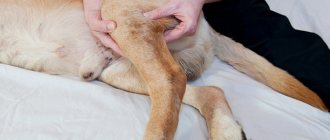
Otoscopy makes it possible to visually evaluate both parts (vertical and horizontal) of the ESP and the tympanic membrane. It is necessary to check the ESP for the presence of foreign bodies, assess the size of the lumen, the nature of the damage and exudate. A dull, convex, dark or perforated PD is a sign of otitis media. Due to pain and swelling in the ESP, a correct otoscopic examination is not always possible. In this case, one or two weeks of anti-inflammatory treatment (prednisolone 0.5–1.5 mg/kg/day) will help relieve pain and “open” the ear canal. Often, otoscopy requires sedation of the animal. Although in the case of chronic otitis media, even after sedation and rinsing of the ear canal, PD is not always visible. Fiber optic video otoscopy, thanks to its very bright illumination and good magnification, greatly facilitates visualization of the ear canal, and the resulting image can be shown to the pet owner and stored in a database.
Microscopic examination of earwax is carried out to exclude or confirm suspicion of parasitic (Otodectes cynotis, Demodex canis) otitis media. To do this, place the material taken from the ear canal on a glass slide, add a couple of drops of mineral oil and cover with a coverslip. For research, low (×40, ×100) magnification is used. If Demodex mites are suspected, it is better to close the aperture, creating greater contrast. If mites are not found in the specimen (provided that a sufficient amount of material was taken and it was carefully examined), then the parasitic origin of otitis media can usually be excluded.
Cytological examination is performed primarily to detect bacterial and/or fungal colonization or infection. In the clinic, it is most convenient to use so-called quick dyes (for example, Leukodif). The material for coloring is taken at the border of the vertical channel with the horizontal one or from the horizontal channel. Lightly touching the glass slide, we “roll” the cotton swab with the collected material over it. We first examine the colored preparation at a magnification of ×100, then, having found the field of view of interest, we use a magnification of ×1000, having previously applied a drop of immersion oil to the glass.
The presence of a small number of cocci (Staphylococcus, Streptococcus) and Malassezia yeast fungi is considered normal for the ear canal. But in the presence of clinical signs of external otitis, even a few cocci and/or Malassezia fungi give reason to suspect their active participation, already as a pathogen, in the inflammatory process. In the presence of neutrophils and especially phagocytosis, regardless of the number of bacteria or Malassezia detected, we are dealing with a true infection.
Cytological examination of ear discharge should be done both at the first visit and at all subsequent follow-up visits to assess the effectiveness of the chosen treatment.
Bacteriology and antibiogram in the case of external otitis are not very informative, since local antibiotics used to treat OI, as a rule, differ from those offered by the laboratory, and the concentrations of the local drug are significantly higher than those used to determine sensitivity.
Additional diagnostics that may be needed to find the primary cause of otitis: elimination diet, thyroid studies, skin biopsy of NSP, etc.
| Photo 5. Hyperpigmentation in chronic allergic otitis media | Photo 6. Copious purulent discharge with pseudomonas otitis |
| Photo 7. Erosive lesions in pseudomonas otitis | Photo 8. Skin hyperplasia and complete closure of the NSP in chronic otitis media |
Treatment
Can be divided into 3-4 important stages: cleaning/rinsing the NSP, local treatment, systemic treatment (not always necessary for OI), finding and eliminating all causes of otitis.
Thorough cleaning/rinsing of the ESP from earwax and exudate allows it to be examined correctly and provides the necessary access of the local drug directly to the skin of the ESP. In addition, the exudate contains bacterial toxins that can inactivate some local antimicrobial drugs (for example, polymyxin, gentamicin). It is best to carry out the first cleaning/rinsing in the clinic. The procedure may require a preliminary anti-inflammatory course of prednisolone and/or sedation. If there is a suspicion of a perforated PD, anesthesia and intubation are indicated to avoid aspiration pneumonia, and the use of, for example, warm saline or a 2% vinegar solution as a lavage solution. With intact PD, you can use a lotion to clean the ears; the choice will depend on the consistency of the ear discharge (more viscous, oily for ceruminous otitis and liquid for purulent).
Local treatment is prescribed based on each specific situation and cytology results. For external otitis, it is most convenient to use ready-made drops, which usually contain two or three components: a glucocorticosteroid, an antibiotic and/or an antifungal drug. For secondary cocci infections, polymyxin and fusidic acid are most often used as antibacterial treatments. For infection with rod-shaped bacteria - neomycin, enrofloxacin, gentamicin, ciprofloxacin, tobramycin. When choosing an antifungal agent for the treatment of Malassezia otitis media, preference is most often given to clotrimazole or miconazole.
Topical corticosteroids are very often indicated for OI and can be used as a single agent (for example, hydrocortisone aceponate) in the treatment of allergic and/or ceruminous otitis media. Including for long-term maintenance therapy, 1–2 times a week.
Systemic antimicrobial treatment is ineffective for otitis externa. Systemic treatment uses either antiparasitic treatments (in recent years, oral drugs from the isoxazoline group have been the most popular) or glucocorticosteroids at the beginning of treatment.
The average duration of treatment for OI is about 2–3 weeks.
It is necessary to regularly, approximately every 7–10 days, monitor the effectiveness of treatment by repeating the examination and cytological examination of the discharge, and change treatment if necessary.
Finding and eliminating all causes of otitis externa is especially important; without it, complete recovery is impossible.
Literature
1. Gross TL, Ihrke PJ, Walder EL, et al. Discoid lupus erythematosus. In: Gross TL, Ihrke PJ, eds. Skin diseases of the dog and cat. 2nd ed. Blackwell Science 2005, pp. 52–55.
2. Muller and Kirk's Small Animal Dermatology, 7th Ed. Miller W, Griffin C, Campbell C. WB Saunders, 2012.
Doctors interested in veterinary dermatology are welcome to Svetlana Belova’s website: www.vetderm.eu.
Here you will find information about the School of Veterinary Dermatology in Tartu, about webinars, upcoming interesting events, and you can also look through the dermatological atlas and subscribe to the blog.
SVM No. 1/2017
Rate material
Like Like Congratulations Sympathy Outrageous Funny Thoughtful No words
1
Factors contributing to the appearance of otitis media
The reasons for the development of otitis media can be completely different. Some occur due to congenital predisposition, while others occur as a result of parasitic infections or improper care.
Fur in ears
Excessive growth of hair in the ears leads to disruption of the natural ventilation of the ear canal. The wool is saturated with natural ear secretions and glued together. This makes it difficult to remove earwax. Excess sulfur is a favorable environment for the development of inflammatory processes.
Curves of the ear canal
Congenital, pronounced bends of the ear canal also make it difficult to remove earwax.
Active work of the secretory glands
The active work of the secretory glands contributes to the excessive formation of earwax, which does not have time to be removed from the ear canal and clogs it, preventing natural ventilation.
External parasites
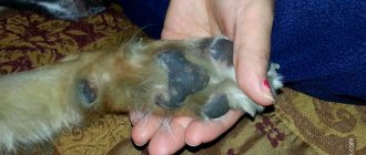
The most famous of them is the ear mite Otodectos cynotis. The appearance of parasites is accompanied by itching and the formation of black-brown crusts and plaque. Settling in the ears, the arthropod parasite feeds on blood, skin particles, and sulfur. The mite eats away at the skin, causing unbearable itching and pain. To escape from ear scabies, the dog scratches its ears until it hurts and causes an infection. Dark discharge, dead skin particles, and black grains appear in the ear canal. All these are waste products of the tick. As a rule, otodecosis affects both ears at once.
Decreased immunity
Hypothermia, long-term treatment, frequent stressful situations, and an incorrectly selected diet can cause a decrease in immunity. A weakened animal’s body ceases to resist various infections and viruses. Diseases such as bacterial or fungal otitis, demodicosis, and so on appear.
Neoplasms
Just as in the case of abundant growth of hair in the ears, there are various neoplasms that can grow both on the surface of the ears and in the ear canals. They prevent air from entering and natural sulfur separation.
Papillomas, tumors and cysts can cause otitis media in a quadruped.
Atresia
Atresia, or congenital/acquired overgrowth of natural openings and canals. It is most often found in dogs with a breed characteristic such as increased formation of skin folds. Shar peis, bulldogs, and chow chows are at increased risk. Intensively growing folds can block the ear canal and, as a result, the dog develops an inflammatory process and otitis media appears.
Allergies
Often, otitis in a dog can develop against the background of an allergic reaction of the body. Otitis media is the most common symptom of food allergies in dogs. In 80% of four-legged animals with tolerance to a particular product, this symptom first appears.
At the peak of allergy development, your dog experiences excessive earwax production. If the allergen is not identified and eliminated in time, otitis media develops.
Injuries, foreign objects
Often the inflammatory process occurs due to the presence of a foreign object in the ear canal. Plant seeds, insects, and grass that get into the ear can cause the development of the disease. Sometimes situations occur when children's games lead to small toys getting stuck in the ears, which injure the animal's ear and lead to inflammation.
Unprofessional trimming can also damage the ear and cause inflammation.
Water remaining in the ear after swimming
Sometimes, after taking water treatments, water may remain in the ears of your tailed friend. Together with earwax, this mixture is an excellent source of nutrition for pathogenic bacteria and microorganisms.
Hormonal disorders
Otitis in dogs can develop against the background of hormonal imbalances due to problems with the thyroid gland, abnormalities in the functioning of the adrenal glands, the genitourinary system and other vital organs. Such failures are often accompanied by inflammation, dry skin, itching, dandruff, and the appearance of tumors.
Improper care
Excessive care over a pet is no less harmful than its complete absence. By interfering too often with the ear canal and frequent cleansing with ear sticks, you remove the natural protective layer of sebum and wax.
Excess sweets
Excessive feeding of sweets to dogs leads to the production of earwax mixed with sugar particles. This formation of “sweet sulfur” is an excellent food for bacteria and fungi.
If the cause of otitis in a dog is not identified and timely treatment is not started, the eardrum may rupture or be dissolved by purulent secretions.
How to treat otitis media in dogs? Answer to the question
Ears on the top of the head: treating otitis Oksana noticed that her collie named Curie began to scratch behind the ear too often. The girl looked into the pet’s ears and discovered that there was a lot of sulfur in them. She began to clean them every other day and rinse them with warm herbal infusions. In addition, the girl purchased ear drops from a veterinary pharmacy and used them strictly according to the instructions. After a week, the dog no longer tore at his ears. The inflammation seemed to have gone away. But the satisfied housewife did not rejoice for long. After another week, the collie began shaking her head desperately and whining pitifully. When someone tried to touch her ears, she immediately growled and ran away, whining. There was nothing left to do but take the dog to the vet. He immediately made a diagnosis: acute otitis media and ordered several examinations. Oksana’s family budget could barely cover such expenses, and she had to spend money set aside for her vacation on medicine. What did the girl do wrong? Why did otitis media return? And how can you protect your dog and cat from this scourge?
Amazing anatomy The ear of a dog and a cat has a rather complex structure. This is an advanced device for capturing distant noises and the smallest rustles. The auricle is designed so as to easily “collect” all sounds coming to it. In this case, the cartilage can rotate in different directions, which makes the task easier. A separate muscle group is responsible for its work. At the base of the concha there is the auditory opening and the external ear canal, which ends in the eardrum. Its internal part is quite tortuous, so the membrane is reliably protected. In general, the passage consists of cartilage tissue, but from the inside it is lined with skin. It contains the same glands that secrete earwax. This substance has a very complex composition: proteins, fats, carbohydrates, acids, salts. On the one hand, it lubricates the ear canal. But its main function is protective. Bacteria and fungi die in an acidic environment. If the ear is healthy, then the wax, along with dead cells, sebum, dust and microorganisms taken by surprise, leaves the ear without outside help. Despite the wax produced, the outer ear is the most unprotected. It is not surrounded by bones, unlike the middle and inner ear. In addition, it is constantly in contact with the external environment. Therefore, otitis media primarily affects it. Without proper treatment, they penetrate into the middle and then the inner ear, where the pet’s sensitive hearing aid is located.
At risk In dogs, otitis media is more common than in cats. Unlike purrs, they are forced to constantly be outside - in the rain, in the cold, and in the snow. However, the disease can occur not only due to hypothermia. Ear inflammation can be caused by a variety of microorganisms: streptococci, staphylococci, E. coli, microscopic mites and even yeast. Most of them are in the bodies of our pets throughout their lives and are inactive for the time being. Allergies, colds, metabolic or hormonal disorders, poor diet, decreased immunity - all this can “wake up” them. Animals with erect ears are least susceptible to otitis. So cats rarely get sick. The only people at risk are Scottish Fold cats and, oddly enough, Sphynx cats. In the latter, sulfur is released very intensely - this is a breed trait. So a lot of dirt collects in the ears of “naked” cats. Among dogs, the situation is not so rosy. All fold-eared dogs have very poorly ventilated ears. In warm conditions, bacteria multiply more actively. In addition, dirt is more likely to stick to hanging ears. Otitis often affects poodles as well. These dogs have too narrow ear canals. But shepherd dogs produce a lot of sulfur, which also causes many difficulties.
Painful symptoms Due to ear inflammation, the animal feels severe discomfort. This is visible to the naked eye. The pet becomes restless and avoids contact. He may constantly scratch his ears, rub them against various objects, and shake his head. It happens that otitis media affects only one ear. Then the pet constantly tilts its head towards the sore ear. Sometimes the pain appears suddenly. The peacefully dozing animal quickly breaks away from its place, screams pitifully and looks around timidly. When pain does not allow the pet to relax, he tries to press his ear to the bedding. If the pain is not expressed, the pet can allow the ear to be examined. But if you put a little pressure on the base, he will yelp in pain or quickly jerk his head back. In an inflamed ear, the skin becomes red and sometimes crusty due to discharge. In this case, the pus that has accumulated inside the middle ear may squish, and an unpleasant odor will come from the pet. In advanced cases, the animal’s body temperature rises. Sometimes a pronounced distortion of the lips appears. This signals that inflammation has paralyzed the facial nerve.
Note! Without timely treatment of otitis media, the animal can be seriously harmed. The infection can infect the blood, spread to the brain, or cause deafness. In addition, otitis media can develop into a chronic disease. Then the pet will need surgery. But surgery does not guarantee a complete recovery.
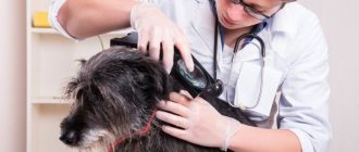
Without self-medication At the first signs of otitis media, even the slightest redness of the outer ear, you need to show your pet to a doctor. Self-medication is inappropriate here for two reasons. Firstly, many products sold in veterinary pharmacies simply relieve allergies and soften skin irritation. Visible symptoms will disappear for a while, but microorganisms will continue their destructive work. Secondly, otitis may be a symptom of another, more serious disease. For example, allergies, due to which the pet constantly scratches its ear. No matter how much you treat the inflammation, it will still return when your pet rips open the ear and introduces another portion of bacteria. Sometimes tumors and endocrine disorders are to blame for the return of otitis media. In short, you can’t do this without the help of a veterinarian. Before visiting a doctor, do not clean or treat your ears with medications under any circumstances. This can make examination difficult and make diagnosis more difficult. And if a disease occurs, it is already useless to clean your ears - this is a preventive, not a curative procedure. To prescribe treatment, one examination is not enough. You will have to take a swab of ear discharge, take various tests, and sometimes even do an MRI and X-ray.
Caring for the ears If the disease has not yet taken hold and seriously harmed the animal, then ear drops and ointments will be sufficient for treatment. In addition, the doctor will prescribe a drug that will destroy the causative agents of otitis identified during the analysis of ear discharge. Antibacterial, anti-mite, antifungal agents or glucocorticoids for the endocrine system will be prescribed. Carefully follow all instructions for use of the drug. The ointment should be distributed evenly over the skin of the ear canal. When re-treating the ear with ointment, the remnants of the previous product must be removed. If your pet has been prescribed ear drops, be sure to ask your veterinarian about the dosages. The fact is that average values are indicated on the packages. But for a small lap dog and a huge Great Dane, the amount of medicine is different. In severe cases of the disease, the pet is prescribed several medications, including antibiotics, painkillers, anti-inflammatory drugs and novocaine blockades.
Note! Do not buy medications that are not prescribed by your doctor, even if the salesperson at the pet store strongly recommends them. Different drugs have different principles of action. So, if your pet has a perforated eardrum, you cannot drip medications with antibiotics, glycerin, mineral oil, or chlorhexedine. They will harm the middle ear.
PREVENTION It’s good if your pet has never had otitis media. It is in your power to do everything to prevent the disease from affecting him. To do this, you need to follow a number of simple rules.
1. Clean your pet's ears periodically. Your pet's ears should be cleaned at least once a month. Just don't stick the cotton swab deep into your ear canal. It is enough to wipe the shell of the ear and the auditory opening. You can drop a special product inside. Just make sure it doesn't contain antibiotics or hormones. Also, don’t drip hydrogen peroxide. It is only needed to disinfect the ear. Be sure to remove any remaining product to avoid irritation.
2. Check your ears regularly. This procedure can be combined with monthly cleaning. Look for signs of inflammation, scratching, bites, and excessive discharge. The main thing is that your tailed friend does not resist. Therefore, it is better to accustom young pets to such examinations. As a last resort, you can distract him with something or ask someone to hold the animal while you look at his ears.
3. Protect your pet from the cold Make sure that your pet's bed is in a warm and dry place in the apartment. Avoid frequent drafts, especially after water treatments and walking outside. When bathing, make sure that water does not get into the ear. It is better to cover the ear canal with a cotton swab.
4. Observe quarantine Since otitis media are often caused by parasites, be sure to purchase a protective collar for your pet for the warm season. Keep your animal away from other dogs and cats with otitis media. If it is tick-borne in nature, then the disease will quickly spread to a healthy individual.
5. Visit the veterinarian Once a year, bring your animal for a routine examination. Since the veterinarian pays attention to the pet’s ears, he will be able to recognize the disease in the bud.
What breeds are at risk?
Due to the structural characteristics of the body, some dog breeds are at risk. These are mainly quadrupeds with increased folding of the skin, long-eared watchdogs and animals with excessive accumulation of fur inside the ears. These are breeds such as: Shar Peis, Spaniels, Chow Chows, Dachshunds, Collies, Basset Hounds, Toy Terriers, Bulldogs and other similar breeds.
When buying a puppy, you need to know in advance about possible problems and be more attentive to the hygiene of your four-legged friend’s ears.
Symptoms of inflammation
There are some basic symptoms that can help identify the disease. Depending on the causes and degree of damage, veterinarians also identify individual clinical signs. Let's look at them.
General symptoms of otitis media
Sick quadrupeds may experience the following behavioral and physiological abnormalities:
- The dog starts shaking its head.
- There is a constant desire to scratch your ears.
- The presence of scratches, scratches, bruises and other damage.
- Frequent and intense scratching leads to the formation of bald patches.
- Excessive discharge of earwax (more than usual).
- Redness and swelling of the ears.
- The ears become “hot” to the touch.
- Unpleasant odor from the ears.
- Bonding of hair in the ears, formation of crusts.
All these symptoms of otitis media can be observed with inflammation of the external ear canal.
If the condition worsens, when the inflammation affects the middle ear, the following symptoms are added to the above signs:
- Sweetish, putrid odor from ears.
- Copious formation of scabs in the ears and at the exit of the ear canal.
- Painful touch (the animal does not allow the ears to be examined).
- Increased body temperature.
- Lack of appetite, depressed state.
- Steady tilt of the head to one side.
- Crooked eyes and sunken eyes.
The advanced form of the disease is accompanied by:
- Facial nerve paralysis.
- Enlarged lymph nodes.
- Hearing impairment.
- Loss of coordination of movement (chaotic movements and frequent falls).
- Nausea.
- The inflammatory process also affects other organs, conjunctivitis appears.
- Complete refusal to eat due to unbearable pain.
Of course, all of the above symptoms can be caused by any other disease. In any case, they are all serious enough to warrant a visit to the veterinarian.
Individual clinical signs of otitis media
In addition to the general symptoms, there are some individual clinical signs of otitis media, which appear depending on the specific causative agent of the disease:
- With allergic otitis in dogs, signs of individual intolerance, as a rule, appear on other parts of the animal’s body. These can be various rashes, redness, itching and irritation of the skin, and so on.
- Otitis caused by ear mites is accompanied by dark brown discharge with a consistency similar to grains. The disease affects both ears.
- With otitis media caused by excessive hair growth, many hairs can be found in the ear discharge.
- In case of otitis media that occurs as a result of neoplasms or a foreign object entering the ear, upon examination the specific causes of the disease are revealed.
- With otitis caused by water getting into the ear, the ear discharge is always of a liquid consistency.
- Fungal and bacterial otitis is accompanied by an increase in the animal's body temperature and purulent discharge.
Symptoms of otitis media in dogs
Depending on the type of disease, its symptoms may vary. However, there are a number of common symptoms of ear inflammation in dogs. Among them:
- The pet often scratches its ears and shakes its head.
- Noticeably excessive sulfur production.
- An unpleasant odor appears, possibly the discharge of pus or other secretions.
- Crusts form around the ear canal.
- Signs of inflammation are noticeable: redness, swelling, pain, the skin is hot.
- The pet feels unwell, may whine, and does not allow its ears to be examined.
- With a long-term inflammatory process, the submandibular lymph nodes enlarge.
- Open wounds, scratches, and bruises may be observed on the shells.
- Due to intense scratching, hair falls out and bald patches appear.
There are also signs of otitis media in dogs that are characteristic of certain types of disease:
- When infected with ear mites, black-brown, grainy discharge appears, and barely noticeable open wounds can be found. In most cases, both ears are affected at once.
- With bacterial or fungal otitis, the body temperature rises, and purulent discharge is always observed.
- When water gets in and stagnates, there is discharge from the ear (can have different forms: cloudy, purulent, serous).
- When the cause of otitis is a foreign body, polyp or other neoplasm, diagnosis with an otoscope is necessary. Without a doctor, you can only find out about a problem if an object or tumor is visible from the outside.
- When inflammation is the result of an allergy, the reaction occurs in other areas of the body. Urticaria, dermatitis, itching, and conjunctivitis may occur.
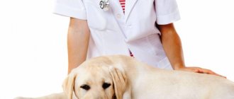
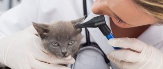
Diagnosis of the disease
As a rule, to determine the cause of otitis in a dog and identify the causative agent of the disease, the veterinarian conducts the following studies:
- A visual inspection of the ear canal is performed.
- Taking ear secretions.
- Microscopy of skin scraping.
- Blood analysis.
- If, after collecting an anamnesis, it is not possible to make an accurate diagnosis, an additional x-ray or tomography is prescribed to determine the depth of the lesion.
After making a diagnosis, the doctor decides whether treatment at home is permissible.
It is not recommended to clean your ears 2-3 days before your upcoming visit to the veterinarian. A sufficient amount of exudate should accumulate in the ears to be taken for laboratory testing to identify the causative agent of the disease.
How you can help your pet at home
Have you discovered that your dog has a red ear inside, how to treat it and how to help at home? The first and simplest home remedy for otitis media is cleaning the ear itself. It is important to carry out cleaning carefully. Avoid deep penetration into the auricle to avoid damage to the eardrum. Use cotton swabs; for small breeds you can use cotton swabs. You can massage your ear before cleaning. Use hydrogen peroxide (can be diluted if necessary), chlorhexidine, etc. For acute pain, you can give your dog painkillers such as Solpadeine, Ibuprofen or simple Analgin. The doctor will prescribe a more in-depth treatment, but if you have the appropriate skills, you can do novocaine blockades yourself.
Treatment of otitis media in dogs
In most cases, the dog is prescribed medications for otitis media and sent for home treatment. The owner is only required to strictly follow the veterinarian’s instructions and the instructions for the medications.
The therapy is aimed at relieving pain, destroying the pathogen, removing discharge from the ears and removing pathogenic bacteria not only from the source of inflammation, but also from the entire body.
If otitis media in a dog is caused by a foreign object entering the ear canal, tumors, warts, or overgrowth of the ear canal, then surgical intervention is required. The pet will have to spend some time in a hospital setting, under the supervision of specialists. After the pet’s condition improves, it is sent for home treatment.
How to prepare your pet's ears (therapeutic manipulations)
Before you start using medications, you need to properly prepare your pet’s ears:
- Examine your dog's ears for wounds, growths, scratches, or foreign objects. When inspecting, do not use any foreign objects under any circumstances.
- Treat any wounds found with chlorhexidine or 3% hydrogen peroxide solution.
- Next, it is necessary to remove purulent or copious sulfur discharge from the Barbosik’s ears. To do this, you need to put prophylactic drops or lotions into your ears, which dissolve the wax accumulated in the ear canal and soften the crusts. Drops such as Otipax or Otinum are absolutely safe. They do not have an antimicrobial effect, but they are good at soaking dried secretions and waste products of ear mites.
- After you have dropped 3-4 drops of any of the above drugs into the ears, you need to massage the base of the four-legged ears and wait a while until the ear contents soften.
If the dog wants to shake its ears, do not prohibit it from doing so. Soaked wax will move closer to the passage, and it will be easier to remove.
- Well, then take a regular cotton pad and carefully remove excess discharge from your ears.
- Now, after cleaning your ears, you can use the ear drops prescribed by your veterinarian.
- If there are wounds and abrasions in the ears, wound healing ointments are prescribed.
List of drugs
Here is a list of the most popular drugs prescribed for the treatment of otitis media in dogs.
The course of therapy, in most cases, is 5-7 days. But even if the furry friend’s condition improves earlier, the treatment does not stop.
All of the above medications are prescribed individually, depending on the diagnosis.
Diagnosis of otitis in dogs
Inflammation of the ear canal may occur periodically. Before starting treatment, it is necessary to determine the causes and severity of the disease. It is very important to first make a correct diagnosis, otherwise you can worsen the situation or provide ineffective help to your pet. Diagnosis of otitis in dogs is carried out in three directions:
- Visual inspection - using an otoscope, a full examination is performed, including the cavities of the outer and middle ear, eardrum, skin, and ear canal. And also the emphasis is on the degree of swelling and the presence of foreign objects in the canal;
- Laboratory tests - a blood test is necessary to assess the general condition of the body and check for the presence of allergic reactions. If there are grounds for hypothyroidism, then an additional blood test is performed showing the concentration of thyroxine and triiodothyronine. If hypothyroidism is confirmed, then a test is also done to activate thyroid-stimulating hormones; cytological tests to determine the tumor, the state of the microflora, hyperplasia of the sulfur gland, sensitivity to antibiotics; microscopy - using a microscope, skin scrapings and exudate of the ear gland are studied;
- X-ray – prescribed if it is impossible to determine the disease in a laboratory; the prerequisites may be suspicion of tumors or polyps of the nasopharynx;
- Computed tomography and magnetic resonance imaging help to most accurately determine the condition of the middle ear and inflammation of the brain tissue.
Only a well-made diagnosis will allow you to prescribe effective treatment. You should not self-medicate without identifying the causes. Some procedures can be performed using anesthesia.
It is used if the dog is in severe pain and behaves aggressively. With secondary otitis, things are a little more complicated. It is necessary to first determine the root cause, and then begin treatment.
By origin
Based on its origin, otitis media is divided into:
- Primary, develops independently, causes may be hypothermia, infection, or ingress of foreign objects. With primary otitis, it is easier to find the cause;
- Secondary, manifests itself as a consequence after an illness or during a hormonal imbalance.
By localization
Based on localization, otitis media in dogs is divided into:
- Otitis externa;
- Middle ear;
- Internal - considered the most dangerous because it can affect the brain and create a threat of sepsis.
By the nature of the discharge
Based on the nature of the discharge, otitis media is divided into:
- Purulent, characterized by yellow-green discharge with an unpleasant odor;
- Exudative, accompanied by copious secretion of sulfur.
With the flow
Along the course, otitis is divided into:
- Acute otitis media, accompanied by severe pain and rapid development of the disease;
- Chronic otitis is associated with periodic manifestations of symptoms of the disease.
According to the source of inflammation
Based on the source of inflammation, otitis media is divided into:
- Fungal;
- Bacterial otitis;
- Malassezia - belongs to the category of fungal types of otitis, manifests itself when immunity decreases, often a bacterial infection begins to develop along with it;
- Allergic;
- Verrucous - characterized by the appearance of warts and growths that eventually block the ear canal.
Complications and consequences
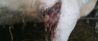
The most dangerous is advanced otitis media in dogs. If treatment is not started in time, due to the abundance of purulent discharge, the eardrum may dissolve or rupture. The pus enters the inner ear and causes serious complications: hearing loss, inflammation of the meninges, purulent meningitis...death of the animal.
In addition, when trying to save a dog by performing surgery on inflamed and festering tissues, the doctor may touch the nerve endings responsible for motor function. The four-legged one will remain disabled.
Why might a dog's ears pop?
It is necessary to take a responsible approach to the treatment of otitis in dogs, since hearing is their main tool for communicating with the outside world. Due to the anatomical features of the auricle, animals experience increased temperature and humidity in their ears. A closed ear space leads to intensive growth of bacteria. Often a dog's ears squelch due to:
- hypothermia;
- water entering the ear;
- poor hygiene;
- spread of infections from another area;
- the appearance of ear mites;
- bruises;
- scratches, abscesses in the passage.
Important! When a dog's ear is squelching and brown discharge is combined with an unpleasant fetid odor, this is considered a sure sign of infection. In this case, an urgent visit to the veterinarian is required.
A sick animal becomes restless, refuses to eat, does not allow its ear to be touched, and when you press on the hearing organ, a characteristic squelching sound is heard.
First aid
If it is not possible to immediately go to a veterinary clinic, you can alleviate your pet’s condition yourself with the following simple manipulations. The main thing, when providing first aid, is not to further worsen the four-legged condition. To do this, you need to know what you can do and what actions will harm your pet.
How to treat a pet if the diagnosis is unknown
After preliminary cleaning of the ears from purulent discharge and crusts (as described above), you can use combined ear drops for otitis media with analgesic, anti-inflammatory, antimicrobial, antiparasitic and antifungal effects.
These are drugs such as Surolan or Aurikan. There will be no harm from them. But, these remedies can be used only in mild cases of the disease.
If after a course of treatment there is no improvement, you should seek veterinary help.
What not to do
But these actions can harm the four-legged:
- Do not use cotton swabs or any other objects. This can damage your already inflamed ear canals. Plus, there is a possibility of damage to the eardrum. There may be cotton left in the ear canal, or you may push dirt and unsoaked wax inside even deeper. Use only cotton pads.
- Warming up is strictly prohibited. A warm environment will increase inflammation due to the growth of pathogenic microorganisms.
- Remember that alcohol-containing preparations can only be used to treat (wipe) the outer surface of the ears. Under no circumstances should they be placed in the ears. Reacting with bleeding wounds and purulent secretions, peroxide begins to hiss and foam. This causes pain and deafening noise in the ears, which can frighten the pet and cause even greater suffering.
- It is not uncommon for pet owners to develop their own treatments for otitis media in dogs. Occasionally they help, but in most cases they are not only useless, but can also cause harm. Kerosene, sunflower oil, garlic, tea leaves, human ear drops - all these products are not able to have a therapeutic effect, especially if the pet has neoplasms, fungal or bacterial otitis.
Avoid using medications unless absolutely necessary without consulting a veterinarian. How to treat otitis in a dog depends on its origin. And this can only be determined by a doctor after an examination and test results.
Prevention of otitis media
Of course, otitis in dogs can occur completely unexpectedly. But in order to protect your pet as much as possible from this disease, it is important to observe the following preventive measures:
- Check your furry friend's ears regularly.
- If the Barbosik is genetically predisposed to increased hair growth in the ears, regularly pluck or cut off excess “vegetation” and visit a groomer.
- There is no need to rinse, clean or treat healthy ears again, as this will disrupt the natural protective bacterial balance. A visual inspection is sufficient.
- Pay attention to the smell from your ears.
- Avoid getting large amounts of water into your ears.
- After taking water procedures, use a cotton pad to remove excess moisture from your ears.
- Regularly treat your pet against parasites.
- During the cold season, protect your tail's ears from hypothermia.
- To prevent your ears from getting blown out by the wind, don't let your curious nose stick out of the window while driving.
- Feed your dog only high-quality and healthy food. If you discover an allergy to a particular product, immediately eliminate it from your diet.
Otitis media in dogs is a fairly common occurrence. But if you regularly carry out preventive measures and examine your pet’s ears, then even if any problems are detected, you can quickly eliminate the inflammation.
Good luck and take care of your pets!