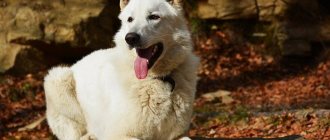
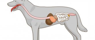
There are two types of gastrointestinal tract obstructions. An upper obstruction is a situation where a “blockage” forms in the esophagus or stomach. With lower obstruction, the place of stagnation is the small or large intestine. The latter option is called intestinal obstruction. This is a very serious disease; without timely and competent treatment, the dog dies within a few days. Therefore, if you notice symptoms in your pet that indicate intestinal coprostasis (cessation of movement of food masses through the digestive canal), you should seek qualified help as quickly as possible.
Causes of coprostasis
Intestinal obstruction in a dog can occur:
- When the intestinal lumen is blocked by a foreign body. According to statistics, obstructive ileus (as this pathology is called) is the cause of coprostasis in 80% of cases .
- Due to the accumulation of dead worms in the intestines as a result of deworming.
- In the presence of an intestinal tumor. Of all types of neoplasms, coprostasis is most often caused by carcinoma growing into the intestinal lumen.
- Due to blockage of an area of the intestine with fecal stones. Such stones often form in the intestines when feeding a dog roughage with low nutritional value.
- With intussusception (pressing one intestinal segment into the lumen of the adjacent one). This phenomenon is commonly called volvulus. A typical site of intussusception is the site where the small intestine enters the colon.
In much more rare cases, the cause of coprostasis is strangulated hernia, paralysis or congenital stricture (narrowing) of the intestine.
Symptoms
The most pronounced symptoms of intestinal obstruction in dogs manifest themselves when the upper part of the gastrointestinal tract is affected - the stomach or duodenum. With coprostasis of the colon, the symptoms of the disease are usually less pronounced.
Clinical signs of intestinal obstruction in dogs include:
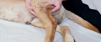
- Bloating, a noticeable increase in its volume due to the lack of gas discharge;
- Tense abdominal muscles, pain in the abdominal wall even with light pressure on it;
- Forced unnatural pose;
- Scanty liquid stool or its complete absence. The dog often and unsuccessfully tries to empty the intestines, while emitting characteristic groans;
- Refusal of food and water;
- The urge to vomit or vomit with foam, vomit may contain feces;
- Temperature below 38 degrees.
Attention! If a dog exhibits symptoms of intestinal obstruction, it is strictly forbidden to take any measures on your own; the animal must be taken to a veterinary clinic as quickly as possible. You cannot feed him, force him to drink, give him sorbents, laxatives, antiemetics, give him an enema, or perform gastric lavage. If the owner knows how to give injections, you can give the dog an injection of painkillers to make it easier for him to endure a trip to the doctor.
Symptoms of intestinal obstruction in dogs
Only ultrasound or x-ray can reliably identify obstruction. Symptoms in each individual case can be either sluggish or with vivid manifestations.
The main signs of intestinal obstruction in dogs:
- Weakness, poor health. The mucous membranes become pale and sometimes even blue.
- Body temperature sometimes drops below 37 degrees.
- Rejection of food and drink.
- Vomit. It can be abundant or rare, in small quantities. But if feces come out along with it, then the situation is critical. The intestines are full and can rupture at any moment.
- The dog has been trying to relieve himself for more than a day without success. At the same time, she moans and an unpleasant smell of rotting is heard from her mouth.
- Tension of the abdominal muscles and their soreness on palpation are observed.
- When the intestines rupture, internal bleeding occurs and hematomas form. In this case, the dog is unlikely to be saved.
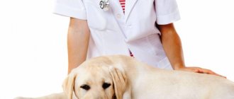
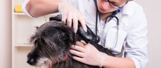
Diagnostics
To make a diagnosis, anamnesis is studied, a clinical examination is performed with bimanual palpation of the abdomen, and if necessary, blood and/or urine tests can be performed. When intestinal obstruction is suspected, hardware diagnostic methods are considered the most informative.
X-ray examination.
X-rays are taken in direct and lateral projections, this allows you to accurately determine the location of the “congestion” in the intestine, or the formation of narrowing, displacement, or intussusception in it. Often, for better visualization, x-rays are done using contrast agents (for example, barium salts), which are administered orally into the dog's gastrointestinal tract in the form of a solution.
Ultrasound diagnostics.
With the help of ultrasound, it is possible not only to accurately establish the zone of stagnation, but also to determine its cause, as well as obtain information about the presence of fluid in the abdominal cavity, even when the symptoms of ascites do not yet appear. In addition, an ultrasound examination can assess the intensity and nature of intestinal movements: with coprostasis they become pendulum-like.
Magnetic resonance and computed tomography are rarely used to diagnose coprostasis in dogs. Since the condition for their implementation is complete immobility of the patient, MRI and CT scans are performed on animals under general anesthesia and the supervision of an anesthesiologist.
Stomach and intestinal surgery
1.
DO NOT FEED OR WATER categorically!! This is the first and main commandment of first aid to an animal with increasing vomiting. Anything your pet eats or drinks in this state will only make the vomiting worse.
2
. DO NOT DO AN ENEMA!! In 99% of cases, obstruction occurs in the small intestine, which is inaccessible for washing, and the thinned intestinal wall at the site of blockage can simply burst under water pressure.
3.
DO NOT GIVE LAX!! Forceful contractions of a blocked intestine can also cause it to rupture.
4.
DO NOT ADMINISTER ANTIEMETICS (cerucal, metoclopromide)!! These drugs help normalize gastrointestinal motility, thereby doing your pet a disservice. Contractions of the intestinal wall contribute to the advancement of a foreign object and aggravation of injury, and as a result, aggravation of the pain reaction.
Difficulties in diagnosis.
To make a diagnosis of intestinal obstruction, a careful clinical examination and detailed information about the animal’s condition over the next few days may be sufficient. Survey X-rays in 2 projections (lateral standing and dorsoventral) give good results (Fig. 1).
Figure 1. X-ray (lateral view) - Foreign body in the thoracic esophagus.
If these images cannot reveal gastrointestinal obstruction, contrast fluoroscopy is used (Fig. 2) and the images are repeated several times in the lateral projection until the contrast agent passes through the entire intestinal tube.
Figure 2. – X-ray with contrast agent.
But this does not always allow one to accurately determine the presence or absence of intestinal obstruction, especially if it is incomplete. Ultrasound also does not have sufficient information to detect intestinal obstruction, although, like an x-ray, there are certain signs that can be used with a high degree of probability to indicate the presence of intestinal obstruction.
We believe that 90% of animals with suspected intestinal obstruction should undergo DIAGNOSTIC LAPAROTOMY.
This operation allows you to actually examine the entire intestines and stomach and, if necessary, immediately remove the obstruction. If intestinal obstruction is detected, the surgeon immediately begins to eliminate it. The operation is performed in the dorsal position of the animal under general anesthesia and local anesthesia. An incision in the abdominal wall is made parallel to the linea alba with retraction of the rectus abdominis muscle (this preserves its integrity), behind the xiphoid cartilage, for 8-12 cm. Having opened the abdominal cavity, the doctor examines all parts of the gastrointestinal tract and finds the site of obstruction. If it is a foreign object, then it is removed and sutures are placed on the intestinal wall; if it is a volvulus or intussusception, then they resort to resection of the dead area. The incision in the wall of the stomach or intestine is closed with a continuous two-story suture (the suture of the first floor is applied to all layers of the wall), the second - only to the seromuscular layer, and the abdominal wall, as in any other operation. When signs of peritonitis develop, drainage is inserted into the abdominal cavity. In advanced cases, the death of the animal occurs due to the development of peritonitis and severe intoxication.
Exploratory laparotomy and operations on the stomach and intestines can be planned or emergency. In our clinics, they are carried out after a preoperative examination of the animal, which includes ultrasound, ECG, General and Biochemical Blood Analysis, R-studies (survey images, images with a contrast agent). Before the operation, the doctor and the owner draw up a document on informed consent for the operation, after all the risks of these activities are explained to the owner.
It is very important that the owner brings a warm blanket to the operation; it will be needed to warm the animal after the operation, disposable absorbent diapers, and napkins.
ATTENTION!!!
In addition to surgical intervention, the most important role in the treatment of intestinal obstruction is played by competent and adequate fluid therapy: without replenishing the loss of moisture, salts, and in some cases amino acids, the most successful operation can end in failure. If the animal is brought to the clinic in serious condition, intensive infusion therapy must be started before surgery.
How to care for an animal during the postoperative period?
In all cases, it is recommended not to feed the animal for at least 5 days and not to give water for at least 1 day. At the same time, intensive infusion therapy (droppers 2 times a day) is prescribed for a period of 5 to 10 days, as well as antibiotics and metronidazole, etc. depending on the condition. It is best to leave the operated animal in the hospital for several days; the specialists on duty will perform all assigned procedures in a timely manner.
For feeding in the postoperative period, RECOVERY dietary food from Royal Canin has proven itself well. This diet can be diluted with water to any consistency.
What objects most often cause obstruction in dogs and cats?
1. Fragments of plastic toys, plastic bags, sausage casings;
2. Threads, stockings and other textiles;
3. Tinsel, “rain” (Most dangerous for cats, since they cannot get rid of it on their own, they swallow it. Rain can cut through the intestinal wall in several places and lead to peritonitis. In our practice, there have been cases when rain tied on the root tongue and enters the stomach, small and large intestines); 4. Eraser, rubber plugs, pieces of shoe soles.
Dear owners!!!
When providing emergency surgical care on the gastrointestinal tract, the clinician has to face a number of difficulties. Due to frequent vomiting, sequestration of fluid in the intestine, intestinal perforation with the development of peritonitis and sepsis, animals may be in a state of severe dehydration, and their cardiovascular system may be unstable. Vomiting or regurgitation may be complicated by aspiration pneumonia.
The sooner you contact a veterinary clinic, the more favorable the outcome of your pet’s illness will be.
Treatment
Depending on the cause of intestinal obstruction, the dog may be prescribed: conservative treatment, removal of the blockage using gastroscopy, or surgery.
Therapy methods
Conservative treatment is usually used in cases of partial functional obstruction caused by a gastrointestinal disease (eg, parvovirus enteritis). Without the use of a scalpel, it is often possible to get rid of the “plug” of dead worms or soft fecal stones.
In such cases, the doctor may prescribe:
- Laxatives (castor, vaseline or sunflower oil, lactulose).
- Enemas with a 1% solution of table salt or with salt and vegetable oil. However, when using laxatives and cleansing enemas, the doctor must be sure that the intestinal blockage is not complete, otherwise, when the intestine fills and its muscles contract strongly, intestinal rupture may occur.
- Antibiotics or antiparasitic agents.
- Antispasmodics and painkillers.
- Probiotics (cultures of beneficial microflora) and/or prebiotics (growth stimulators of beneficial microflora).
- In case of severe intoxication or dehydration, the dog is given drips with glucose or saline solution.
Endoscopy
Sometimes the only chance to save an animal by quickly removing a foreign object that has entered the esophagus, stomach or adjacent part of the duodenum is endoscopy. The procedure is performed under general anesthesia and is carried out using a special instrument on a long thin tube that is inserted through the mouth. No preparation is required for this operation.
Surgical treatment
In many cases, most intestinal obstruction can be eliminated only through abdominal surgery, which is performed under general anesthesia. The technique depends on the location of the foreign body, tumor or area of intussusception. If an area that has undergone necrosis is identified, resection (removal) of the affected part of the intestine is performed.
It is important! After abdominal surgery, the dog must be constantly monitored during the first 24 hours and given only water. On the second day, you can feed the dog a small amount of broth or liquid porridge. Then the doctor will tell you what diet to follow during the recovery period (it usually takes 1-2 weeks).
Carrying out the operation
Before the operation, a fasting diet is prescribed. The operation is performed as planned in a hospital setting. In this case, the animal is rigidly fixed, and a surgical incision of the required size is made. Next, the affected tissue is removed and the intestine is connected with an internal suture. If necessary, a feeding tube is installed in emaciated animals.
The period of the patient's stay in the hospital is determined by the doctor depending on the situation. In the postoperative period, bactericidal drugs and antibiotics are necessarily prescribed. Droppers are prescribed to replenish the loss of electrolytes and fluids, and intravenous nutrition.