My dog has joint pain, what and how to treat it? Quite often, many pet owners ask this question, since joint pain is a common painful manifestation of various diseases of the musculoskeletal system of dogs. In order for treatment to be much easier and more effective, it is very important to detect joint disease at an early stage of development and correctly determine the cause of its occurrence. Therefore, if you notice the first signs of abnormalities in your pet’s well-being, you should contact a veterinarian. The specialist will make an accurate diagnosis and answer the question of how and how to treat this unpleasant disease.
Causes of pain
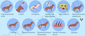
There are a huge number of causes of joint pain. As a rule, they can be associated with wear and tear of cartilage tissue, or with the progression of inflammatory diseases. The most common joint diseases in dogs are:
- Arthrosis is a severe and intractable chronic joint disease caused by wear and tear of intra-articular cartilage. Over time, in addition to cartilage, other components of the joint undergo changes. Quite often, arthrosis occurs due to the aging of cartilage tissue cells. The disease develops slowly, and obvious signs may not be noticed for a long time. The cause of this disease can be injuries of various types, excessive loads, heavy weight of the dog, and others.
- One of the causes of joint pain may be arthritis. This is an inflammatory disease of cartilage and joints. The course of this pathology can be acute or chronic. Not only older dogs are susceptible to arthritis; this pathology can also develop in the body of a young dog. There are several varieties of this disease depending on the cause of their development, the age of the dog and the degree of involvement of the elements that form the joint in the inflammatory process. The main factors contributing to the occurrence of arthritis are: injuries of various origins, physical overload of the joint, genetic predisposition, the consequences of complex infections, functional failure of the immune system and others.
- Common joint diseases include osteochondrosis, which manifests itself in ossification of cartilage with subsequent destruction of the joint. Osteochondrosis affects dogs regardless of age and breed, but large dogs or obese dogs are more susceptible to this disease. Nutrition and genetics play a significant role in the development of this pathology.
Common joint diseases in dogs
Arthritis of a purulent nature.
Arthrosis.
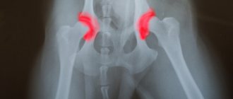
Hip dysplasia.
Elbow dysplasia.
Dislocation.
Stretching.
- arthrosis;
- purulent arthritis;
- dislocation;
- hemarthrosis;
- hip dysplasia;
- joint wounds;
- stretching, distortion;
- synovitis;
- chronic osteoarthritis.
These pathologies arise for various reasons, but they create many problems for both animals and owners.
Predisposing factors
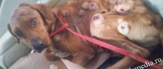
Naturally, each individual pathology has its own causes, but there are common factors of occurrence. Diseases of inflammatory origin suggest the presence of injuries, bruises or dislocations. Dogs are very active and fights often occur, resulting in injuries.
Injuries are possible due to fights.
- Hereditary factors play a significant role in the origin of joint pathologies . As a result of selection errors, congenital anomalies arise, which subsequently affect the development of the skeleton. Natural predisposition plays an equally significant role. Some breeds are characterized by natural fragility of joints, which subsequently leads to the progression of various diseases.
- Pathologies often result from the negligence of owners who do not properly care for their pets. For the proper development of the animal, a feeding and maintenance regime is necessary. Poor nutrition and lack of vitamins and nutrients also lead to joint destruction.
Characteristic symptoms
Quite often, the first symptoms of joint disease are so mild that even the most attentive owners may not notice them in their pet. They intensify gradually and become more visible.
The main symptoms of joint damage in dogs are:
- refusal to walk, play;
- lethargy;
- unsteadiness of gait;
- shortening the stride;
- lameness caused by pain;
- whining when getting up and starting to move;
- squeals when touching a sore joint;
- loss of appetite;
- weight loss.
There are characteristic differences for each of the diseases. For example, with arthritis, an increase in body temperature, swelling, and visible joint deformities may occur.
Typically, all symptoms are worse in cold, wet weather.
Prevention
To prevent the development of arthritis, it is necessary to follow preventive measures:
- frequent but gentle walks;
- warm and soft sleeping place;
- weight control (light diet, dry food with chondroprotectors);
- a balanced diet and the inclusion of vitamins (after consultation with a veterinarian);
- swimming and massage.
Arthritis cannot be cured completely, but long-term remission is quite possible. This is one of many diseases that threaten the health of a pet, but it is less dangerous than viral enteritis, gastritis or eclampsia in a bitch after giving birth.
Compliance with preventive measures, timely visits to the veterinarian, and keeping the animal warm on soft bedding will make your pet’s life easier and relieve him of constant joint pain. How to treat arthritis in a dog, even if it is not in an advanced form, should be decided by a veterinarian.
Additionally, check out the video about the manifestations of arthritis in dogs:
Drug treatment
Drug treatment of joints in pets should be started only after the cause of the pain has been determined. Under no circumstances should you use medications on your own without consulting a veterinarian, since the choice of medications and their dosage are selected exclusively individually. Incorrectly selected treatment can lead to dire consequences.
Non-steroidal anti-inflammatory drugs may be prescribed to reduce pain. The most popular drug in this group is Rimadyl. It is available in the form of tablets and injections. The drug has analgesic and antipyretic properties. The active ingredient of the drug is a strong analgesic carprofen, which can eliminate pain in an animal for a long time (10-12 hours). Rimadyl is contraindicated in pregnant and lactating dogs, as well as those with individual intolerance to carprofen. The drug is prescribed with extreme caution to animals with chronic diseases of the cardiovascular system, liver, and kidneys.
An equally popular non-steroidal anti-inflammatory drug is Meloxidil. Available in the form of a suspension. The drug can be used not only orally, but also mixed with food. A significant advantage of Meloxidil is that it has a minimum of side effects and is easy to use. Contraindicated in animals with high sensitivity to the main component of the drug, meloxicam. Prohibited for dogs with diseases of the digestive system, kidney and liver failure.
Hormonal medications (corticosteroids) quite effectively reduce inflammation and significantly improve the condition of your beloved pet. The most commonly used in veterinary medicine are Prednisolone and Hydrocortisone. Corticosteroids have a suppressive effect on the immune system, blocking the production of substances that cause inflammatory reactions.
Chondroprotectors are one of the key veterinary drugs for combating joint damage in dogs. Thanks to glucosamine and chondroitin sulfates, drugs slow down the destruction of cartilage tissue and promote its restoration. The preparations also contain vitamins and minerals that have a beneficial effect on the animal’s musculoskeletal system. These products include: Katrofen, Stride, Arthroplex, Chondroitin, Teraflex, Stoparthritis and others.
In the treatment of joint diseases, broad-spectrum antibiotics are used, such as: Amoxicillin, Lincomycin, Cephalexin, Gentamicin and others.
Nialando
What is joint degeneration?
Joint degeneration in dogs is a condition characterized by the gradual deterioration of the joint integrity of the hip, shoulder, elbow or knee. There are known diseases such as hip dysplasia, degenerative joint disease, arthrosis, osteoarthritis, rheumatoid arthritis, and all of them can lead to the same symptoms, such as late loss of cartilage integrity, inflammation and stiffness. Although there are many names used for these diseases, there are two main types of joint disease, or arthritis: rheumatoid arthritis and osteoarthritis. Among people with arthritis, rheumatoid arthritis and osteoarthritis occur in approximately equal proportions, and most cases of arthritis occur in companion animals, especially in dogs. Osteoarthritis in dogs is a disorder of the synovial joint characterized by degeneration of articular cartilage and the formation of osteophytes. Hardening of the underlying subchondral bone may in some cases be associated with osteoarthritis, and, in some cases, synovial inflammation may be present to varying degrees as the disease progresses.
Possible mechanism underlying joint degeneration.
Connective tissues in dogs, particularly large breed dogs, are constantly subject to stress and strain from mechanical forces, which can result in diseases such as arthritis, joint inflammation and stiffness. The causes behind rheumatoid arthritis and osteoarthritis differ. Rheumatoid arthritis is characterized as an autoimmune disease, i.e. Loss of the ability to differentiate between the body's own substances and foreign substances results in an immune response directed at the body's own tissue, which affects both joints and systemic immune functions. Osteoarthritis (degenerative joint disease), on the other hand, can be caused by high levels of mechanical forces on the joints, combined with age, resulting in damage to the articular cartilage, and then local inflammation of the joints. In more detail, increased stress directed at the joints results in loss of cartilage integrity, and the resulting damage causes accelerated cartilage breakdown and excessive release of cartilage plate components into the synovial fluid. Connective tissues are designed to repair themselves by producing and transforming vast amounts of proteoglycans and collagen, the main components of connective tissue. At the same time, cartilage restoration is a very long process, and with age, the ability to restore and synthesize normal collagen structures deteriorates. As a result, this creates pain, deformity and limited mobility of the joint. Such inconveniences occur mainly with fast-growing large breed dogs, because the forces acting on the joints of these dogs are more powerful.
Treating connective tissue disorders in dogs is a challenge: simply reducing the stress that connective tissue is subject to is not the answer. Consequently, treatment is often aimed at controlling the symptoms rather than the causes of the disease, regardless of the stage of the degenerative process.
In veterinary medicine, the drugs most commonly used to treat these problems relieve pain and swelling associated with diseases stemming from connective tissue problems, and almost all drugs lose effectiveness over time and may even have unwanted side effects, as well. damage the chondrocyte and homeostasis matrix. The obvious effect of some non-steroidal anti-inflammatory drugs on the development of degenerative joint disease can be attributed to the inhibitory effect on the synthesis of prostaglandins, which are also responsible for their side effects in the gastrointestinal tract. Unfortunately, many nonsteroidal anti-inflammatory drugs promote the generation of catabolic cytokines, causing chondrocytes to degrade matrix, or prevent the release of anabolic cytokines, resulting in the progression of arthritis.
Joint diseases
The most common diseases in dogs are exudative, septic and purulent diseases of the joints.
Aseptic serous synovitis. It is characterized by serous exudation, as a result of which the joint cavity overflows, which is accompanied by some expansion of the joint space and protrusion of the anterior and posterior eversion. Pressure from the latter is transferred to the front, and vice versa. In this case, a painful reaction occurs. The dog's movements are accompanied by moderate lameness.
Treatment. The exudate is sucked out with a syringe, and 1-2 ml of a 3% solution of novocaine with a broad-spectrum antibiotic and hydrocortisone is injected into the joint. Apply a pressure alcohol-drying bandage. If more than one or two weeks have passed since the onset of the disease, paraffin applications are used, changing them after 12-24 hours.
Serous-fibrinous synovitis. Accompanied by abundant serous exudation and release of fibrinogen from the vessels. The latter turns into fibrin flakes, settling in the lower part of the articular cavity. The protrusion of synovial ectropions is well expressed. Their lower part is doughy when palpated. Slow flexion of the joint may be accompanied by weak crepitus. Fibrin flakes are found in the punctate of the joint. Lameness and pain are more pronounced than with serous synovitis.
Treatment. It is the same as with serous-fibrinous tendovaginitis. Fast movements and especially jumping are unacceptable.
Fibrinous synovitis. Accompanied by severe lameness and pain upon palpation. Synovial ectropions are not protruded. Passive flexion is painful and accompanied by crepitus, audible at a distance. The joint cavity is filled with a network of fibrin, its overlap with the synovial membrane is significant. The general condition of the dog is depressed, the temperature is not elevated or subfibrile.
Treatment. It is the same as with fibrinous tendovaginitis. After the acute signs of inflammation have subsided, short stepping movements are recommended, followed by a massage with snake venom paste.
Purulent synovitis. Occurs when the joint capsule is injured and infected with pyogenic microorganisms or carried with blood. In this case, closed synovitis occurs.
Open wound synovitis is accompanied by the discharge of pus from the synovium from the joint. With prolonged suppuration, not only the layer of the synovial membrane is involved in the process, but also other layers of the capsular bag and articular cartilage, which transforms synovitis into chondroarthritis. At the same time, cartilage cells are found in the smears of pus; there is no sharp protrusion of synovial eversion, since the pus more or less freely leaves the joint.
Treatment. The hair around the wound is cut off, the skin is lubricated with an alcohol solution of iodine. A tampon soaked in a 0.3% chlorhexidine solution is inserted into the wound. Then the hair around the joint is shaved, the shaved skin is dried and treated with an alcohol swab. Remove the tampon and insert a needle into the eversion of the joint (in the opposite direction to the wound). Wash it with a 0.02% solution of chlorhexidine or other solutions. Then proceed in the same way as in the treatment of tendovaginitis. Additionally, the joint wound is subjected to surgical treatment, sparingly excising the damaged edges, and depositing it with a complex powder. Apply an alcohol-drying bandage soaked in iodized 1:1000 or ichthyol alcohol to the joint area. In the following days, this procedure is repeated daily until the separation of pus stops. It is advisable to combine local treatment of the joint with subcutaneous or intravenous injections of a 0.25-0.5% solution of novocaine with ampulated gentamicin, or inject a solution of novocaine with norsulfazole sodium into a vein at the rate of 0.25-0.5 mg/kg of dog weight.
Closed purulent synovitis. The disease is more severe. In a shorter period of time, 2-3 days after the onset of suppuration, the articular cartilage is affected, and the pressure in the joint increases on the second day, which is accompanied by protrusion of the eversion of the capsular bag. In the following days, the protrusion increases and becomes very tense. Significant collateral edema occurs in the joint area. On the radiograph, with closed purulent synovitis, the joint space is widened.
Treatment. In principle, it is the same as for purulent tenosynovitis, and is aimed at the earliest possible release of the joint cavity from pus and suppression of infection. The joint is washed through the posterior inversion needle with a warm solution (36-38 °C) for 2-3 minutes until a clean rinsing solution appears from the needle inserted into the anterior inversion. The fluid is then forced out of the joint by blowing filtered air. To do this, attach a rubber tube with a sterile cotton filter to the posterior eversion needle and blow through the joint cavity with a syringe. Syntomycin liniment is injected into the joint and an alcohol-drying bandage is applied. The treatment process is repeated for two to three subsequent days, combined with intravenous administration of novocaine-gentamicin or norsulfazole-sodium solution. This treatment is effective at the onset of purulent synovitis and does not always provide recovery for purulent chondroarthritis and especially purulent osteoarthritis.
Purulent arthritis. It occurs as a complication of prolonged purulent open and closed synovitis. Cartilage cells are found in the pus. The cartilages become narrower and thinner as a result of degeneration and purulent melting. On the radiograph, the surface of the composite cartilages is unclear, the joint space is narrowed.
Treatment. The same as with open purulent synovitis. Additionally, novocaine-gentamicin or novocaine-sulfacyl-sodium solution (10 ml of 0.25% novocaine solution and 0.25-0.5 g of sodium sulfacyl) is injected into the main vessel. Novocaine blockades and vitaminization are indicated. Unfortunately, treatment is ineffective and the process often becomes chronic.
Purulent osteoarthritis. It is characterized by purulent-necrotic lesions of the osteoarticular plates. They undergo purulent-refractive necrosis. Their surface, facing the joint cavity, becomes “eaten away”, lacunar, which is clearly visible on an x-ray; the joint space is almost invisible.
All layers of the capsular ligament are drawn into the purulent-necrotic process, a breakthrough occurs, which contributes to the development of periarticular phlegmon. Osteophytes and exostoses (periarticular periarthritis) form in the area of attachment of the capsular ligament. At high general and local temperatures, the limb is motionless. Signs of a preseptic state may be detected. The outcome is unfavorable.
Treatment. It is carried out against the background of antiseptic therapy. A wide arthrotomy is performed using anterior eversion of the joint. Maximum extension exposes the affected bone plate, and removes the necrotic areas of the bone with a curette. Then the joint is generously covered with a complex powder containing sodium sulfacyl and semi-synthetic penicillin; The surgical wound is partially sutured and a bandage with Vishnevsky liniment is applied for 3-5 days. Further treatment is symptomatic. The outcome is ankylosis - fusion of articular bones.
Panarthritis. This is a complication of purulent osteoarthritis as a result of perforated purulent-lacunar necrosis of the bone articular plate. In addition to the described clinical and morphological changes, symptoms of epiphyseal osteomyelitis additionally occur in the joint area. The epiphyses are swollen and thickened due to periosteal, rarefied bone layers (sequestral box). In smears of pus, in addition to bone sequestra and cartilaginous cells, droplets of fat are found, stained black by Sudan.
Treatment. High amputation against the background of antiseptic therapy.
Rheumatic polyarthritis. Refers to infectious-allergic diseases of the connective tissue system - collagenosis. Occurs during hypothermia, after a long pursuit of game or catching it from cold water, after significant overheating with simultaneous high mechanical stress on the joints. The background of the disease is sensitization of the body by streptococcal antigen. The general temperature suddenly rises and severe pain appears in several joints. Upon palpation, an increase in local temperature and a rather sharp pain reaction are detected. More often, rheumatic arthritis is acute. There are three stages of the disease:
the first is inflammatory. Characterized by serous exudation in the joint cavity and moderate collateral very painful swelling;
the second is the formation of granulomas (accumulation of macrophages, fibroblasts, mast and other connective tissue cells, as well as vasogenic cells in the subsynovial and synovial layers of the capsular bag). The first and second stages are accompanied by an attack of severe pain in the joints involved in the process;
the third is sclerotic. Fibrous tissue forms in the granuloma area. The periosteum in the area of attachment of the capsular bag is involved in the sclerotic process. As a result, the articular ends of the bones thicken, taking the shape of the ends of drumsticks. At this stage, the attack of pain and general temperature gradually decrease. Remission occurs, painless mobility in the affected joints is restored, and general condition improves. And suddenly an attack occurs again in the same joints, but often in others. With repeated attacks, rheumatism takes a chronic course.
Treatment (see “Muscular rheumatism”). Additionally, a light massage is performed in the joint area with pain-relieving liniments: ointments of bee or snake venom and warm wrapping of the joints. It is advisable to inject novocaine-antibiotic solutions into the joint during an attack.
Chronic serous synovitis. Characteristic signs: almost painless swelling (protrusion) of synovial joints, mild lameness, aggravated by prolonged movement. On an x-ray, the joint space may be slightly enlarged. Fibrous tissue grows in the fibrous and subsynovial layers, which complicates the resorption of exudate and reduces the elasticity of the joint capsule.
Treatment. Aspirate the exudate, inject 2 ml of a 1% solution of novocaine with 0.5 ml of an ampoule solution of lidase and apply a pressure bandage. The procedure is carried out daily. The best results are obtained with injections after 3 minutes of ultrasonic exposure to the joint area. The course of treatment is 19 days. Then massages are performed with resorbing ointments in combination with warm wrapping. Repeated course, if indicated, after 2-3 weeks.
Periarticular fibrositis. It occurs against the background of macro- and microdamage to the joint capsule. As a result of frequently repeated microtraumas or single stretches of the joint capsule during dislocations, fibroblasts and other connective tissue cells proliferate into its layers. This is accompanied by the growth of fibrous tissue and sclerosis of the joint capsule, it shortens, bringing the articular bones closer together, stiffness occurs in the joint, it becomes thickened, barrel-shaped. Finally, mobility in it almost stops, and fibrous ankylosis occurs. Subsequently, the fibrous capsule undergoes calcification and ossification, and the barrel-shapedness of the joint increases (ossifying ankylosis).
Treatment. In the initial stage - point cauterization with rubbing of resorbing ointments; ultrasonic phonophoresis. At the stage of ossifying ankylosis, treatment is ineffective.
Periarticular fibrositis. It is characterized by the proliferation of fibroblasts and the replacement of loose connective tissue surrounding the joint with fibrous tissue. As a result, the skin in the joint area becomes sclerotic, thickens, folds poorly, and the joint capsule fuses with the surrounding tissues and tendons. The joint area is thickened. Stiffness occurs in the joint. On palpation there is a weak pain reaction, the local temperature is not increased.
Treatment. Use spot cauterization of the first, preferably the second degree, rub in potassium-iodide ointment or iodvasogen and apply a warming alcohol-ichthyol compress; Ultrasonic phenophoresis with glycerol-Lugol suspension or glycerol-lidase suspension is advisable. Phonophoresis is carried out daily for 12 days with a repeat course after 2 weeks.
Head of the Soviet Veterinary Hospital Mikhail Sergeevich Kalishyan
Joint diseases in dogs. Treatment of joint diseases
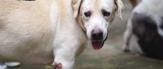
Joint diseases have become common among dogs kept in the city. This is facilitated by a sedentary lifestyle, excess feeding and, accordingly, excess weight, congenital characteristics of many dog breeds. The most common manifestations of joint diseases are decreased mobility, lameness, difficulty moving up stairs, and stiff movements, especially after sleep or rest. Mostly joint diseases occur in the second half of life.
Surgical operations on joints and ligaments are a kind of aerobatics, which, of course, affects their cost, so often the only available option for many dog owners is drug treatment, which must be comprehensive.
Drug treatment for joint disease in dogs has become much more successful due to the introduction of new nutritional supplements and medications, but hereditary joint disease cannot be completely prevented by drugs. Specially selected diets, moderate exercise, nutritional supplements, anti-inflammatory drugs and pain relievers can reduce the progression of inflammatory and degenerative joint diseases, but birth defects such as dysplasia will not disappear.
Any treatment, surgical or drug, will be more successful if the dog is not overweight. Convincing owners to feed and pamper their dogs less is the most difficult task in treating joint diseases.
Your dog's exercise should provide a variety of movement and exercise the muscles, but not place excessive stress on the joints. Walking on a leash, swimming, walking on a treadmill, jogging slowly, and going up and down stairs are excellent exercises. The selection of an exercise program should be individual for each dog, depending on the severity of the joint disease, weight, and general condition of the dog. Lack of exercise is even more harmful than excess, but choosing the wrong type of exercise can cause harm. Physical activity should be uniform and regular. A situation where a dog sleeps on the sofa all week, and on weekends the owners give it a full load, is unacceptable. Only your veterinarian can determine the best exercise program for your dog.
In the treatment of joint diseases, physiotherapy is successfully used - magnetic, laser, infrared effects. Just like massage, physiotherapy is prescribed only by a veterinarian.
Arthritis symptoms tend to get worse in cold, wet weather. At such times, it is better to put a woolen overall on the dog.
Recently, special orthopedic mattresses for dogs have appeared that reduce the load on the joints. In any case, the place where the dog sleeps should be warm and away from drafts.
For large breed dogs, we recommend purchasing special stands for bowls so that the dog does not have to bend down for food and food.
Health to you and your pets!
Diet and physiotherapeutic treatment
For more effective treatment of any joint disease in dogs, a special diet is prescribed. The diet should not contain dry food containing large amounts of carbohydrates and starch. It is also necessary to exclude cereals and potatoes from the diet, since such products can provoke an exacerbation of the existing disease. It is important to diversify the dog’s menu with cartilage, greens and fatty fish. You should not feed a sick animal fatty meats, which include pork. It is better to give preference to beef and poultry.
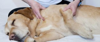
Physiotherapeutic treatment is widely used in the fight against this disease. For example, massage improves blood circulation in the affected limb, reduces pain, relaxes muscles, and promotes the resorption of exudate in the joints.
To stimulate restoration processes in cartilage tissue, heating is used. This procedure has a beneficial effect on the ligaments of the joints, thereby increasing their elasticity and improving mobility.
Diagnosis and treatment
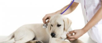
If signs of arthritis appear, you should contact your veterinarian. The doctor will examine the dog, assess the condition of the joints, and check their mobility.
The veterinarian will prescribe general clinical tests (general and biochemical blood tests) to determine the type of arthritis.
For further diagnosis, it is necessary to take an x-ray of the affected joints. Sometimes a diagnostic puncture is used.
Treatment of arthritis in dogs is most often medicinal in nature, with the following drugs prescribed:
- Non-steroidal anti-inflammatory drugs, for example Metacam, Onsior. They relieve pain and help reduce inflammation.
- Antibacterial drugs. Arthritis should be treated with antibiotics if it is caused by a bacterial infection.
- Chondroprotectors. Prescribed if the appearance of arthritis is associated with degenerative changes, as well as with arthrosis.
In addition to drug treatment, physical therapy is also indicated. A number of exercises can be performed at home. Any load should be soft, gentle and gradual. Veterinarians recommend water treatments, such as visiting a swimming pool with your dog.
Treatment of bursitis in dogs is also medicinal and involves the use of painkillers and anti-inflammatory drugs.
Prevention
A set of preventive measures should begin from the “childhood” of the pet.
- To avoid irreparable changes, it is necessary to control the puppy’s physical activity from a very early age.
- Systematically carry out a routine medical examination, and in no case ignore medical examination. This is especially true for dogs that are intended for breeding.
- It is mandatory to carry out X-ray monitoring of the skeleton.
- The pet's diet should consist of a full range of vitamins and minerals that are necessary for the normal development of bone tissue.
- Avoid overcooling the animal and avoid dangerous situations associated with injuries.
- Do not use artificial fabrics for bedding.
- Promptly carry out disinfection, deworming, and keep the dog in decent sanitary conditions.
- It is unacceptable to allow an animal to overeat to avoid obesity. This fact also contributes to the development of the disease. If your pet is gaining weight, it is worth reviewing its menu and creating a new diet that allows you to control body weight as much as possible.
- It should be noted that therapeutic measures will not give the desired effect if excess weight is an obstacle.
A routine medical examination is a preventive measure.