Etiology of pneumothorax in animals
Air can enter the pleural cavity through a wound in the chest wall.
Secondary pneumothorax occurs when the pulmonary pleura is ruptured, it happens in the case of a rib fracture, a sudden increase in intrapulmonary pressure during coughing or heavy work, through the rupture of pulmonary abscesses, gangrenous or tuberculous foci.
In dogs, pneumothorax often occurs due to trauma (bites by large dogs, car injuries, strong blows to the chest area, gunshot wounds, puncture wounds to the thoracic area).
Pneumothorax may be a consequence of purulent-putrefactive pleurisy.
In cattle, pneumothorax was observed due to the penetration of gases from the forestomach through traumatic reticulophrenitis (traumatic reticulitis with damage to the diaphragm). That is, various pathological processes in the gastrointestinal tract can lead to pneumothorx in cattle.
Types and varieties of pneumothorax in cats. Their reasons
There are closed (internal) and open (external) pneumothorax.
There are also several more varieties, and they all have different etiologies. With closed pneumothorax, the pleural cavity communicates with the external environment through the respiratory tract: in particular, ruptures of the lungs, ruptures of the trachea or bronchi, penetrating injuries to the esophagus (extremely rare); and with open pneumothorax - through damage to the chest wall: wounds from blows, bites or gunshot wounds.
The following types of pneumothorax are distinguished:
- valve (in some sources it is designated as tense);
- traumatic;
- spontaneous.
Valvular pneumothorax develops when the chest is damaged, when, when inhaling, air is sucked into the pleural cavity, but when exhaling, it cannot come back out due to the closure of the edges of the wound. As a result, excess pressure is created in the chest cavity on the lungs, and they are compressed. This condition prevents the flow of venous blood to the heart.
The causes of traumatic pneumothorax are automobile injuries, gunshot or stab wounds of the chest wall, injuries during intubation and thoracentesis, etc.
Spontaneous pneumothorax develops in middle-aged or elderly animals with diseases of the lungs and pleura associated with the destruction of the lung parenchyma, the formation of pathological cavities and necrosis of the visceral pleura. Such diseases include: dirofilariasis, invasive thymoma, oncology or lung abscess, congenital lung cysts, etc.
Pathogenesis
There are open, closed and valve pneumothorax.
With open pneumothorax in animals, when inhaling, air enters the pleural cavity through a wound in the chest wall, and when exhaling, it comes out. Gradually, the pressure in the pleural cavity equalizes with atmospheric pressure, which leads to collapse of the lungs. On the damaged side, the mediastinum shifts to the healthy side, and intrathoracic pressure increases. The healthy half of the lungs takes over the function of the damaged one, so vicarious emphysema may occur. In horses, the pleural cavities are connected, so they have a bilateral pneumothorax, and if the amount of air in them increases significantly, then death occurs from asphyxia.
Pneumothorcus in horses is an extremely dangerous pathology!
Closed pneumothorax in dogs and other animal species is characterized by a similar development, but after the hole is closed, the air resolves and all symptoms disappear. Closed pneumothorax also occurs in cases where gas in the pleural cavity is formed from putrefactive exudate.
Valvular pneumothorax occurs in cats, dogs and other animal species. This type of air accumulation in the pleural cavity is characterized by the fact that air enters the pleural cavity with each breath, but its exit is difficult, so the air pressure in the lungs can exceed atmospheric pressure.
Pneumothorax is accompanied by compression of the lungs, difficult gas exchange in the lungs, can lead to death from asphyxia, and is complicated by pleurisy and pneumonia.
View this post on Instagram
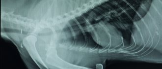
A dog, German Spitz breed, age 1.5 years, was bitten by another dog. Clinical signs: lethargy, cyanosis, subcutaneous enphysema. X-ray: pneumothorax, fracture of ribs 5-6-7-8-9 on the right. A thoracotomy, examination of the chest cavity (pulmonary contusion), and drainage of the chest cavity were performed. The patient is in stable condition. #SPb#Peter#trauma#pneumothorax#bitusanarana#wound#thoracotomy#surgeon#doctor#veterinarian#veterinarian#doctors#surgeons#surgery#X-ray
Publication from Khabarov Nikita Vladimirovich