Lymphoextravasat
Lymphoextravasat is a closed tissue injury accompanied by rupture of lymphatic vessels and accumulation of lymph in the newly formed cavity.
Etiology. Lymphatic extravasation develops when damaged by blunt objects acting on tissue in an oblique direction. They arise from sliding blows from horns, hooves, falls, or from forcible movement of a lying animal by dragging. In horses, lymphatic extravasation often occurs in the area of the lateral slopes of the withers, due to tissue displacement by a saddle or saddle, and in cattle - in the area of the anterior slope of the withers due to compression by the restrictive pipe in front of the feeders.
Classification. With simultaneous damage to blood vessels and blood admixtures in the lymph, hemolymph extravasates develop. Under the influence of mechanical force, separation of tissue layers occurs with simultaneous rupture of lymphatic and small blood vessels. Depending on the depth of tissue damage, lymphatic extravasates can be superficial, subcutaneous, deep, interfascial, intermuscular.
In cattle, lymphatic extravasation most often develops in the groin, thighs, perineum, abdominal and chest walls, and in horses - in the withers and back of the head. Lymphatic extravasates are not formed in pigs.
Clinical signs. Initially, a slight swelling of the tissue occurs in the damaged area with the presence of an abrasion on the skin. After the edema resolves, on the 3rd – 4th day after the injury, a limited fluctuating swelling is detected, when pressure is applied to it, fluid moves from one part to another. The skin in the area of swelling is not tense and therefore it seems that the volume of the cavity is much larger than the liquid contained in it. Over time, the swelling can take on different shapes and sizes, sometimes reaching a volume of up to 10–15 liters. Weak blows to the wall of the lymphatic extravasate cause wave-like movement of the contents (undulation).
In cases of deep lymphatic extravasation, swelling is detected much later than superficial ones. At the same time, it does not have a sharp outline and its outer wall is tense. Unlike hematomas, lymphatic extravasation develops slowly over several days and even weeks after injury. When hemolymphoextravasate is formed, fibrinous crepitus is detected by palpation, the punctate has a red or pink color, while with lymphoextravasate it has the appearance of a yellow opalescent liquid. In most cases, with lymphatic extravasation there is no pain, no increase in local and general body temperature.
Forecast. For small lymphatic extravasations it is usually favorable, for large ones it is cautious (due to poor thrombus formation in the vessels).
Treatment. In order to reduce lymphatic drainage, the animal must be given complete rest, since active movements increase lymph flow by 5 times. Cold and heat are contraindicated, since cold can cause necrosis of exfoliated skin, and thermal procedures significantly increase lymphatic drainage. To reduce lymphatic drainage, moderate pressure bandages with camphor, boric or ichthyol alcohol are used in the first 1–2 days.
Lymphoextravasate in cats: basic information, diagnosis and treatment
Subsequently, emptying punctures are carried out followed by the introduction of a 1–2% alcohol solution of iodine into the cavity of the lymphatic extravasate.
In order to increase the coagulability and resorption of lymph, it is advisable to inject autologous blood into the lymph extravasate cavity (after it has been emptied). More effective is the administration (after emptying puncture) of hydrocortisone, which, acting on the intima of the lymphatic vessels, causes a decrease in lymphatic effusion. If the above treatment is ineffective, they resort to opening the cavity of the lymphatic extravasate with a linear incision in its wall. After opening, fibrin clots are removed from the cavity, tissue soaked in lymph is excised, the walls of the cavity are treated with antibiotics, the exfoliated skin is sutured to the underlying tissues with vertical loop-shaped sutures with rollers, the skin incision is closed with an interrupted suture.
If it is not possible to apply sutures, the lymphatic extravasate cavity is drained with gauze soaked in a 1–2% alcohol solution of iodine or 10% iodoform ether. The drainage is removed after 2 days and further treatment is carried out as with a regular wound. However, according to research (E.I. Veremey, V.M. Lakisov, etc.), the most effective is to use gauze soaked in a 2.5% solution of vagotil in 70% ethyl alcohol or ayatin for draining the cavity of lymph extravasate. After a single use of this solution, lymphatic drainage completely stops.
A blister is an element of a rash; it belongs to the primary ephemeral (existing no more than a few hours) elements. Typically, blisters appear against the background of the appearance of red spots (skin hyperemia). Non-specialists often confuse blisters with other primary elements of the rash (papules, vesicles).
The most common mistake that cat lovers make is when they are looking for treatment for burn blisters. They mistakenly consider the liquid-filled element formed on the pet’s skin due to a burn to be a blister. This is actually a post-burn blister. Sometimes even skin metastases of malignant neoplasms are called blisters.
Treatment of lymphoextravasate in cats
Treatment of the disease is carried out by opening the edematous cavity, the liquid inside is pumped out, and rinsing with an antiseptic solution is carried out. Instead of opening by dissection, in unadvanced cases, the swelling is punctured and the exudate is pumped out using a syringe and needle. A complication after such a procedure may be a recurrence of the disease, despite complete pumping of the fluid. If the affected area of the cat is large, veterinarians do not recommend puncture treatment; surgical intervention is performed: under anesthesia, the skin is dissected, the fluid is removed, the cavity is washed, exfoliated, damaged tissue is excised, at the end of the procedure the wound is sutured and covered with a sterile bandage.
Description of blisters and spots
A blister or urtica is a cavityless element that occurs as a result of acute inflammatory edema of one of the layers of the dermis. It disappears without a trace and quite quickly (within several hours). The appearance of urticarial rash is characterized by an acute onset, the rash appears almost instantly. Dense pink areas appear on the skin, rising above the general surface of the skin. The rashes are round or irregular in shape.
A spot or macula is a change in the color of the skin. Spots are a vascular reaction of an inflammatory or non-inflammatory nature. They appear and pass quickly enough.
Causes of blisters and spots
Blisters usually occur as an allergic reaction to irritants and disappear quickly. Irritants can be:
- endogenous;
- exogenous.
Most often, blisters in a cat occur as a reaction to an insect bite or a food allergy (urticaria), or toxidermia.
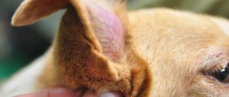
Hives in cats refers to a mild reaction to insect bites. Very often it develops from a flea bite. In this case, along with the bite, the animal develops a red hot spot and a blister, and the lips may swell (because the cat licks itself and flea saliva and feces get into its mouth). The skin around the eyes and neck may also swell. Cats often have completely swollen faces.
When bitten by poisonous insects (wasps, bees) or snakes, the animal develops a large red spot at the site of the bite and a large blister, and the skin temperature rises at the site of the bite. The bite site may be painful.
Blisters on the skin can appear due to food allergies.
If the cat has lymphatic extravasation, treatment
In this case, small blisters usually appear throughout the animal's body. The blisters are very itchy and the animal itches intensely.
When the animal's body comes into contact with hot objects (batteries), vascular pink spots may appear on the skin, which quickly disappear. Redness may also occur when the skin comes into contact with ice.
A blister is one of those elements of the rash that can appear at the initial stage of development of any type of dermatitis. But they should not be considered a characteristic sign of dermatitis. They quickly pass, giving way to blisters, papules, ulcers, and fodder. Red spots on the skin of an animal with dermatitis are usually present along with other elements of the rash.
An allergic reaction to food may initially appear in the form of scarlet spots on the animal’s skin. This reaction is accompanied by itching and loss of fur at the site of the spot. This may include food allergies and contact dermatitis.
Ringworm in cats can also begin with the appearance of a pink spot on the skin. Hair falls out at the affected area. The animal may be bothered by itching. Then other elements of the rash appear.
Symptoms of hematoma in dogs
At the site of hemorrhage, a hematoma is characterized by swelling, which appears immediately after the injury, has well-defined contours and quickly increases in size. An ear hematoma in a dog looks like a pillow or hot water bottle filled with water.
At first it causes pain to the dog, but after a few days the pain goes away and the swelling may begin to smooth out.
Usually the local temperature of the organ increases in the area of the hematoma, sometimes it can increase near
Treatment
It is best to show the animal to a specialist. He may prescribe antihistamines to help relieve itching and swelling.
If blisters appear as a result of a food allergy, it is advisable to change the animal's food. If he eats natural food, then he will have to tinker with eliminating the allergen, eliminating one product after another until it becomes clear what exactly the cat is reacting to.
If blisters or spots on the skin are the result of an insect bite, you can apply a cold compress to the bite site. This will reduce itching and relieve swelling.
If fleas are present, antiparasitic treatment of the animal is required.
If lichen is detected, specific therapy is aimed at destroying colonies of parasitic fungi.
Causes of auricular hematoma in cats and dogs
So, as excessive shaking of the head and excessive trauma to the auricle with the front and hind paws is the main root cause of the occurrence and development of lymphatic extravasation in cats
and dogs, you need, first of all, to understand the cause of the itching and discomfort in this area. This is important both for diagnosis and for treatment and prevention.
Often the cause of soreness is an obvious injury, bruise or wound to the ear, but more often it is an infection or a severe allergic reaction. The ear of a cat or dog is carefully examined using an otoscope; The contents of the auricle are cleaned out and a small amount is taken for microscopic examination for the presence of bacteria, yeast or mites. Any of these reasons should be considered for the diagnosis of lymphatic extrovase, since any of them causes irritation of the delicate skin of the ear canal.
If a specific microflora is detected, it is necessary to immediately determine its sensitivity to antibiotics and antifungal drugs; if a tick infestation is detected, then it is necessary to develop a scheme for acaricidal treatment of the animal, causing the least irritation of the ear canal.
If the roots of this problem are of allergic origin, then, if possible, it is advisable to establish what exactly it could develop into. If its cause is discovered, it is necessary to exclude this allergen, be it an environmental factor or a nutrition-related factor.
Particular attention should be paid to dogs with long ears. It is important to check and clean them regularly to keep your ears and ear canal clean and not overly wet. This prevents stagnation in them and the proliferation of opportunistic microflora. After bathing and hygiene procedures, it is advisable to wipe the animal’s ear dry and let it dry.
leave a comment
Lymphatic extravasates can be localized in various areas of the body. In cattle they most often occur in the area of the hips, perineum (Fig. 93), abdominal and chest walls, and in horses - in the area of the withers, poll and dewlap. In the first hours, a slight, painless inflammatory swelling of the tissues occurs at the site of injury with a slight increase in local temperature. After resorption of the edema, a sharply demarcated swelling clearly appears, when pressure is applied to it, a wave-like movement of fluid from one section to another is felt - undulation. Lymphoextravasate is characterized by slow and prolonged development of swelling. It reaches its maximum size several days and even weeks after the injury. Despite the significant accumulation of lymph in the cavity formed by it, there is no skin tension in the affected area. It seems that the volume of the resulting cavity is much greater than the volume of the lymph contained in it. It is also characteristic of lymphatic extravasates that the inflammatory reaction and pain with them are mild, there is no local increase in temperature and no general reaction of the body. When the swelling is punctured, a clear or slightly opalescent lemon-yellow liquid is obtained - lymph, sometimes mixed with fibrin. The presence of blood in the punctate indicates hemolymph extravasation. Forecast. With lymphatic extravasation, the prognosis in most cases is favorable, since animals usually recover even if they have extensive lymphatic extravasation.
Ear diseases in cats
In cases of complications of lymphatic extravasation by the development of microbes in them, the prognosis depends on the type of microorganisms that caused the complication. Treatment. The sick animal is given rest, which is especially important to reduce the secretion of lymph and accelerate the organization of a blood clot in damaged lymphatic vessels. It should be borne in mind that conservative treatment methods for lymphatic extravasation are ineffective, and some of them, such as the use of cold and heat, are even contraindicated. The latter is due to the fact that cold can cause skin necrosis, and thermal procedures increase lymph circulation and promote lymphatic effusion. For the same reasons, massage is not used for lymphatic extravasation. Considering the above, for small superficial lymphatic extravasations, a conservative-operative method of treatment is used. It is as follows. During the first 24 hours, moderately pressing wet-dry dressings with camphor alcohol or an alcohol solution of ichthyol are used. Subsequently, emptying punctures are used, followed by the introduction of a 1-2% alcohol solution of iodine into the lymphatic extravasate cavity and the application of a pressure bandage. These procedures usually have to be repeated several times. The most reliable method of treatment for lymphatic extravasation is surgery. The wall of the lymphatic extravasate is dissected with a small linear incision near its lower border, the contents of the cavity are removed, and the cavity itself is drained with gauze, abundantly soaked in a 1-2% alcohol solution of iodine, 10% iodoform ether or 1% alcohol solution of formaldehyde. The drainage is removed after two days. Subsequently, liquid Vishnevsky ointment, sulfonamides and other antiseptics are used. With this method of treatment, recovery occurs in 2 to 3 weeks. To speed up healing, I. E. Povazhenko recommends bringing the wall of the lymphatic extravasate cavity closer together by applying a suture with rollers.
Tags: veterinary medicine, treatment, injuries
Diagnosis and treatment of auricular hematomas
Usually, this problem is quite clearly visible visually and a veterinarian, upon examination, can easily diagnose lymphatic extrovase in a cat or dog. If the swelling is soft in consistency and warm, this indicates a small hematoma, but if it is tense and hot, then most likely this hematoma fills a large area of the auricle.
Treatment of hemolymphoextrovasation of the auricle in cats and dogs includes not only a solution to relieve swelling, but also determining the true cause of its occurrence and resolving this causative factor. There are several different options aimed at reducing the volume of the hematoma, and the first of them is pumping out the exudate using a syringe and needle. The surface of the hematoma is treated with an antiseptic antibacterial solution that prevents the spread of infection, and a syringe is used to puncture the skin on the inside of the ear at an angle of 90° or 45° and, if possible, completely pump out the contents of the pocket. Quite often, without removing the needle, a second syringe is connected to it, with a special solution of antibiotic, hormonal and anti-inflammatory drugs that promote the development of adhesive inflammation; and is introduced in a small volume into the cavity of the pocket. This is the simplest and most inexpensive way to solve the problem, but it has many disadvantages.
The most common consequence of puncture of the hematoma may be its fairly rapid refilling, despite complete pumping of the exudate. As mentioned above, there is also a fairly high risk of introducing opportunistic microflora and developing inflammation. If, when the skin of the inner surface of the auricle is punctured, a blood vessel is additionally damaged, then blood clots may form in the hematoma cavity and, during its healing, quite powerful and rough scar tissue may develop. If rapid healing does not occur, then the clots will have to be evacuated through a massive incision.