Ascites is dropsy of the abdominal cavity in cats. The idea that this is a separate disease is erroneous. Ascites develops against the background of a chronic pathology that has developed in the body, and is characterized by the accumulation of fluid in the abdominal cavity, which exceeds normal levels.
Excess intrauterine fluid puts pressure on the internal organs, making it difficult for them to function. The process is dangerous because dangerous complications, including death, will arise in the pet’s body.
Why does ascites develop?
Absolutely any animal can suffer from ascites, but cats that lead a sedentary lifestyle are most susceptible to this pathological condition. A small volume of serous fluid is always present in the abdominal cavity, ensuring satisfactory functioning of the internal organs.
Indicators exceeding the norm pose a danger to the body.
Excessive accumulation of fluid in the abdominal cavity is provoked by the following factors:
- cardiovascular pathologies;
- renal failure;
- liver diseases;
- decreased albumin levels;
- hepatitis;
- diabetes;
- tuberculosis;
- peritonitis;
- infections, parasites, fungi;
- weak immunity;
- overweight;
- hormonal imbalances;
- oncology;
- abdominal injuries that provoked an inflammatory process in the abdominal cavity;
- bladder rupture;
- nephrotic syndrome;
- excess salt in the diet, economy class food and, as a result, an increase in sodium concentration;
- violation of water-salt and protein metabolism.
This is not a complete list of the reasons leading to abdominal dropsy in cats.
Symptoms of ascites
Intra-abdominal fluid in a cat accumulates gradually over some time. The manifestation of external signs occurs with the development of changes in the internal organs. Signs of ascites are:
- the cat's unnaturally large belly;
- tousled, dull coat;
- difficulty breathing, shortness of breath;
- decreased motor activity, the cat lies most of the time;
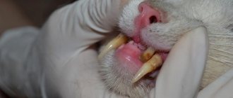
- yellow tint of the gum mucosa;
- rapid heartbeat;
- refusal of food, rapid exhaustion.
INTERESTING TO KNOW: Symptoms and treatment of rhinotracheitis in cats
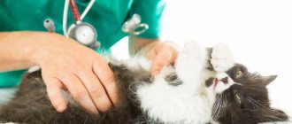
Ascites in a cat can be determined by its moving abdomen. It shifts when the animal’s body position changes. When palpating the abdomen, one feels the movement of fluid under the fingers. But in order to accurately determine the presence of fluid in the cat’s abdominal cavity, you need to contact a veterinarian. The true cause of ascites can be determined through a diagnostic examination of the animal.
Clinical picture
The initial stage of the pathological condition is virtually asymptomatic, since the accumulation of fluid in the abdominal cavity is a gradual process. Characteristic signs can only be noticed when the volume of fluid exceeds normal levels. Ascites can be suspected by a noticeably swollen abdomen and stretching of the sides when the animal is active.
Symptoms:
- the abdominal cavity is swollen, hard to the touch;
- fluctuation - when pressing on the abdomen, you can feel the liquid inside it swaying;
- the belly takes on the shape of a pear when the cat stands on its hind legs, and inflates and rounds when taking a sitting position;
- dullness and disheveled fur;
- yellowish tint of mucous membranes;
- paws, ears, crotch, sternum swell;
- vomiting, nausea;
- lack of appetite;
- constipation or diarrhea;
- breathing problems, hoarseness, shortness of breath;
- weakness, apathy.
It is very important to distinguish ascites from ordinary overeating.
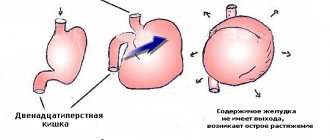
Abdominal dropsy. Ascites
Abdominal dropsy, ascites (Ascitis) is a chronic secondary disease associated with difficulty in resorption of peritoneal fluid into the blood and lymph circulation system and the accumulation of this fluid in the abdominal cavity.
All types of farm animals and birds suffer from ascites, but the disease is most often recorded in dogs, sheep, pigs and less often in large animals.
Etiology . The cause of ascites in animals is obstruction of the outflow of peritoneal fluid. The outflow of fluid depends on the state of the portal circulation and normal heart function. The disease in animals is registered with diseases of the liver, heart, kidneys, lungs and blood vessels located in the abdominal cavity of the animal. In case of chronic diseases of the heart, liver, kidneys (liver cirrhosis, amyloidosis), tumors, fascioliasis, echinococcosis and other organs, animals may develop general venous stagnation. With venous stagnation in animals, transudate leaks into the subcutaneous tissue and all body cavities, causing dropsy. Ascites in piglets occurs due to edematous disease. The occurrence of the disease is facilitated by increased permeability of the walls of blood vessels, hydremia, and a lack of vitamins K and C in the body.
Pathogenesis . The pathogenesis of ascites is based on functional liver failure, disruption of water-salt and protein metabolism, as well as damage to the vascular system of the peritoneum and its mesothelial cover. A large accumulation of fluid in the abdominal cavity also leads to difficulty in the functioning of the circulatory organs, both in the portal and systemic circulation, causing restriction of the movement of the diaphragm, as well as inhibiting the motor activity of the stomach and intestines.
Clinical picture . For abdominal dropsy, a characteristic symptom is a gradual, over several months, symmetrical increase in the volume of the abdomen. When conducting a clinical examination of an animal with ascites, the veterinarian is struck by a symmetrical bilateral protrusion of the lower and lateral surfaces of the abdominal wall, sometimes the retraction of the iliac fossae, and the back of the sick animal bends. The animal experiences rapid fatigue, swelling of the limbs and emaciation of the animal. Sick animals with ascites reluctantly change their position, and small ones either tend to lie down or sit.
On clinical examination, the visible mucous membranes are pale at the beginning of the disease, then become cyanotic (cyanotic). Pulse and breathing are rapid and weakened. When percussing the area of ascites, we obtain a dull percussion sound that has a horizontal line of dullness; when the position of the body in space changes, the line of dull percussion sound changes.
When auscultating the intestines, bowel sounds are not audible or are greatly weakened. In the absence of complications, the temperature of a sick animal is always normal. A sick animal periodically has cases of tympany and constipation, the animal refuses or is reluctant to eat the offered food. When palpating the area of ascites, we register fluid fluctuations. Upon test puncture, we obtain a straw-yellow liquid that contains a small amount of protein. The appearance of jaundice in a sick animal is a sign of an unfavorable outcome of the disease.
Pathoanatomical changes . During a post-mortem examination of dead adult animals, we find a large amount of yellowish, rarely reddish fluid in the abdominal cavity (up to 20 liters in dogs and up to 100 liters in horses). The fluid contains few blood cells and some protein (1-20). At the beginning of the disease, albumin predominates in the fluid, and globulins predominate in the final stage.
The surface of the peritoneum was not changed upon examination. We find damage to the liver, heart, kidneys and other organs, which are the main cause of ascites in animals.
Diagnosis . Veterinary specialists make a diagnosis of ascites based on the clinical symptoms of the disease, which we establish during a clinical examination (palpation, percussion and auscultation). Carrying out a diagnostic puncture of the abdominal wall in the area of ascites, ultrasound, x-ray, and urine analysis.
Differential diagnosis . A veterinarian must differentiate ascites from pregnancy and bladder enlargement, which can be ruled out by performing a rectal examination. We exclude chronic peritonitis and internal bleeding (by examining the transudate with a test puncture).
Flow. The course of ascites in sick animals is chronic; ascites lasts for months.
Forecast. The prognosis for ascites is directly dependent on the presence of the underlying disease in the sick animal that caused ascites and the possibility of its elimination. If the underlying disease begins to progress in the animal, then exhaustion and death occur.
Treatment . Treatment for ascites should be aimed at the underlying disease that led to ascites. It is necessary to exclude liquid food from the feeding diet, reduce, and sometimes eliminate, the supply of table salt, and reduce the amount of water. We carry out symptomatic treatment with the aim of resolving transudate from the abdominal cavity, using for this purpose diuretics (diacarb, mercuzal, aldactone, veraminaron, furosemide lasix, torasemide) and sweatogenic drugs, as well as agents that enhance salivation in animals. Sick animals are given a laxative internally. To maintain cardiac activity, sick animals are injected with a 20% sodium caffeine solution and digitalis preparations are prescribed in standard doses. To reduce the permeability of the walls of blood vessels, sick animals are injected intravenously with a solution of 10% calcium chloride in generally accepted dosages, vitamins K and C.
If a sick animal has accumulated a large amount of transudate, we do, in compliance with the rules of asepsis and antisepsis, puncture the abdominal wall and release the accumulated transudate, but this gives a temporary effect.
When the fatness and productivity of farm animals decrease, it is advisable to cull them.
Prevention . Animal owners must promptly treat patients with hepatitis, hepatosis, liver cirrhosis, as well as diseases of the cardiovascular and urinary systems.
Diagnostics in a veterinary clinic
The task of diagnosis is to differentiate otitis from other diseases with similar symptoms, for example, from exudative peritonitis. Thus, with peritonitis, an increase in temperature, pain when pressing on the abdomen, and a significant number of leukocytes and proteins in the exudate are observed.
And finally, the most obvious sign of peritonitis is the transience of the disease. With ascites, as mentioned above, the disease has a long development.
Necessary diagnostic methods for suspected ascites include:
- anamnesis;
- analysis of clinical symptoms;
- palpation of the abdominal cavity;
- urine and blood tests;
- X-ray of the peritoneum, sternum;
- Ultrasound of the chest and abdominal cavity;
- fluid examination;
- endoscopy;
- biopsy.
Differential diagnosis helps to identify the underlying pathology that caused ascites. Without determining the provoking factor, it is impossible to prescribe adequate treatment, since the amount of fluid will increase with each relapse.
Differential diagnosis of ascites
When diagnosing ascites, it should be differentiated from exudative peritonitis, the external manifestations of which are similar to those of ascites. Distinctive signs of ascites are:
- Normal body temperature. With peritonitis it increases significantly.
- Painless condition. With peritonitis, a cat reacts painfully to touching its stomach.
- Various chemical composition and specific gravity of fluid in the abdominal cavity. The main difference is that with peritonitis, the exudate contains a large amount of protein and leukocytes.
- Ascites develops gradually and over a long period of time, while peritonitis develops rapidly over several hours.
Can ascites in cats be treated or not?
Treatment of ascites is carried out comprehensively. Therapy is aimed at eliminating the underlying disease, alleviating pain symptoms and strengthening the immune system.
To reduce the amount of fluid in the abdominal cavity, laxatives and diuretics are indicated (Furosemide, Temisal, decoction of bearberry leaves, liver and kidney teas).
To stabilize the functioning of the cardiovascular system, drugs such as Olitorizide, Strophanthin, Digitoxin, Cardiovalen are necessarily prescribed.
If there is an infection in the body, antibiotic therapy using drugs from a number of cephalosporins is indicated.
To strengthen the walls of blood vessels and reduce their permeability, the veterinarian prescribes intravenous solutions of calcium chloride (10%).
To pump out the fluid, a puncture is done when the veterinarian pierces the abdominal wall. The procedure is performed at least twice a week. In this case, the removal of ascitic fluid must be compensated by reinfusion of ascitic transudate or administration of an albumin solution. These procedures provide a chance for additional remission and prolongation of the animal’s life.
The course of treatment depends on the condition of the animal and the amount of fluid. This is a long process that must continue until the clinical signs of dropsy completely disappear.
As for the prognosis, with adequate and, most importantly, timely therapy, it is favorable. If you do not seek help from a specialist in time, the volume of fluid will increase to two liters per day or more. This pathological condition is fraught with pressure on internal organs and disruption of their functions.
Disease prevention
The best method of dealing with any disease is its prevention. Preventive measures against dropsy are available to any cat breeder, the main thing is not to forget about it:
- Compliance with the diet (when preparing a diet, you can contact a veterinarian).
- Walks in the open air.
- Fortification (you can select good vitamins with the help of a veterinarian).
- Avoidance of stressful situations (cats under stress have reduced immunity).
- Preventive vaccinations, as well as treatment for worms, fleas and other parasites.
- Preventative examinations at the veterinarian.
Ascites (abdominal dropsy) is an accumulation of free fluid in the abdominal cavity, but this phenomenon cannot be called an independent disease. Typically, ascites occurs against the background of another disease as a symptom. This disease is the cause of dropsy. Therefore, treatment must be comprehensive. Veterinarians prescribe groups of drugs aimed at combating symptoms, and the underlying disease is treated separately.
What should the owner do
Under no circumstances should you play Aibolit or give your pet medications without consulting a veterinarian. Only a specialist can prescribe medications and set their dosage, based on the severity of the pathology, general condition and age of the animal.
Uncontrolled use of medications can lead to potassium being excreted from the body along with urine. This will worsen the cat’s well-being and lead to complications, even to the point where the animal dies. Human diuretics are especially dangerous in this regard.
Increasing their dosage independently can lead to disturbances in the level of electrolytes and leaching of fluid from the microcirculation network. This is fraught with the possible development of encephalopathy.
If the owner does not have the opportunity to immediately take his four-legged pet to a veterinary clinic after identifying dangerous signs, this can alleviate his condition with the help of birch decoction. To do this, dry birch leaves are brewed with boiling water in a ratio of 1:10, leave for at least 5 hours. Give your cat the prepared infusion twice a day.
However, you need to understand that this folk remedy can only alleviate symptoms, but it cannot replace full-fledged drug treatment.
As soon as possible, you should take the animal to the clinic so that the doctor can examine it and prescribe appropriate treatment.
A cat with abdominal hydrops is prescribed a therapeutic diet that involves limiting the consumption of water and any other liquid. The food of a sick pet should be nutritious, balanced in vitamins, minerals and carbohydrates and consist mainly of proteins.
The diet includes meat, poultry, sea fish, cottage cheese, and kefir. You can add salt to your food, but the amount should be minimal.
Treatment
In the meantime, symptomatic treatment is necessary, especially if the case is serious and the general condition of the animal suffers. The following symptomatic therapy may be used for some (but not all) animals with ascites. It can reduce the intensity of symptoms and make life easier for your pet. However, this non-specific treatment is not a substitute for definitive treatment that addresses the disease underlying the animal's current condition.
The most important aspect of treating ascites is to determine how quickly the ascites is developing and the clinical condition of the animal. If ascites develops slowly and the animal is strong enough, then emergency care is not required. If ascites develops quickly, which is often associated with loss of strength in the animal, emergency care is necessary. Treatment until a diagnosis is made may include:
Therapeutic abdominocentesis. If a lot of fluid accumulates in the abdominal cavity, it can put pressure on the diaphragm, making it difficult to breathe. A needle is inserted through the abdominal wall and the fluid is drained to relieve pressure and make breathing easier for the animal. As soon as the animal feels better, the needle is removed. You cannot remove all the fluid, as this can lead to a shift in homeostasis in the body and shock.
Diuretics are prescribed to remove fluid from the body. They increase the excretion of fluid in the urine. Diuretics are more effective at removing fluid from tissues rather than from body cavities, so their effect on ascites is limited. The most popular drug is Lasix (Furosemide).
Oxygen therapy is often required to stabilize the animal's respiratory failure. Oxygen may be provided through a mask, nasal oxygen cannula, or oxygen chamber. Typically, once some fluid has been removed from the abdominal cavity, oxygen therapy is no longer required.
Rapidly accumulating ascites requires intravenous fluids to maintain tissue perfusion and prevent shock. If an animal has reduced total protein in the blood (due to low albumin), colloidal solutions (liquids whose osmotic pressure is equal to the pressure of the blood plasma) can be used to slow the progression of ascites.
For ascites due to bleeding into the abdominal cavity, transfusion of blood or blood components is used. If a transfusion is necessary, the animal is usually very weak and has a reduced hematocrit (a blood indicator that characterizes the degree of anemia).
If infection is suspected, intravenous antibiotics may be administered until a definitive diagnosis is made. When infected, ascites requires immediate treatment.
Diagnostics
Typically, abdominal ascites in cats is a consequence of some changes in the internal organs. This may be a consequence of diabetes mellitus, diseases of the cardiovascular system or liver cirrhosis in the final stages. Peritonitis (fungal or bacterial) can also cause water to collect in the animal's abdominal cavity.
Overfeeding or eating large amounts of smoked and over-salted foods contributes to the occurrence of a disease such as ascites in a cat. In order to help your animal in time and not miss the first signs of the disease, you should periodically take tests and visit a veterinarian.
How to diagnose ascites
The veterinarian will examine your pet and ask you a series of questions to make an accurate diagnosis.
Your pet will then be sent for blood and urine tests and an ultrasound examination of the internal organs - the chest and abdomen.
If necessary, the cat will undergo an x-ray of internal organs and a biopsy.