Description
There are 2 forms of hyperparathyroidism:
- Primary - characterized by increased levels of parathyroid hormones. The disease occurs due to dysfunction of the gland itself. This form of pathology is very rare. Typically, primary hyperparathyroidism is diagnosed in older cats with parathyroid hyperplasia or enlarging tumors.
- The secondary form is the most common. Its development is caused by kidney disease, in which the balance of electrolytes in the body is disturbed. As a result, the amount of phosphorus increases and the level of calcium in the blood decreases. To correct the imbalance, the body begins to intensively produce parathyroid hormone. Against the background of such disorders, there is a decrease in the level of calcitrol, which is the active form of vitamin D. It is produced in the kidneys and helps improve the absorption of calcium from the intestines. Its deficiency in the body leads to disruption of the process of bone mineralization.
A nutritional form of hyperparathyroidism, caused by excess phosphorus in the diet, is common in kittens. For normal formation of bone tissue, the ratio of calcium and phosphorus in the skeleton and blood must be constant.
An imbalance occurs as a result of the kitten's intake of predominantly meat food, which is rich in phosphorus but contains little calcium. A cat is a predatory animal that, under natural conditions, eats its prey along with the bones, thus replenishing its body with calcium. Pet owners often feed them beef, chicken, liver, and sausage. Introducing cottage cheese and milk into the diet does not save the situation, since the level of phosphorus in dairy products is 2 times higher than the amount of calcium.
Kittens are susceptible to the disease due to the fact that the growing body requires tens of times more calcium compared to an adult. Most often, Maine Coons, Siamese, British and Scottish kittens suffer from hyperparathyroidism.
The progression of the disease leads to the formation of multiple pathological bone fractures - both folded and microfractures, which are not visible on x-rays. Damage to the pelvic bones causes severe deformation of the pelvis and disruption of the bowel movement. Fractures or cracks in the spine cause neurological disorders.
Pathophysiology and differential diagnosis
Hypoparathyroidism is a state of absolute or relative deficiency of parathyroid hormone (PTH), which may be permanent or temporary. Hypocalcemia and clinical signs related to low calcium ionization are hallmarks of progressive hypoparathyroidism. It is an uncommon cause of hypocalcemia in dogs and cats (Table 1), but it is the only condition that requires acute and chronic treatment to reduce the clinical signs caused by hypocalcemia. Hypoparathyroidism in dogs is mostly idiopathic or primary. Surgical removal or damage to the parathyroid glands during thyroidectomy to correct hyperthyroidism is most common in cats.
| Table 1. Diseases in dogs and cats associated with hypocalcemia* |
| Common Hypoalbuminemia (with normal amount of ionized calcium) Chronic renal failure Postpartum tetany (eclampsia) Acute renal failure Acute pancreatitis Undetermined cause (mild hypocalcemia) Random Hypoparathyroidism Primary Absence or destruction of parathyroid sacs Idiopathic-spontaneous, immune Bilateral thyroidectomy After sudden cessation of chronic hyperthyroidism calcivmia ( atrophy of the remaining parathyroid glands) Suppression of PTH secretion (without destruction of the gland) Ethylene glycol intoxication Phosphate enema After ingestion of NaHCO3 Soft tissue injury, acute necrosis of skeletal muscles Rare Laboratory error Inappropriate anticoagulant for the sample (EDTA) Rapid intravenous infusion of phosphates Rapid intravenous infusion in the absence of calcium (diluted ) Intestinal hypoabsorption syndrome, severe fasting Hypovitaminosis D Blood transfusion (anticoagulant citrate) Hypoparathyroidism due to malnutrition Infarction of parathyroid adnoma (in dogs) Hypomagnesemia Parathyroid gland damage by canine distemper virus In humans ** Sepsis/critical illness Drug-induced hypoparathyroidism ( aluminum, asparaginase, doxorubicin, cytosine arabinoside, cimetidine, ethanol) Drugs that interfere with absorption (estrogen, plicamycin, calcitonin, bisphosphonates) Pseudohypoparathyroidism Parathyroid agenesis Osteoblastic bone neoplasia Hypercalcitonism Damage from reactive iodine |
| * Based on total serum calcium ** Comparative causes of disease in humans (no documented data for dogs and cats). PTH - parathyroid hormone EDTA - ethylenediaminetetraacetic acid. |
PTH levels that are too low cause hypocalcemia, hyperphosphatemia, and decreased levels of 1,25-dihydroxycholecalciferol (calcitriol). Hypocalcemia is due to increased urinary calcium excretion (hypercalciuria), decreased bone mobility, and decreased intestinal calcium absorption (secondary effect) during periods of low PTH levels. The cause of hyperphosphatemia is a decrease in the excretion of phosphorus in the urine (hypophosphaturia), which overcomes the decrease in bone mobility and absorption of phosphorus in the intestine during the period of decreased PTH levels. PTH is a strong stimulant and phosphorus is a strong inhibitor of the 25(OH)-cholecalciferol-1a hydroxylase system in the renal tubules; As a result, the absence of PTH and the presence of hyperphosphatemia contribute to a decrease in calcitriol synthesis in the kidneys. Decreased calcitriol levels contribute to hypocalcemia mainly due to decreased intestinal calcium absorption. Part of the hypocalcemia not associated with low PTH levels may result from increased calcium supply to bone following rapid correction of long-standing hypoparathyroidism or hyperthyroidism, both of which are associated with loss of bone calcium before treatment (“hungry bone syndrome”).
Patients with hypoparathyroidism can be divided into three groups: (1) with absence or destruction of the parathyroid glands, (2) with sudden correction of chronic hypercalcemia, (3) with suppression of PTH release without destruction of the parathyroid glands. The most common group of hypoparathyroidism in dogs and cats is that associated with the absence or destruction of the parathyroid glands.
Causes
Causes of primary hyperparathyroidism include:
- Malignant and benign tumors of the parathyroid gland.
- Congenital disorders in which the parathyroid gland produces excessive amounts of parathyroid hormone.
Symptoms of the primary form of the disease are more common in older animals, with cats getting sick less often than dogs.
The secondary form of pathology develops due to:
- Calcium deficiency, poor absorption of this substance by the body.
- Kidney failure.
- Gastrointestinal diseases.
- Diseases of the thyroid gland.
- Excess in the diet of foods rich in phosphorus.
- Accelerated growth of kittens aged 1-7 months.
Symptoms
At the initial stage of the disease, the cat becomes lethargic and apathetic, constantly lies in one place, gets nervous, and tries to run away when picked up.
Sinusitis
Cystitis
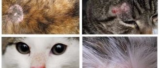
Atopic dermatitis
Over time, the animal develops severe pain in the bones, the pet becomes aggressive, and when trying to pet or pick it up, it hisses and may bite.
When the disease enters the active phase, its symptoms become noticeable even to the most inattentive owners. The cat experiences pain when walking and limps. With sudden movements (for example, jumping from a chair), fractures and microcracks form in fragile bones. The pet's gait changes: it moves slowly, on widely spaced paws.
There are also the following symptoms:
- Deformation of the pelvic bones and sternum.
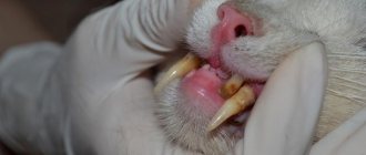
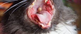
- Impaired tooth growth, tooth loss.
- Problems with urination and bowel movements (retention or incontinence).
- Development of complete or partial paralysis of the paws.
- Abdominal bloating.
- Curvature of paws.
Progression of the disease and lack of treatment leads to the development of arthritis caused by pathological changes in bone tissue.
Laboratory diagnostic methods
When examining the urine of animals suffering from hyperparathyroidism, a slight decrease in calcium levels is noted. The amount of phosphorus in the blood and urine does not deviate from the norm. Changes indicating reduced skeletal mineralization are detected only in advanced stages of the disease.
X-rays are used to confirm or refute the diagnosis. The bones of sick animals in the photographs look transparent, have thinned walls, and low density. The boundaries between soft tissues and bones are erased, and already healed fractures can be detected.
X-rays help assess the degree of bone deformation. The prognosis for a successful recovery is made based on the condition of the spine, thoracic and pelvic bones.
Cats with neurological disorders are carefully examined for spinal injuries. The pictures may show cracks, curvatures, deformation of the vertebrae, and hernias.
If an X-ray examination reveals a full intestine or bladder, the animal is given immediate assistance.
To obtain high-quality x-rays, the pet must remain motionless for several minutes, which causes difficulties during this procedure. If, due to severe pain and anxiety, the cat does not want to lie still, you need to be patient and calm the animal. The effectiveness of further treatment largely depends on the quality of the images obtained and the accurate diagnosis.
Treatment
Treatment depends on the severity of the disease, its type and existing complications. In both primary and secondary forms of the pathology, the pet is prescribed a diet that includes calcium-rich foods. Food containing a lot of phosphorus is given in moderate doses. To clearly control the amount of mineral compounds your cat eats, the veterinarian recommends switching to special balanced food. When buying finished products for animals, you must remember that most feeds contain 4 times more phosphorus in relation to calcium. You can only give a sick cat food that contains more calcium and is low in phosphorus.
If treatment is started in a timely manner, adjusting the diet will quickly restore the animal’s health. If the bones have undergone deformation, no medicine will help return them to their previous shape.
First aid
If there is severe pain, the animal is placed in a special container that limits its movement. This helps prevent further injury to the bones and speed up their healing. The pet spends 3-4 weeks in such a container. During this time, he is given medications to strengthen bones, as well as vitamin preparations.
The following are used as drug therapy:
- Chondartron is an injection solution that has anti-inflammatory, regenerating, chondoprotective and analgesic effects. Used in the treatment of the musculoskeletal system in cats and dogs. The drug promotes normal skeletal formation in fast-growing animals, prevents degenerative changes in cartilage tissue and joint deformation.
- Travmatin - has anti-inflammatory properties, accelerates regeneration processes in case of injuries, and has an analgesic effect. The medication is available in the form of an injection solution.
- Calcium gluconate 10% - administered intravenously every day in the presence of muscle weakness and cramps.
- Miacalcic is used in severe cases to restore mineralization of bone tissue and reduce pain. The drug is administered 2 times a week, intramuscularly.
- Calcium Lactate - Suitable for breastfed kittens only. The enzyme that promotes the absorption of calcium lactate is not produced after switching from breast milk to a normal diet.
- Calcium hydroxyapatide and calcium citrate are well absorbed by the body of adult cats and kittens. To treat pets, you can use both the indicated types of calcium in pure form and as part of vitamin complexes.
For severe pain, painkillers are prescribed. In the presence of pathological fractures, fixators and staples are used. To improve the absorption of nutrients contained in food, the cat is given complex vitamins.
Basic treatment
If the cause of the disease is kidney failure, treatment is aimed at normalizing kidney function and reducing the amount of phosphorus in the blood. This disease cannot be cured because damaged kidney tissue is not able to recover. Food for an animal suffering from renal failure should contain easily digestible proteins and a minimum amount of protein. Food is given to the cat in small portions, 7-8 times a day.
If tumors are present, a surgical operation is performed during which the damaged lobes of the parathyroid gland are removed from the animal. Surgery can also reduce the production of parathyroid hormone and prevent the absorption of calcium from the bones into the blood. After the operation, the cat is under constant supervision of a veterinarian for 7 days.
Treatment of renal failure and other causes of hyperparathyroidism is carried out in parallel with the use of Chondartron, Travmatin, calcium supplements, and painkillers.
What is hyperparathyroidism?
Pets are susceptible to a number of metabolic diseases, including hyperparathyroidism. The disease affects both dogs and cats. Excessive production of parathyroid hormone is observed in the body of sick animals. The substance redistributes calcium ions from bones and regulates kidney function. As a result of metabolic disorders in the body, the concentration of calcium in the blood increases while the level of phosphorus decreases.
Active removal of calcium from bone tissue and saturation of the blood with it leads to bone destruction and thinning. Violation of the metabolism of minerals in the body is accompanied by the development of osteoporosis and urolithiasis. In advanced cases, the digestive system is involved in the pathological process.
We recommend reading about when a dog lacks calcium. From the article you will learn about the causes of calcium deficiency, symptoms of hypocalcemia, treatment with medications and diet correction.
And here is more information about the causes and treatment of urolithiasis in dogs.
Prevention
To prevent your cat from developing hyperparathyroidism, it is necessary to give her a balanced diet with the correct ratio of phosphorus and calcium. Kittens are given only special food intended for their growing bodies. If the cat eats regular food, its diet must be enriched with vitamin and mineral supplements.
Cat owners need to remember that the problem does not appear due to calcium deficiency, but due to an excess of phosphorus. Owners of purebred cats—British, Maine Coons, and Siamese—should be especially careful when choosing a diet.
If suspicious symptoms appear - lameness, nervousness, you must immediately show your pet to a veterinarian. Correct and timely treatment will prevent the development of severe consequences and maintain the health of your furry pet.
Primary: diagnosis
The primary disease appears as a result of certain diseases, namely:
- cancer;
- adenomas;
- glandular hyperplasia.
Diagnosis and symptoms
The disease is diagnosed in the clinic, a blood test is performed and, if necessary, an x-ray is taken.
Symptoms in the early stages are mild. Typically, the animal is lethargic, eats little and does not move much. For some animals this behavior is the norm, while for others it is the opposite. Knowing his pet, the owner will certainly be able to determine whether everything is normal with him.
Peculiarities
In the mildest cases, the peculiarity is that treatment as such is not required. It is enough to switch to proper nutrition, preferably premium. Your veterinarian will tell you which food to choose in this particular case.
Treatment
The primary stage is easily treatable with proper feeding.
Attention!
After just a few months of proper nutrition, the balance of phosphorus and calcium in the body is restored.
If the disease has developed, for example, due to an adenoma, then surgical intervention will be required.
