Owners of small breed dogs often encounter the problem of luxating patella.
Pathology is also detected in large animals and can be caused by various reasons. If left untreated, lameness develops over time.
It is important to take your pet to the veterinarian as soon as possible. In dogs, patellar luxation can occur as a result of trauma to the knee joint.
Description
A knee dislocation is a displacement of the bones that damages the integrity of the tissue.
When injured, blood vessels and cartilage are also destroyed. The pathological process may involve the ligamentous-articular apparatus and tendons. When a luxation occurs, the kneecap begins to shift.
The pathology in most cases is detected in small dogs, but it also occurs in large breeds (the hind legs suffer).
While running or walking calmly, a ripple effect becomes noticeable, that is, the cup moves from its correct position.
As the disease progresses, the animal's attacks become more pronounced and frequent . If left untreated, your pet may become debilitated and lose the ability to move.
basic information
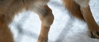
Most often, this pathology occurs in dogs of small and dwarf breeds, as they are genetically predisposed to it. The “dislocation” itself refers to the situation when the kneecap moves from its usual place and “goes” towards the abdomen (lateral luxation of the patella in dogs) or in the opposite direction (medial dislocation). However, dogs of large and especially large breeds are also not spared this misfortune. They have common problems with rear leg alignment.
When walking and running, a kind of “wave effect” occurs, due to which the kneecap simply “knocks out” of its place. For “giants,” this problem is age-related and develops due to wear and tear of the condyles and other parts of the pelvic girdle due to the effect described above. In addition, dachshunds have a specific type of dislocation, the pathology of which is due to the particularly “unsuccessful” structure of their body, artificially bred through long-term selection.
Attention! How can you determine that you and your dog are faced with this particular pathology? Firstly, it is distinguished by suddenness and spontaneity. So, the dog may suddenly whine and squeal in pain, and after a couple of minutes he will play again. These are very specific signs.
Gradually, the attacks will become more frequent, the pain will become stronger and, in the end, the dog will not be able to get back on its feet. This is what a luxated kneecap is. By the way, “technically” this pathology is explained by the fact that the cup simply jumps off the condyles. The latter, roughly speaking, play the role of a kind of “guides” on which it should be attached.
Types of dislocations
There are the following types of dislocations in dogs:
- congenital (a puppy is born with a pathology, which can occur if it is not presented correctly during intrauterine development or during labor);
- acquired arise as a result of injuries, fractures, ligament ruptures.
Depending on the displacement of the knee cap, veterinarians classify dislocations into two types.
Medial
In this case, the shift occurs to the inner side in relation to the limb.
Medial dislocation is diagnosed more often in small breed dogs. The tibia rotates proximally inward.
The process may involve the hip joints. The development of osteochondritis of the femoral head is noted, the angle of the position of the hip bone changes.
Lateral
This species is characterized by an outward rotation of the bone. Lateral dislocation is diagnosed in large animals.
Symptoms of a luxated patella
Patellar instability is one of the causes of lameness and gait disturbance in dogs. In cases of severe traumatic origin, the support function suffers more severely, up to complete inability to use the limb.
There are lateral and medial dislocation of the patella. With lateral displacement, the kneecap moves to the outer side of the limb, and with medial displacement, it moves to the inner side. Lateral dislocation is diagnosed mainly in large dogs, and medial dislocation is diagnosed in representatives of small decorative breeds, such as English Toy Terrier, Chihuahua, Yorkshire Terrier, Pomeranian Spitz, Toy Poodle. The appearance of lameness as a result of displacement of the patella is observed after an unsuccessful jump, fall, or blow.
The diagnosis is made by orthopedic examination. The degree of dislocation is determined by palpation of the affected knee joint. For radiological confirmation, photographs of the pelvic limbs are taken in two projections. In addition to determining the exact position of the kneecap, x-rays will help exclude fractures of bone and cartilaginous elements.
There are 4 degrees of patellar dislocation:
- 1st degree – the patella returns to its place on its own when the limb is straightened;
- 2nd degree - the displacement of the patella is almost constant in any position of the limb. The dog limps when moving and protects its limb. When the patella is realigned, a clicking sound can be heard;
- Grade 3 – the patella is permanently displaced and cannot be realigned. Severe lameness of the supporting type is detected, or the dog holds the limb in a bent position;
- Grade 4 – the patella is permanently displaced and fixed in an abnormal position. The dog does not rely on the affected limb or uses it periodically.
Often, the displacement of the patella is accompanied by a rupture of the ligament between the patella and the femur. In this case, the affected area is inflamed and swollen, and movements in the joint and attempts to straighten the patella are very painful. The development of hemarthrosis (filling of the joint cavity with blood) is possible. Sometimes severe damage to the knee is accompanied by separation of osteochondral fragments with complete loss of limb function.
Stages
Depending on the severity of the pathological process, knee dislocation goes through 4 stages.
First
The dog moves without any difficulty.
With increased physical activity, periodic limping is possible. Displacement of the cup occurs after the limb relaxes . When it returns to its original position, no crunching is heard.
The deviation of the tibia bone is minimal. It may be slightly moved to the side.
Second
Dislocations occur quite often. When exposed to loads, the kneecap always shifts; it can easily be moved back into place. Support on the injured limb occurs only after loading.
At this stage, the tibia is minimally rotated, with the apex pointing toward the inside.
Third
The dislocation is permanent. The bone rotates almost 50° relative to the vertical line of the paw. The dog no longer leans on her.
Fourth
This degree is considered the most difficult. The femur deviates inward by 90°. During movement, the dog does not rely on the limb . A dislocation cannot be corrected at home.
Patella luxation
Dislocation of the kneecap (patella) is its displacement relative to its normal position on the block (special depression) of the femur. Small breeds of dogs and, less commonly, cats are most predisposed to luxating the patella. In large breeds of dogs, patella dislocation is also possible, but as a consequence of other diseases (valgus antetorsion). In small breeds of dogs, dislocation can be congenital or acquired. Congenital dislocation can be detected immediately after birth, but, as a rule, animals older than four months are seen by a veterinarian. Often the cause of acquired dislocation is a minor injury in combination with an existing predisposition in the form of a poorly developed block. In veterinary medicine, it is customary to distinguish 4 degrees of dislocation depending on severity:
- Grade 1 - the dog moves normally, sometimes the kneecap may come out of the block, but easily snaps into place when the limb is stretched.
- 2nd degree - dislocations occur frequently, the kneecap easily returns to its normal position and its displacement occurs just as easily. At this degree, the femoral block becomes almost flat.
- 3rd degree - dislocation of the calyx is almost permanent, the dog tries not to step on the sore paw. When the kneecap is adjusted, it immediately returns to the wrong position.
- 4th degree - the kneecap is constantly in a state of dislocation, the dog does not lean on the affected limb, and it is not possible to straighten it into the femur block by hand.
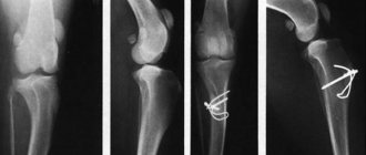
Dislocation of the kneecap in a dog on the left. On the right is a normal joint.
X-ray of a dog with medial luxation of the patellas on both limbs.
Diagnosis of a patella dislocation is based on a clinical examination; an additional X-ray examination is performed to exclude secondary changes in the joint.
Video 2. Clearly visible 2nd degree luxation of the kneecap.
Treatment of patella dislocation is only surgical and consists of increasing (deepening) the block of the kneecap of the thigh, strengthening the direct ligament of the kneecap due to the transposition of the large roughness of the tibia.
X-ray of a dog after surgical treatment of a luxated patella.
As a rule, the prognosis after surgery is good, the animal can again fully use its paw and upon external examination it cannot be said that this animal experienced such a serious problem. Given that luxating patella is mostly a congenital disease (or there is a genetic predisposition to it), veterinarians do not recommend breeding from animals suffering from this disease.
Symptoms and manifestations
The owner of the dog may suspect problems if certain symptoms appear:
- the pet does not use the affected paw when walking or standing up, lameness is observed;
- if the forelimb is dislocated, the animal will try to pull it towards itself;
- If the hip joint is damaged, problems arise when getting up after sleep.
The anatomical configuration of the damaged musculoskeletal system changes. This occurs due to tissue swelling. The joint swells.
With any, even minor, mechanical impact, the pet will experience a pronounced pain syndrome, which is accompanied by whining, growling and squeals.
Diagnosis of pathology
To determine that a dog has a dislocation and not a subluxation, it is necessary to show the animal to a veterinarian. The specialist conducts a visual inspection of the limbs and palpates the damaged area.
The nature of the lameness is assessed and the way the dog places its paws is analyzed . At the onset of the disease, this symptom is periodic, and diagnosis can be difficult.
To clarify the pathology, an additional examination is prescribed.
Palpation makes it clear in which direction the joint has been displaced, and to determine the degree of likelihood of the cup being realigned.
X-ray examination is carried out in two projections. In the lateral position, it is clear that the knee is in place, and with a direct projection, its shift is clearly noticeable.
An X-ray of the hip area helps clarify the diagnosis and is necessary to confirm or exclude Legg-Calvé-Perthes disease.
What to do?
The diagnosis for this disease is all clear - it is made based on a combination of clinical signs, “to be sure” by performing an ultrasound and/or x-ray of the paw. The signs are very specific, and it is not difficult to determine the pathology.
But what is the treatment for a luxated kneecap in a dog? The most important thing to remember is that any therapy should be aimed at preserving the animal’s quality of life. What is better - to cut off the dog’s sore paw, or to make sure that even with an advanced form of dislocation, the cup remains in its “rightful” place almost all the time? We think the answer is obvious.
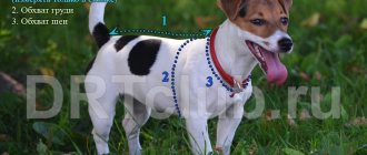
Important! The first task of the owner of a sick animal will be to maintain a normal weight for his pet. Less weight will automatically relieve the dog's joints. Ideally, your pet should have only muscle with a minimal amount of subcutaneous fat. This condition is natural for a dog. Of course, a certain walking regime should be created. You shouldn’t get carried away with training, but the dog’s muscles should be in good shape. They will “clamp” the cup in its proper place.
Several years ago, many veterinary publications recommended limiting the movements of a sick dog. This theory has now been completely debunked. It has been proven that even in the second stage of dislocation, but with good muscle tone, which is maintained by regular and long walks, the cup comes off the condyles much less often. So it is in your interests to walk your pet more often and farther. This is especially true when treating medial patellar luxation in dogs.
Treatment methods
The choice of treatment depends on the breed of the dog, the stage of development of the disease and the accompanying clinical picture. Conservative and surgical methods are used.
Non-surgical
Treatment without surgery is possible only when diagnosing the first or second stage of the pathological process; it is often used for small breeds.
The veterinarian prescribes drugs from the group of chondroprotectors, which are added to the food .
It could be Chondroitin or Glucoamine. To achieve maximum effect, medications are prescribed whose action is aimed at collagen synthesis. This promotes rapid healing of the damaged limb.
Painkillers are used to relieve pain.
Alternative medicine
In such situations, folk remedies are ineffective.
When is surgery required?
Surgery is a priority for veterinarians because patellar luxation is a mechanical problem. The choice of method for realigning the joint will depend on its changes and the reasons that provoked this condition.
If the dislocation occurs as a result of injury, then the operation is aimed at eliminating the consequences that affect the restoration of the limb.
For example, osteosynthesis is performed for a fracture of the tibia. If the capsule ruptures, it is stitched up.
Wedge-shaped plastic is used when there is no groove, or it has a slight depression.
A lateral suture is applied if the tibia has excessive rotation.
If the tuberosity of the bone is located incorrectly, then the task of surgical intervention is to move it to the correct position.
A screw or knitting needles are used for fastening.
Corrective osteotomy is aimed at correcting a deformed tibia or femur. The operation is performed after the dog stops growing. This avoids re-deformation of the bones.
If the dislocation is caused by more than one provoking factor, then several methods of surgical intervention can be performed simultaneously.
Regardless of the treatment tactics, the result will be favorable if the disease is detected in a timely manner, which prevents the development of serious complications and maintains joint mobility.
Methods of surgical treatment of luxating patella in dogs
Since a luxated patella is a mechanical problem, surgical stabilization of the patella is the preferred treatment option. The method of stabilizing the patella depends on the pathological changes in the knee joint that cause it.
In the case of a traumatic origin of the patella dislocation, a surgical operation is performed to eliminate the cause of the dislocation, for example, in case of a fracture of the tibial tuberosity, osteosynthesis is performed (the tuberosity is secured with the help of knitting needles), in case of a rupture of the joint capsule, the joint capsule is sutured, etc.
In case of genetically determined patella dislocation, stabilization is carried out:
- In case of deformation of the femur and tibia, a corrective osteotomy of the femur or tibia bones is performed. This operation is performed after the dog has finished growing to avoid subsequent deformation.
- If the tibial tuberosity is located incorrectly, the tuberosity is transferred to the anatomically correct position. The tuberosity is secured with a knitting needle or screw.
- If there is no or shallow gutter, the gutter is deepened using various surgical methods, for example, wedge plastic surgery of the gutter.
- If there is excessive rotation of the tibia, an operation such as a lateral suture is performed.
Sometimes, when performing surgical treatment to stabilize the patella, a combination of several techniques is required, since there can be several reasons for the luxation of the patella.
With any type of treatment, early diagnosis of patella dislocation is first necessary in order to avoid irreparable consequences of the dislocation and help your pet lead an active and healthy lifestyle.
Rehabilitation period
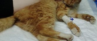
After the operation, the animal is brought out of anesthesia within three hours. All this time the pet is in a special chamber that maintains body temperature.
Post-operative care is simple.
To ensure that the internal organs work without disruption, IVs are placed in the morning and evening. This speeds up the process of removing the anesthetic substance from the body.
The dog is prescribed antibiotics and the sutures are treated.
To make the recovery process faster, you must follow all the veterinarian’s recommendations. It is worth limiting the dog’s mobility for some time, even if the limb has returned to normal. The pet will be able to stand on its paw 4-5 days after surgery.
How quickly does a dog recover from kneecap surgery?
Immediate postoperative period
After surgery, we remove your pet from anesthesia, which usually takes about three hours, during which he is in an oxygen chamber on a heating pad to maintain body temperature, and IVs are performed to maintain internal organs. You will need to give saline drips twice a day to speed up the elimination of waste products from the anesthetic drug, give antibiotics, and clean sutures. On the third day, we recommend showing up for a follow-up post-operative appointment and taking blood tests in order to objectively understand how the body endured the operation and anesthesia. The stitches are removed on the eighth day.
Possible complications
If the joint is positioned incorrectly, it wears out faster. Against this background, degenerative arthritis often develops.
With increased sensitivity to novocaine after the introduction of anesthesia, there is a risk of death.
Wound infection is also considered dangerous .
The possibility of re-dislocation cannot be ruled out. This happens if the animal is too active.