| Increased intraocular pressure and optic nerve atrophy in glaucoma |
| This is how a healthy animal sees |
| Narrowing of the peripheral visual field in early glaucoma |
| This is how a dog with advanced glaucoma sees a butterfly. Sharp narrowing of visual fields. |
| Advanced glaucoma. Vision is practically absent. |
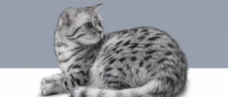
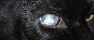
| Dog 8 years old, mixed breed with glaucoma. The eye is enlarged in size, the cornea is swollen and blue, the vessels of the sclera are tortuous and dilated. Vision is lost forever. |
Glaucoma is a group of diseases associated with a constant or periodic increase in intraocular pressure and impaired blood supply to the eye.
These disorders can gradually lead to damage (atrophy) of the optic nerve (the nerve fibers that carry information from the eye to the brain), which in turn leads first to loss of peripheral and then central vision and irreversible blindness. What makes this disease one of the most dangerous and insidious in veterinary and humanitarian ophthalmology. The main cause of damage to the retina and optic nerve in glaucoma is an increase in intraocular pressure, which causes compression of the blood vessels that supply oxygen to the nervous tissue. Which in turn leads to hypoxia and atrophy of the nerve fibers responsible for transmitting nerve impulses to the visual areas of the brain. The narrowing of the visual fields occurs imperceptibly, but quickly enough and the animal can completely lose vision in literally 2-3 months.
Main forms of glaucoma
There are three main types of glaucoma: congenital, primary and secondary.
- Congenital glaucoma is caused by congenital defects in the development of the drainage system of the eye, located in the area of the anterior chamber angle. If developmental defects are pronounced, then the disease manifests itself immediately after birth; if minor, then the disease may appear later, in adolescence (juvenile glaucoma).
- Primary glaucoma develops without any previous eye disease. Depending on what local pathological factor causes difficulty in the outflow of aqueous humor from the eye.
- As a result of some eye diseases, such as inflammation, trauma, neoplasms, cataracts, lens luxation, etc., secondary glaucoma can develop.
Glaucoma in dogs: causes and types
- Glaucoma in dogs is classified according to the following criteria:
- The cause of the disease;
- Position of the anterior chamber angle.
According to some classifications, the disease is divided into two types: primary and secondary.
Primary glaucoma in dogs is still poorly understood. It appears suddenly in a healthy body; there were no particular preconditions for its appearance.
Secondary glaucoma in dogs involves a pre-existing eye disease, such as a misaligned lens in the eye.
The angle of the anterior chamber can be narrow, open or closed.
Such classifications exist together and inextricably, complementing each other.
.
It is worth highlighting several main reasons why the primary disease appears in dogs:
Hereditary form and genes - glaucoma in dogs
Genetics plays a big role in this disease
, therefore, if one eye is already sick (and it is impossible to stop and identify this in advance), then it is worth starting an examination of the second eye too, even if it seems absolutely healthy after the illness.
It is also worth listing some breeds that are more susceptible to the disease than others.
. In general, there is no division into large and small breeds, and gender does not play a role here. And so, the main breeds are approximately the following: huskies (including Siberian), terriers and fox terriers (Boston, smooth and wire-haired), Akitas, Shibas, cocker spaniels (English, American and regular), Shihtzu, poodles, bulldogs, sharpeis , Giant Schnauzers, Dalmatians, Hounds, Chow Chows, Mastiffs, Italian Greyhounds, Schnauzers.
As you can see, there are really a lot of breeds and they are all diverse. Of course, just because your pet's breed isn't listed here doesn't mean he won't be susceptible to the disease.
Primary form of glaucoma with open angle
Primary form of glaucoma with open angle. This form is also called hereditary
, poodles and beagles are susceptible to it. The pressure in the eyes or eye rises slowly because the disease becomes chronic. Often vision persists for a long time and the disease is not immediately noticeable.
Goniodysplasia - glaucoma in dogs
Goniodysplasia is characterized and found in primary glaucoma
, whose angle is low. It is found in Labradors, bulldogs, spaniels, hounds and some others. The disease begins quite suddenly and is characterized by swelling, dilation of blood vessels, redness and, if left untreated, blindness. If the disease has begun, then the second one must also be examined and treated, by analogy with a hereditary disease. The pressure in the eye is reduced, but the angle may close and then vision will be lost.
Secondary glaucoma
All that is said about primary glaucoma is heredity or some unknown factors that veterinarians have not yet been able to understand and discover. Secondary glaucoma develops during an infectious disease, and the signs of the disease are slightly different. For example, the pressure in a dog’s eye increases , the blood vessels become very dilated, the eyes become cloudy , and the pupils most often become narrow.
Unlike the primary disease, the disease is accompanied by pain
, because of which
the dog whines , stops sleeping and eating normally , tries to scratch his eyes and, in general, behaves extremely restless and tense. Plus, the animal becomes apathetic, stops playing and shows interest in walks. The dog also begins to avoid bright places and mostly sits in corners and dark secluded places.
Secondary glaucoma in dogs, cause
If we talk about the cause of the secondary disease
in more detail, doctors believe that almost all eye diseases lead to glaucoma, ranging from tumors, keratitis and ending with conjunctivitis. In dogs, secondary glaucoma most often occurs as a consequence of an illness. Also, various bruises and injuries lead to eye disease. A traumatic situation with a dog’s head is often not without consequences, especially for such sensitive organs.
Glaucoma in dogs, symptoms
Like any other disease, it is better to detect glaucoma in the early stages, before it develops into a more serious form that is much more difficult to treat. Therefore, if there is any suspicion or hint of glaucoma, you should immediately contact
see
a veterinarian ophthalmologist who will prescribe the necessary treatment.
- Main symptoms of glaucoma in dogs, which are isolated in both the secondary and primary stages:
- Copious amounts of tears;
- Puffiness and red eyes of the pet;
- The animal’s apathy, reluctance to play and “communicate” with humans and other pets, sometimes aggression;
- Lack of appetite;
- The membrane of the eye becomes cloudy;
- The desire to leave the light, to hide in a dark corner;
- The eye becomes large;
- Pain and inflammation in secondary glaucoma;;
- The animal is lost in space.
Of course, it all depends on the individual characteristics of each dog. Indeed, depending on the breed, age, gender, as well as the phase and general form of severity of the disease, the signs and symptoms depend. They are often mixed and matched and vary from situation to situation.
It sometimes happens that the symptoms of primary glaucoma, which is caused by heredity and genes, do not appear during most of the pet's life to the extent described above. Only an experienced veterinarian can identify the disease based on other signs. For example, by the total pressure inside the eye. Also, with primary glaucoma, swelling of the cornea of the eye occurs, pressure increases, mydriasis and hyperemia appear.
If the disease has developed and nevertheless begins to act, then visible swelling in the dog appears , the eye increases in size, pressure increases inside, mydriasis becomes slower, the lens inside the eye can shift and be dislocated.
The signs of the secondary disease coincide with the symptoms of the primary form of the disease, which has progressed to a more acute stage. In this regard, it is worth determining the reason why the disease appeared. Otherwise, the symptoms are similar, but the treatment should be slightly different. So, for some time, incorrect treatment will not aggravate the situation, but precious time, especially when it comes to glaucoma, will be lost.
If the case is very acute and critical, then the signs become more widespread and terrible: the eyeball increases in size several times, loss of vision and disorientation , the animal feels severe pain.
The optic nerve becomes pinched, deformed and deteriorated, making it very difficult to restore vision.
Symptoms and causes of glaucoma
In most cases, glaucoma is asymptomatic in the initial stages of the disease, which seriously complicates early diagnosis. An animal may not show any visible causes associated with visual impairment for a long time, since it “gently” adapts to reduced vision and quite successfully compensates for this condition with other senses, such as smell, hearing and touch. Only in cases with an acute attack of glaucoma, the symptoms of the disease are extremely pronounced.
These symptoms are:
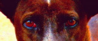
- Increased filling and tortuosity of scleral vessels (cobra syndrome)
- Corneal clouding and increased lacrimation
- An increase in the volume of the eyeball (“buvthalm” - bull’s eye)
- Sudden loss of orientation, depression, lack of appetite.
However, if intraocular pressure is normally 18-25 mm Hg, and with a sharp rise it can increase to figures of more than 50 mm Hg, the risks of permanent vision loss are extremely high, if not guaranteed!
Due to vague symptoms, pet owners rarely consult a doctor at the early stage of the disease. There is only one way out of this situation - to be more attentive to the condition of your pets’ eyes and undergo an eye examination if any vision problems appear, regardless of the form in which they appear.
The causes of the development of primary glaucoma are often due to age-related changes in the body, therefore, after 6 years, all animals are recommended to have their intraocular pressure checked once a year. It should be borne in mind that diseases such as hypertension, diabetes mellitus, diseases of the cardiovascular system, diseases associated with taking hormonal drugs increase the risk of developing glaucoma.
Timely detection of glaucoma allows you to manage this disease using drug treatment. In the vast majority of cases, blindness does not occur.
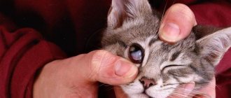
Glaucoma in cats
Typically, glaucoma in cats can be recognized in the initial stages by clouding of the cornea. This condition is preceded by an increase in intraocular pressure, which appears due to the abundant production of intraocular fluid or due to a violation of its outflow from the chambers of the eyeball.
Glaucoma may be caused by:
- eye injuries;
- hemorrhages in the chambers of the eyeball;
- congenital anomalies;
- corneal ulcer;
- displacement of the lens towards the anterior chamber of the eyeball.
The most dangerous complication of glaucoma is blindness. The retina is very delicate, even slight pressure on it can cause detachment. To avoid this condition, the animal needs urgent veterinary care to reduce intraocular pressure.
Glaucoma usually affects only one eye, sometimes both. With unilateral development, the pathology is easier to notice against the background of the contrast between the left and right eyes.
Clinical signs of the disease:
- increase in the size of the eyeball;
- whitening of the cornea;
- swelling of the eyelid;
- redness of the blood vessels of the sclera;
- squinting of the eye.
The affected apple is painful, this can be understood by the restless behavior of the animal that protects it, sometimes scratching it. The diagnosis will be made by a veterinarian at our clinic after examination and instrumental examination.
Diagnosis of glaucoma
The ophthalmology department of our clinic has all the necessary diagnostic equipment that allows you to conduct a complete ophthalmological examination and make a diagnosis of glaucoma.
A comprehensive examination usually includes the following main items:
- intraocular pressure examination;
- fundus examination;
- biomicroscopy of the anterior part of the eye;
- assessment of the condition of the optic nerve head
Glaucoma is a disease that can accompany an animal for the rest of its life. However, timely treatment and constant qualified observation can preserve vision and ensure an adequate quality of life for animals with glaucoma.
Drug treatment of glaucoma
Drug treatment of glaucoma is primarily aimed at reducing intraocular pressure. Treatment usually begins with the prescription of eye drops, the regular instillation of which reduces intraocular pressure to a level acceptable to the patient.
Modern eye drops are effective, easy to use, have minimal side effects, and are usually well tolerated by sick animals. There are several groups of drugs in drop form that reduce intraocular pressure by improving the outflow of intraocular fluid and reducing the production of aqueous humor. When prescribing drug treatment, the ophthalmologist selects a combination of drugs that is optimal for each individual patient. However, regular instillation of antiglaucomatous eye drops only in 50% of cases allows stabilizing the glaucomatous process and preserving visual functions.
Useful video
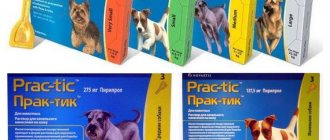
Modern drops for glaucoma and cataracts are designed to reduce intraocular pressure and prevent clouding of the lens. In severe cases, their combined use is allowed. All drugs in question are dispensed from pharmacies only with a doctor's prescription.
Author's rating
Author of the article
Alexandrova O.M.
Articles written
2029
about the author
Was the article helpful?
Rate the material on a five-point scale!
( 1 ratings, average: 5.00 out of 5)
If you have any questions or want to share your opinion or experience, write a comment below.
Surgical treatment of glaucoma
If eye drops that reduce intraocular pressure do not give the expected result, or the stage of development of glaucoma has reached the terminal stage, it is possible to perform an operation to introduce a drainage system into the diseased eye, filtering excess moisture out. In cases where surgery to install drainage is impossible, as with painful glaucoma, cosmetic surgery - evisceration - is used to relieve a strong pain component due to complete loss of vision. Evisceration with the introduction of a silicone implant into the eye allows you to achieve an excellent cosmetic effect for your pet, since the implant completely follows the shape of the eye. From a distance of 20 cm it is not possible to distinguish a prosthetic eye from a healthy one. In our clinic we use products from the leader in the veterinary ophthalmology market, the German company Acrivet, a quality standard in Europe.