Causes
In a cat, normal levels of intraocular pressure are considered to be 15-25 mm. Hg Art. If they are exceeded, an enlargement of the eyeball is observed and optic nerve atrophy occurs. These destructive changes cause the animal to lose the ability to see.
Factors that provoke the development of glaucoma include the following:
- congenital anomalies of the structure of the drainage system of the eye;
- ophthalmological diseases (conjunctivitis, uveitis, etc.);
- the use of corticosteroid drugs to treat eye diseases;
- damage to the eyes or skull due to trauma;
- genetic predisposition;
- diabetes;
- elderly age;
- hypertension.
Many experts also argue that glaucoma can develop due to the use of hormonal and steroid medications.
Causes and signs of the disease
Factors for the development of glaucoma in cats:
- pathological disorder in the cornea;
- traumatic damage to the lens, accompanied by inflammation;
- chronic purulent inflammation of the eye;
- getting particles of soil or other debris into the eye.
The primary form of the disease is most often diagnosed in Siamese, Persian and short-haired breeds.
Infectious diseases that cause a secondary form of the disease in cats:
- systemic mycosis;
- infectious peritonitis;
- leukemia;
- toxoplasmosis.
Processes in the eye
The eye resembles a ball filled with liquid, while it constantly circulates, flowing in and out of the vessels. The liquid is under pressure, preventing the balloon from deflating or bursting. The drainage system is responsible for the drainage of fluid from the eye and regulation of IOP. When it is blocked, fluid flows in, but its outflow is inhibited. Eye pressure increases.
Main symptoms
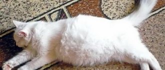
Glaucoma does not make itself felt for a long time. The owner can notice that his pet has red eyes and contact the clinic. Thanks to this, the disease can be detected at an early stage. As the pathology progresses, the cat develops clinical signs, the most obvious of which is mydriasis (dilated pupils, lack of reaction to light).
The pet's eyes water, swell, sometimes turn red, and the eyelids do not close tightly. The eyeball increases in size, the quality of vision decreases. The animal experiences pain and discomfort in the affected eye, squints and looks depressed.
May refuse to eat, tries to hide in a dark place, away from daylight or electric light. A previously active pet does not show activity and is poorly oriented in space. Jumping onto a hill is difficult for him.
If the owner tries to play with the cat with a laser pointer, there will be no reaction from the four-legged pet.
How it manifests itself
In the early stages, the pathology can rarely be recognized, since the symptoms are mild. As the disease progresses, in addition to specific symptoms, a general asthenic condition appears. Appetite worsens, during the day the pet is lethargic and inactive.
Primary glaucoma
In the primary form of glaucoma, when visiting a veterinarian, you may notice increased soreness in the eyes and blurred vision. At home, you may notice clouding of the lens. It can be slow or happen quickly. The sparkle in the eyes disappears. The cat begins to avoid brightly lit places, preferring to be in dark parts of the room.
During an examination, an ophthalmologist may detect atrophy of the retina and optic nerve. Due to impaired innervation, the cornea is less well supplied with nutrients, which is why it becomes cloudy and dull. The pupil increases in size. The reaction to light is weakly expressed; when a special liquid is instilled into the eyes, no narrowing occurs.
The chronic form of the pathology, which is less treatable, is manifested by a pathological increase in the size of the eyeball. It protrudes greatly, which is why it cannot be completely closed for centuries. The cornea becomes inflamed due to dryness. Through the enlarged pupil, you can see the greenish color of the fundus.
The lesion can be unilateral, but in most cases it occurs simultaneously in both eyes.
Signs of hydrophthalmos
Dropsy has similar manifestations and often looks like primary glaucoma. The eyeball becomes larger in size, is not completely covered by the eyelid, and there is increased sensitivity and pain. The vitreous fluid liquefies, causing the cat to go blind. The cornea becomes cloudy and often becomes covered with ulcers. The pupil dilates.
Treatment method, prognosis
Glaucoma is treated with conservative and surgical methods.
Drug therapy is aimed at reducing the size of the eyeball and normalizing fundus parameters. Stabilization of the drainage system of the organ of vision is achieved by using drugs such as Timolol, Mannitol, Dichlorphenamide. Miotics help to narrow the pupils - Phospholip, Physostigmine, Pilocarpine.
Corticosteroids and carbonic anhydrase inhibitors are used as prescribed by a doctor.
A more effective method of combating glaucoma is laser treatment. Surgical options include partial removal of the ciliary body or drainage of the anterior chamber. If the eye is completely blind, it is removed.
In most cases, the prognosis for glaucoma is unfavorable. It is impossible to stop vision loss with medications, and surgeries and laser treatments are very expensive, and not every cat owner can afford it.
Pathology treatment methods
Glaucoma in cats is an incurable disease, but it is possible to stop its progression. To curb the development of pathology, medications are used that lower IOP and reduce the production of intraocular fluid.
The treatment regimen is prescribed by a veterinarian-ophthalmologist. The distinction between the primary form and the secondary form is important when choosing therapeutic agents. Certain drugs that are effective in the treatment of primary eye disease are inadmissible for uveitis:
- Humorsol (Demekarium);
- Pilocarpine.
These drugs worsen the condition, aggravating the course of uveitis.
The therapy has the following goals: reducing the production of intraocular fluid and improving its removal. The following groups of drugs are used for treatment:
- Miotic agents lead to pupil constriction and help improve the outflow of fluid from the eyeball. They are prescribed for the primary open-angle form of the disease. The miotic group of drugs includes Pilocarpine, Phospholin, Humorsol. They are contraindicated in secondary glaucoma that occurs against the background of uveitis, because they contribute to the inflammatory process in the iris of the eye.
- Carbonic anhydrase inhibitors help reduce the production of intraocular fluid by the ciliary body. These drugs include: Diacarb, Daranid, Methazolamide. Treatment with drugs in this group causes side effects in cats. Weakness, vomiting, acidosis, restless behavior, diarrhea are the main symptoms of intolerance to the drug. Preparations for instillation into the eyes (Azopt, Cosopt, Trusopt) do not lead to a significant decrease in IOP. Their side effects are not as pronounced as those for internal use. One of the disadvantages of topical carbonic anhydrase inhibitors is their high cost.
- To reduce the formation of intraocular fluid, adrenergic blockers are used. In cats, the effectiveness of Timolol and Betaxolol has not been proven. In veterinary practice, a 0.5% solution is used, because a reduced concentration is ineffective. The drugs are used for any type of glaucoma; these drugs do not affect the course of uveitis. A combination of Timolol and carbonic anhydrase inhibitors (orally) is considered an effective treatment regimen. For prophylactic purposes, β-blockers are used to prevent glaucoma in the second eye.
- Osmotic diuretics are prescribed for acute forms of glaucoma. They lead to rapid dehydration of the eyeball. Cats are administered intravenously a 20% solution of Mannitol at a rate of 1 g/kg. In case of uveitis, the administration of Mannitol is contraindicated. After administration of the product, limit the pet's fluid intake.
- Sympathomimetics help reduce fluid production and improve its removal. Epinephrine and Dipivefrine are used in combination with other groups of drugs, because when used alone they are ineffective.
- Latanoprost is a prostaglandin analogue. Used topically 1 time per day. Prescribed for primary glaucoma alone or in combination with other drugs. The disadvantage of Latanoprost is its high price.
Treatment
An acute condition requires emergency care. The cat is examined to confirm primary or secondary glaucoma.
If the primary form is confirmed, treatment consists of urgently reducing intraocular pressure. For this purpose, the administration of osmotic diuretics (Mannitol, Glycerin) is used. A carbonic anhydrase inhibitor is prescribed orally. The pressure decreases after 2-3 hours. Mannitol is reintroduced after 4 hours, carbonic anhydrase inhibitors - every 12 hours.
Betaxolol, Timolol, Latanoprost are prescribed locally. If glaucoma is secondary, local anti-inflammatory medications are used.
Surgical intervention
Treatment with drugs in animals is not always effective, so they resort to surgical methods:
- Cyclocryodestruction is performed under general anesthesia. The operation involves cauterizing the ciliary body with a cryoprobe. Freezing is carried out at a temperature of -85°C. The disadvantage of the procedure is possible retinal detachment and the development of secondary cataracts.
- Installation of a valve system is the best method of reducing intraocular pressure. The valve creates a flow of fluid from the eye, opening only if the pressure is exceeded. The valve installation operation takes no more than half an hour. Its advantages include speed, low infection rates, and a reduction in the number of medications prescribed after the procedure.
- Cyclodialysis is a labor-intensive operation that requires care and equipment from the surgeon. The purpose of the procedure is to create bypass pathways for the outflow of fluid. The operation is lengthy, but relatively inexpensive. The advantages of this method are long-term compensation of intraocular pressure and an easy period after surgery.
- Laser cyclophotocoagulation is less traumatic than cryodestruction. The destruction of the ciliary body is carried out with a laser beam. The operation is little studied in Russia; it is used by Western surgeons.
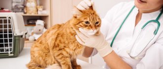
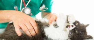
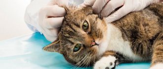
Diagnosis of glaucoma in cats
If you suspect glaucoma in cats, you should contact a veterinary clinic. There, the specialist will prescribe an examination, the main stage of which will be measuring intraocular pressure. The condition of the optic nerve and fundus of the eye is also required to be diagnosed. By and large, this is enough to make a diagnosis.
In a clinical setting, special studies can be performed:
- slit lamp examination;
- ophthalmoscopy;
- measurement of intraocular pressure (tonometry);
- Ultrasound diagnostics of the eye, if there is a suspicion of retinal detachment, a neoplasm in the eye or in the behind the orbital space.
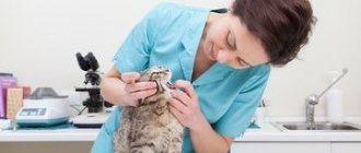
To make an accurate diagnosis, a veterinarian performs a number of examinations:
- study of anamnesis;
- examination of the pet;
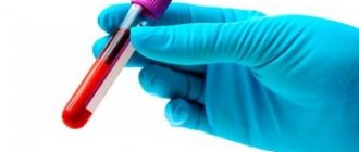
- taking tests;
- tonometry;
- ophthalmoscopy.
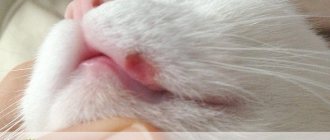
Alas, people turn to veterinarians only at the moment when you can’t even look into the cat’s eyes without fear: the surface of the eyeball is covered with significantly enlarged vessels, the conjunctiva is swollen, one or both eyes protrude terribly from the sockets. Remember that treating glaucoma in cats at this stage is absolutely pointless, since even a slight improvement can take months to achieve.
Naturally, during this time the cat becomes completely blind. Oddly enough, even in particularly advanced cases the eye does not always protrude. Sometimes the eyeball just gets very hard. This is even worse, since with this course the animal experiences very severe pain, from which the cat cannot sleep and eat, quickly loses weight and becomes very nervous and irritable. This is also facilitated by constant deterioration of vision.
Fortunately, glaucoma can be diagnosed at earlier stages, when something useful can still be done for the cat. For this, a special tonometer is used, the appearance of which is very similar to an ordinary pencil. If examination reveals even a slight increase in intraocular pressure, there is every reason to suspect early glaucoma.
Glaucoma is incurable (which should always be remembered), but it is quite possible to reduce pain and reduce the manifestation of other clinical signs. To do this, it is recommended to drip pilocarpine into the eye: in case of glaucoma in a cat, it allows you to maintain a high quality of life of the animal for a long time. Please note that it should only be prescribed by a veterinarian, as the drug has serious contraindications. So, if the course of the disease is complicated by retinal detachment, prescribing pilocarpine is strictly prohibited!
READ How to treat otodectosis in cats
Drugs containing dorzolamide and/or thymol are indicated. These substances allow you to reduce intraocular pressure to a completely acceptable level. To combat inflammation (if it occurs), steroid drugs are often prescribed. Diuretics (drugs that increase urination) are often used, which also help lower blood pressure.
Please note that in this case the cat will have to be somewhat limited in liquids, liquid and canned food. Vitamins are prescribed that help soften and also slow down degenerative processes in the orbital nerve. If the pain of glaucoma becomes unbearable for the animal, painkillers are prescribed.
Here's how to treat this pathology. Of course, as sad as it may be, you won’t be able to completely stop your cat’s vision loss, but all these medications will help greatly delay this process. In rare and especially advanced cases, when intraocular pressure cannot be reduced by any means, and the cat experiences severe pain, a decision may be made to remove the eye. This will help avoid more serious consequences.
To accurately diagnose an animal with glaucoma, clinics conduct a series of ophthalmological and instrumental studies (tonometry, gonioscopy). It is mandatory to measure intraocular pressure and examine the fundus. It is worth noting that, fortunately for breeders of furry purrs, glaucoma can be diagnosed in the early stages of the development of the pathology.
Regardless of the type or type of glaucoma in a cat, both eyes can be treated, even if only one is affected. Therapy is aimed at reducing and normalizing intraocular pressure.
In the treatment of animals, osmotic diuretics, prostaglandins (bimatoprost), which accelerate the outflow of fluid, beta blockers (temolol, betoxolol), calcium channel blockers (amlodipine), anti-inflammatory drugs (diclofenac), restorative drugs, and vitamins are used. Eye drops that narrow the pupil are also prescribed.
Important! Therapeutic therapy is positive only in the case of mild vision loss and the absence of complications.
If drug treatment does not produce results, surgical treatment is resorted to, which consists of surgical drainage or partial removal of the ciliary body. If the eye is completely blind, it is completely removed.
In any case, therapeutic methods and medications should only be prescribed by a veterinary specialist, having a detailed diagnosis in hand.
The prognosis for glaucoma is acute. The outcome of treatment depends on the depth of the lesions and the degree of vision loss. If treatment was started at an early stage, in most cases veterinarians are able to stabilize the condition, but it is impossible to completely restore visual function.
Increased intraocular pressure in a cat is determined by a specialist using a special device - a tonometer. High values of intraocular pressure in combination with clinical signs are the basis for a diagnosis of glaucoma. Diagnostic measures should not end there.
Since the disease is secondary, it is worth looking for the true cause that caused the development of glaucoma. To do this, it is necessary to conduct a full examination of the animal, which will help, if not save his vision, then at least make it possible to make the life of a sick animal more comfortable by eliminating the root cause of glaucoma.
Diagnostics
The diagnosis of glaucoma is made only after confirmation of the disease by measuring intraocular pressure. Before this, the attending physician can only assume the presence of the disease, but diagnoses it on the basis of an increase in pressure.
Similar diseases and their differences:
- Conjunctivitis - redness affects only the conjunctiva of the eye without swelling. Easily treated with local anesthetics.
- Inflammation of the anterior chamber (uveitis) is usually accompanied by decreased pressure inside the eye and miosis (constriction of the pupil).
- Dystrophic changes in the cornea - accompanied by a normal level of intraocular pressure and absence of pain.
- Ulcerative keratitis - pain and discomfort in the eye are relieved with drops of anesthetics; when stained with a special dye with a fluorescent substance, a positive reaction is detected - the damaged areas are painted more intensively.
One of the main methods for assessing intraocular pressure is tonometry. It is performed with local anesthesia - instillation of inocaine or benoxy into the eyes. It is measured with a special device - a Shiotz or Tono-Pen tonometer.
Normal intraocular pressure ranges from 15 mm Hg. Art. up to 25 mm Hg Art. If the difference between the measurement of both eyes is more than 10 mmHg. Art., that is, the basis for diagnosing glaucoma.
Gonioscopy is used to examine the iridocorneal angle. Such a study is carried out to assess the likelihood of disease in the second eye with secondary glaucoma or with a hereditary predisposition to primary glaucoma and assess the risk of occurrence. To carry out the manipulation, special lenses are placed on the cat’s eye and, due to the refraction of light in them, they allow one to see the necessary anatomical formation.
Fundus examination gives the ophthalmologist an idea of the preservation of visual function and visualization of the optic disc. Edema or atrophic phenomena are an unfavorable sign and indicate deterioration with subsequent loss of vision.
Forecast
In the secondary form of glaucoma, the prognosis for a cat is often unfavorable. With early treatment of uvitis, glaucoma can be reversible. The animal manages, although not fully, to retain its vision.
We suggest you read: How many days before vaccination should a cat be wormed?
It all depends on the stage of glaucoma at the time of contacting the clinic. Permanent medical treatment or surgical treatment for glaucoma may be offered if visual function is preserved. However, today any treatment for glaucoma is not a treatment in the sense that we would like to understand this word. That is, we cannot undergo treatment and recover. You will need treatment for the rest of your life, perhaps with varying degrees of success.
In the same case, if at the time of admission the cat has no vision, the eye causes pain, and is enlarged in size, such a patient is offered removal of the eye as a radical but effective measure to relieve pain and the risk of eye perforation. Lifelong medical treatment or a series of surgical operations on the blind eye is unlikely to be a good plan for such a patient and its owner.
Causes of primary (congenital) glaucoma
Why does glaucoma occur in cats (see photo of the animal’s affected eye above)? It is important to understand that each type of this disease has its own characteristics and, of course, causes. Based on this, let's look at what can trigger primary glaucoma.
If a kitten was already born with such a pathology, then most likely its parents had it. Doctors call this a genetic predisposition. Also at risk are those individuals who have been diagnosed with glaucoma in their family. Another cause may be congenital anomalies.
Veterinarians have noticed that there are certain breeds of cats that are more prone to the disease. These include European species, Burmese, Siamese and Persian.
Congenital glaucoma in cats usually presents with the same symptoms described above. The owner can only notice the presence of this pathology by observing the animal. You need to pay attention to how your pet reacts to light. If he avoids bright lighting, preferring shade, then he needs to be examined in a clinic. Also, congenital glaucoma may be indicated by strabismus, with which the kitten was already born.
Treatment of the disease
As we mentioned above, the principles by which the animal will be treated will vary depending on what particular form of the disease the cat suffers from. So, provided that an open-angle variation of this disease is detected, which occurs rapidly and acutely, the animal must be taken to a veterinarian immediately - otherwise death is also possible.
If the disease is in its early stages, therapy will be conservative, using only medications. If the stage is advanced, then surgical intervention may be required.
Sometimes surgery is unavoidable
Conservative treatment
Conservative therapy has several main objectives:
- elimination of disease symptoms;
- improvement of the condition of the affected organ of the visual system.
Comprehensive treatment of glaucoma
To carry them out, drugs are used that have the following effects:
- triggering or enhancing the drainage of fluid accumulated in the eye;
- reducing the production of “water” in the eye;
- constricting pupil;
- diuretic;
- anti-inflammatory (usually hormones);
- pain reliever;
- improving nerve conduction, etc.
Thus, the most popular remedies for combating glaucoma in the field of veterinary medicine are presented in the list below.
Firstly, this is “Pilocarpine” - a drug that:
- constricts the pupil of the eye;
- relieves pain caused by the disease;
- slows down the progression of the disease.
This remedy is given once a day, sold in the form of drops. Each eye of the animal (including the healthy one) must be instilled once a day when the disease progresses slowly; in case of acute progression, the product is used every hour.
Pilocarpine constricts cat pupils
Secondly, Amlodipine is a drug that helps remove moisture accumulated in the eye, seriously improving the condition of the sick animal. This medication is given to the pet once a day, the approximate dosage is approximately 0.6 milligrams. An important nuance: taking the medicine should coincide with meals.
"Amlodipine" removes excess fluid from the eye
The third drug, Mannitol, showed excellent results in the early stages of the disease. The drug is administered inside the animal's body through an injection into a vein. The dosage is calculated as follows: one gram of medicine per kilogram of live weight.
"Mannitol" helps defeat glaucoma in the initial stages
Fourthly, Betoxolol is a drug that helps normalize pressure inside the organ of vision. The medication is administered every 12 hours, it is dripped directly into the animal’s eye.
Betoxolol normalizes intraocular pressure
A mandatory part of drug treatment will also be a limited drinking regime for your pet. In addition, during therapy, food containing various preservatives and additives is excluded from consumption (by the way, this food waste is not suitable for feeding even a healthy animal), and the pet is also given fortified complex preparations that improve its general condition.
Every living organism needs vitamins in the right ratio
Please note the following important fact: each drug indicated in the list above has a number of contraindications. You cannot give it to your pet on your own, since only a veterinarian can judge the potential side effects that may occur.
Here are the most common examples:
- A diuretic should not be given if the cat has a history of urolithiasis;
- Some eye drops can worsen retinal detachment. about the side effects of Ciprovet eye drops in our article.
Surgical intervention
If it is too late to take medications or their use has no effect, veterinarians will recommend surgery to save the animal’s life. Exactly how this intervention will be carried out will depend on how:
- the disease progresses;
- at what stage is the disease;
- whether the animal retained vision in the affected eye.
If medications do not help, the animal will require surgery.
Thus, the operation in which a laser is used as a surgical instrument is considered the most acceptable and has demonstrated a high level of effectiveness. When used, the symptoms of the disease are quickly eliminated, and rehabilitation occurs as quickly as possible. The few disadvantages of this procedure are as follows:
- Not every veterinary clinic has such expensive equipment;
- the operation costs a lot of money, although a loving owner will not consider this circumstance an obstacle to the treatment of pets.
Russian veterinary clinics, as a rule, do not provide laser surgery for glaucoma, but use techniques such as:
- cauterization of the ciliary body at low temperatures;
- implantation of the Ahmed valve;
- cyclodialysis.
Some veterinary clinics offer a laser solution to the problem.
The interventions listed above are aimed at reducing the pressure inside the visual organ affected by the disease. Unfortunately, despite their high effectiveness, they do not protect against:
- postoperative complications;
- re-development of the disease.
If the disease reoccurs, the pet will need surgery again. That is why, if possible, contact a clinic that works with laser methods for treating glaucoma. Thus, you will not only cure your pet, but also save on repeated surgery.
If atrophy of the optic nerve has occurred and it is not possible to restore its functioning, surgical intervention will be performed to eliminate the symptoms of the disease and its consequences, as well as to restore the organ and preserve the appearance of the cat.
The animal is fitted with a prosthesis or undergoes resection of the eyeball
Causes
Each type of glaucoma has its own causes, features and symptoms. Primary and secondary glaucoma are especially different.
Causes of primary (congenital) glaucoma
Congenital glaucoma is most often caused by the genetic predisposition of the animal, as well as congenital abnormalities in the functioning of the ciliary body or drainage system through which the outflow of intraocular fluid occurs in the kitten.
Much attention is paid to the predisposition to the development of the disease of representatives of a particular breed. It is believed that Siamese, Burmese, Persian and European cat breeds are most susceptible to the disease.
Causes of secondary glaucoma
Secondary glaucoma develops as a complication of another ophthalmological or general disease.
Among ophthalmological diseases, glaucoma is most often caused by uveitis, conjunctivitis of various origins, lens dislocations, eye injuries, tumor diseases of the ocular structures, inflammatory processes localized in the iris, ciliary body, choroid and retina, as well as overgrowth of the drainage canal.
As for general diseases, animals suffering from such pathologies as:
- Age-related changes in the body. They provoke destructive processes in the eye.
- Traumatic lesions of the organ of vision. Injuries often affect the ocular structures responsible for the production and drainage of intraocular fluid.
- Diabetes mellitus very often causes complications in the eyes.
- Hypertension. High blood pressure negatively affects the organ of vision.