- When is cytology used What information can cytology provide What information will help the veterinarian in treatment How a sample of cells is taken What are the advantages of cytology What are the limitations of cytology What step follows cytology
Cytology in animals is a morphological diagnostic method based on the microscopic study of body cells. In most cases, cytological studies help determine the presence or absence of malignant tumors and the nature of the tumor process. But also through cytological studies it is possible to assess the nature of the pathological process, evaluate the condition of internal organs, and examine natural physiological and abnormal fluids.
When is cytology used?
A cytological examination is recommended if tumor processes are suspected - any neoplasm can be either benign or malignant, which significantly affects the plan for further therapy and prognosis. Cytology is also performed to examine cysts and detect effusions. Various smears can be examined in animals, for example, for otitis media, to determine what is causing the inflammation (bacteria and what type, fungi - the choice of drugs for treatment depends on this). Vaginal smear cytology is used to determine the optimal mating time.
Analysis methodology
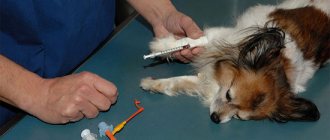
The most commonly used sampling technique for cytological analysis is skin scraping, and if we are talking about a tissue sample from a neoplasm or papule located in the skin, fine needle aspiration.
To obtain a skin scraping, you need: a sterile disposable scalpel or curette (a surgical instrument in the form of a spoon or ring with sharp edges), a microscope with slides, and mineral oil (a biologically neutral liquid obtained from petroleum) to wet the scalpel or curette.
Superficial scraping of the skin is carried out without anesthesia; before performing a deep scraping of the dog’s skin, the veterinarian can treat the desired area of skin with a local anesthetic; most often, chloroethyl is used for this in an aerosol, which “freezes” the dermal tissue for several minutes.
When performing a deep scraping, a scalpel or curette is inserted into the skin until blood appears from the surface capillaries. After this, the resulting material, together with a drop of mineral oil, is applied to a glass slide and covered with a coverslip. The material is visually examined under a microscope using first a small, hundredfold, and then a large, thousandfold magnification.
In some cases, when performing cytoscopy, it is necessary to fix the sample on a glass slide with alcohol or formaldehyde. To detect cellular elements with an irregular structure, the sample is stained with special dyes, which serve as markers of atypical cells. Papanicolaou staining can detect the presence of malignant cells and viruses, while Romanowsky staining is effective in detecting bacteria and fungi.
When studying the biomaterial, it is assessed how the structure of the cells and their shape correspond to the norm. The features of the cytoplasm, changes in the nucleus, and the presence or absence of abnormal inclusions are studied. The laboratory technician documents any detected deviations from the norm on a special form.
The cytological analysis is deciphered by a specialist - all data is entered into the conclusion form in the form of an abbreviation of the Bethesda system, generally accepted in medical terminology, and understandable only to doctors.
The advantage of the cytological examination of the skin of a dog is that the test analysis does not take much time, and its results either provide the information necessary to make an accurate diagnosis or clearly indicate the direction of further diagnostic search. Cytology is also effective as a method of intermediate evaluation of treatment results.
What information can cytology provide?
Cytology is the science of cells. With any cytological examination, we can see the cells of the liquid or smear being examined through a microscope.
With blood cytology, you can see tumor cells in it (in some types of oncology), you can detect blood parasites (babesia, dirofilaria). Even a leukogram, which is included in most cases in a routine clinical blood test, is essentially a cytology of a blood smear, in which the number of different blood cells is counted, which makes it possible to determine whether there is an inflammatory process in the body, to suspect a malfunction of the hematopoietic organs, or the presence of autoimmune reactions.
Urine cytology can provide information about the number and type of urinary crystals that form uroliths. Also, epithelial cells of the urinary tract or even kidneys may be found in the urine, which indicates the need for further diagnosis of the urinary system.
When performing cytology of effusions, you can find out whether it is sterile or whether there are bacteria or tumor cells - all this allows you to make a diagnosis or determine the direction of further diagnosis of your pet.
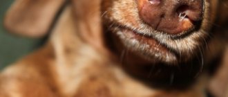
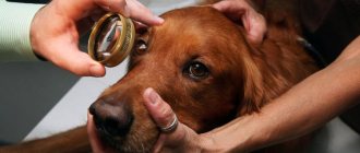
Cytology can also be taken from the surface of the skin - often dermatologists make fingerprint smears and look at what cells are found there, whether there are cutaneous or intradermal parasites. When diagnosing otitis, a smear is required - otitis can be of a bacterial nature, caused by a fungus (usually such otitis is secondary) or begins due to exposure to parasites (for example, otodex).
What types of biopsies are there?
Based on the specific method of obtaining tissue samples for further analysis, there are several main types of biopsy in dogs: puncture, incisional, excisional and trephine biopsy. Each of them has its own individual characteristics, allowing you to determine the best place of application.
puncture
The puncture type involves taking biomaterial in the form of parts of individual tissues by puncturing the skin and muscle layers with a needle. It is effective for collecting material from any internal organ, therefore it is often used when examining the liver, kidneys, prostate, thyroid and pancreas. Based on the specific location of application, the most suitable type of needles is selected:
- aspiration, presented in the form of thin-walled cannulas with tips sharpened at different angles (effective for targeted fine-needle biopsy during cytological examination of tissues);
- modified aspiration, differing from the previous version by a different tip shape (can be used to obtain both cytological and histological samples);
- cutting ones, which are used only in histological studies.
The biopsy technique and the accuracy of the results depend on the type of instrument used, but most often the reliability of the data obtained is estimated at 93–95%, which is comparable to conventional histological examination after classical surgery
Incisional
An incisional biopsy involves taking only a certain part of the tissue of a pathologically changed organ. A puncture biopsy is often regarded as a type of incisional biopsy, since the required sample can also be obtained through a puncture.
In the classic version of the procedure, only a section of the tumor is excised with a scalpel, after which it is sent for detailed laboratory examination. Based on the results obtained, a decision is made on the appropriateness of excision of the entire remaining part of the tumor.
Did you know? The oldest dog breed in the world is the Saluki. The first images of such pets appeared supposedly 5500 years ago.
Excision
This biopsy option is in many ways similar to the previous one, except that instead of part of the tumor, the entire pathologically changed node or neoplasm is subject to resection. Usually this is not only a diagnostic, but also a therapeutic procedure, however, only in the case when it is possible to completely get rid of the problem. For example, if a cancerous tumor is located in only one kidney and has not yet had time to metastasize to other organs, in addition to computed tomography, a biopsy is prescribed to confirm its malignancy.
If, after removal and laboratory examination of the tumor, it turns out to be benign, further treatment will not be required and the problem can be considered solved. In situations where the tumor has already become well established, other types of biopsy that are not associated with classical surgery are usually used.
Trephine biopsy
Trephine biopsy is used exclusively in cases of problems with the spinal cord, when removal of a column of material is required to confirm the doctor’s diagnosis. Subsequently, a histological study of compact cancellous bone substance is carried out to obtain information about the structure of bone tissue and the architectonics of the bone marrow: cellular composition, the ratio of hematopoietic and adipose tissue, and the state of blood vessels.
The main indications for trepanobiopsy in a dog are:
- cytopenia of unknown origin;
- limited information about the bone marrow after a conventional puncture with a needle for aspiration biopsy.
In both cases, this method becomes a decisive step in the differential diagnosis of diseases characterized by secondary damage to the bone marrow and hematopoietic system (for example, chronic infectious diseases or endocrine disorders).
When is a biopsy necessary in dogs?
Removal of biomaterial to confirm the diagnosis is recommended in cases where non-invasive diagnostic methods have exhausted themselves without bringing the expected results. General indications in this case will be the following circumstances:
- the appearance of neoplasms of an unknown nature in the body;
- ailments that are not amenable to standard therapeutic influence;
- the need to verify the diagnosis and prognosis of treatment of the disease in case of its increased cost;
- the need to clarify the diagnosis if a rare and serious disease is suspected.
In the latter case, we are usually talking about eosinophilic gastritis, renal amyloidosis or other autoimmune diseases.
What information will help the veterinarian in treatment?
Cytology is one of the very important diagnostic methods. Depending on whether there are Babesia in the blood or not, a decision will be made on the treatment of piroplasmosis and the use of the drug against this type of parasite. If, when examining a smear from the dog’s ear, no mites were found, it means that the cause of otitis media is something else, different treatment will be required, and so on.
If the fluid taken from the body is sterile, this confirms one diagnosis; if bacteria are found in it, this will be a different diagnosis and a different treatment regimen for the animal.
What Research Shows
The results of the studies largely depend on the specific location of the biopsy. Most often, this method is used to determine the nature of neoplasms on the skin, in the intestines, and the condition of the bone marrow, liver and kidneys. Depending on the structure of the tumor and the health characteristics of the pet, the safest type of intervention is selected.
Leather
In case of skin problems, direct indications for the described diagnostic procedure will be vesicular diseases and the appearance of ulcers that are resistant to drug treatment. Before the procedure, the dog’s hair is cut off in the affected areas, after which part of the surface of the wound is excised under local anesthesia. For greater reliability of the results obtained, it is worth taking several samples, not only from vesicles, but also papules, bullae and crusts of dried scales.
A differential diagnosis is made on the basis of laboratory tissue tests and can confirm or refute the presence of malignant/benign neoplasms, various types of mycosis, autoimmune diseases, such as pemphigoid complex and erythema multiforme.
Intestines
If there are problems with the intestines, a direct indication for a biopsy will be protein-losing enteropathy. Usually, for these purposes, diagnostic laparotomy and exploratory biopsy are prescribed in different places of the stomach, intestines and lymph nodes, although in the latter case there is a high risk due to hypoproteinemia. The endoscopic option for performing all actions has lower risks, but the reliability of its results is also lower.
The resulting differential diagnosis of the examination may indicate the presence of lymphosarcoma, autoimmune disorders, and lymphocytic-plasmacytic enteritis.
Important! 2-3 weeks before the procedure, you need to stop giving the dog corticosteroids, otherwise you cannot be sure of the reliability of the results obtained.
Bone marrow
A bone marrow biopsy is prescribed if the veterinarian suspects non-regenerative anemia, persistent thrombocytopenia, or pancytonemia. Preparing the animal for the procedure involves light sedation or local anesthesia, after which the dog is placed with its chest on the table and its paws are pressed. In most cases, a fine-needle aspiration biopsy is recommended, but if the sample is poorly saturated with cells, a trephine biopsy may be used. The puncture is carried out in the iliac crest, and in small dogs - in the acetabulum. The final diagnosis after the examination may be bone marrow oncology or myelodysplasia.
How is a cell sample taken?
Depending on the object whose cytology is being performed, the method of taking a cell sample will be selected. For skin cytology, a smear or tape test is most often used. Deeper scrapings may be taken. To collect material for vaginal smear cytology, a regular cotton swab can be used.
Blood for a smear is most often taken from peripheral capillary vessels, for example, from the ear; venous blood is not needed here.
If it is necessary to take a sample of cells for cytology of a neoplasm, it is optimal to use a fine-needle biopsy - this method is minimally invasive and practically does not injure the organ being examined and surrounding tissues.
When taking an effusion, the diagnostic procedure is most often combined with a therapeutic one - the fluid is aspirated, and a sample is taken from there for cytology.
Cytological studies
Liquid Based Cytology (LBC) is the “gold standard” of cytological studies.
At the moment, LBC is the most informative technology compared to classical cytology, due to the high level of automation and, therefore, standardization of the preparation and staining processes.
In the modern world, LBC is used in the diagnosis of pathological conditions of various organs and systems:
- Female reproductive system;
- Mammary gland;
- Thyroid gland;
- Lymph nodes;
- Washings and effusions from the abdominal/pleural cavities;
- Formations of the skin and subcutaneous tissue.
The main area of application of LBC is the diagnosis of precancerous lesions of the cervix.
Cervical cancer is one of the most common malignant neoplasms in women and is the leading cause of death from cancer in the age group of 35-45 years. An extremely effective and affordable method of screening for precancerous conditions and cervical cancer is cytological examination.
Restrictions:
The service is available only to medical organizations. We do not perform cytological examinations for private patients.
Advantages of liquid cytology:
1. Improved material quality:
- all material obtained during collection gets into the container with the fixing solution;
- the content of mucus, peripheral blood elements, inflammatory elements, and destroyed cells is minimized;
- cells retain both morphological and molecular biological properties.
2. Long shelf life of the obtained biomaterial: the use of a vial with a fixing solution makes sample logistics more convenient and safe, and also allows for the preservation of cells for a long time under optimal conditions.
3. Fast and high-quality preparation of a standard monolayer preparation is achieved using modern equipment.
4. Several cytological preparations can be prepared from the obtained biological material.
5. Standardized staining techniques.
As is known, one of the main disadvantages of traditional cytological examination of a smear from the surface of the cervix is a large proportion (up to 20-40%) of false negative conclusions associated with errors at the preanalytical stage - during sampling, preparation of the drug and staining.
The UNIM laboratory uses the latest equipment to conduct cytological studies:
- The CellPrep Plus® cytological processor manufactured by Biodyne (Republic of Korea) is a fully automatic system for preparing a monolayer specimen using liquid cytology with a minimum number of manual operations.
- Automatic staining apparatus Leica ST5010 XL
- Digital scanner of microslides Leica Aperio AT2 - for converting cytological slides into digital format.
Duration of the study:
The research period is 3 days, not counting the day of delivery of the material to the laboratory. When ordering a complex that includes an HPV test, the period is 5 days, not counting the day the material is delivered to the laboratory.
How to order a study?
For any questions you may have, you can consult our medical administrator by phone: 8-800-555-92-67 or write to us in any messenger: Viber, WhatsApp: +7 925 740 05 87 or by email [email protected]
You can also contact your personal manager if you are a representative of a client company of the UNIM laboratory.
Conclusion example:
UNIM is the first fully digital pathology laboratory in Russia. Cytology slides are “digitized,” after which the diagnostic team examines them for the presence of atypical cells using the Digital Pathology platform.
UNIM cytopathologists are highly qualified specialists who work according to international standards in compliance with the principle of collegiality, including with foreign colleagues.
You can view a sample conclusion here: Sample life cycle conclusion
Types of research:
| Code | Name of the study |
| 901 | Cytological examination of scrapings of the cervix and cervical canal |
| 901.1 | Co-Test: HPV-PAP test liquid + Qualitative typing of 12 types of human papillomavirus (HPV) types: 16, 18, 31, 33, 35, 39, 45, 51, 52, 56, 58, 59 |
| 902 | Cytological examination of pipell biopsy of the endometrium |
| 903 | Cytological examination of mammary gland punctures |
| 903.1. | Cytological examination of secretion from the nipple |
| 904 | Cytological examination of skin and subcutaneous formations |
| 905 | Cytological examination of thyroid gland punctures |
| 906 | Cytological examination of punctate lymph nodes |
| 907 | Cytological examination of abdominal/pleural cavity washings, ascitic fluid, pleural effusions. |
| 908 | Ki-marker of proliferative activity |
| 909 | Immunocytochemical study of material (1 marker) |
| 915 | Immunocytochemical study of cervical scrapings with determination of p16 and Ki 67 protein |
What are the advantages of cytology?
Cytology is a fairly fast diagnostic method (unlike, for example, histology). It allows you to quickly obtain information important for prescribing treatment.
In most cases, to take cell samples, there is no need to sedate the animal or use any special fixation methods—a swab or taking blood from the ear is practically painless. A fine-needle biopsy may be performed under sedation to keep the patient still. Such a study is most often carried out under visual control - using an ultrasound sensor or endoscope; in some cases, the area of sampling is localized using X-rays.
In some cases, without cytology, it is impossible to correctly prescribe further treatment.
What are the limitations of cytology?
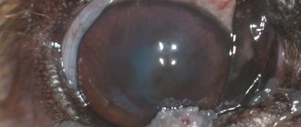
Limitations in cytology mainly relate to the diagnosis of neoplasms. The volume of cells taken for cytology is small; there may be cases when tumor cells simply do not get there or it is not possible to get a complete picture or determine the specific type of tumor.
Since cytology is minimally invasive, there are few contraindications, but if the animal has bleeding disorders and the doctor needs to perform a biopsy of a liver or kidney tumor, it is recommended that the procedure be performed in the operating room under the supervision of a surgeon in order to stop the bleeding if necessary. In the vast majority of cases, a thin needle will not cause bleeding.
Video about cytology in animals - interview with the head of the diagnostic laboratory:
What stage follows after cytology
When diagnosing tumors after surgical removal of the tumor and cytology, histology should be performed. In some cases, when malignant tumors are detected, immunohistochemistry may be required to select the most appropriate chemotherapy.
For all other types of cytology, after the analysis, the diagnosis or direction for further diagnosis is confirmed, and treatment is prescribed.
(c) Veterinary center for the treatment and rehabilitation of animals “Zoostatus”. Varshavskoe highway, 125 building 1. tel.