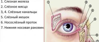
Why does a dog need a third eyelid?
During the process of evolution, the third eyelid in humans has almost completely atrophied, but in dogs it continues to perform important functions that ensure the full functioning of the visual organs.
You can understand why a dog needs a third eyelid by familiarizing itself with its main functions.
- Cleaning the cornea from dust, lint and other contaminants.
- Production of tear fluid due to the presence of the excretory ducts of the Gardner's lacrimal glands.
- Biological protection of the organs of vision from infectious agents, which is provided by immune bodies produced by adjacent lymphoid tissue.
Normally, the third eyelid only protrudes slightly from the inner corner of the eye, but with pathology it can increase significantly and change the color, shape and structure of the tissue.
What is third eyelid adenoma in dogs?
Even knowing what adenoma of the third eyelid in dogs is, it is impossible to distinguish it from pathologies such as hyperplasia and prolapse by external signs. An accurate diagnosis can only be made after a histological examination in the laboratory.
Adenoma is a benign neoplasm formed by glandular epithelial cells. Most often it develops in older animals, although a genetic predisposition is clearly visible in representatives of bracheocephalic breeds.
Third eyelid in dogs
The third eyelid (lunar fold) is located in the inner corner of the eye and is a vestigial human organ, i.e. an organ that has lost its significance in the course of evolution and is in its infancy.
Content
- Protective function of the third eyelid in animals
- Pathologies of the third century
- Breed predisposition
- Treatment
Protective function of the third eyelid in animals
Not every owner knows that for their pets the third eyelid is one of the most important protective and functional structures of the auxiliary apparatus of the eye. When the eye is touched, pressed on, or the animal lowers its head, the third eyelid instantly closes the surface of the cornea, protecting it from damage. In addition, in the thickness of the third eyelid there is an additional lacrimal gland, which ensures the production of 30% of tears. As the third eyelid moves, tears are distributed over the surface of the cornea, simultaneously washing away foreign particles and bacteria from it.
Pathologies of the third century
Pathologies of the third eyelid may be associated with its anatomical features. So, for example, with prolapse (loss) of the third eyelid, its protective shell is no longer held in place by the thin ligament that holds the gland in the orbit of the eye. Most often, this pathology occurs in dogs between 3 and 9 months, when there is active growth of both the animal as a whole and, accordingly, the eyeball and third eyelid. At the same time, in case of prolapse, it is necessary to exclude the entry of a foreign body behind the third eyelid, damage to the eyelid, the presence of eye trauma, neoplasms or disorders of the nervous system.
Breed predisposition
The most prone to third eyelid adenoma (prolapse associated specifically with developmental pathologies) are dog breeds such as bulldogs, pugs, cocker spaniels, Newfoundlands and some others.
Treatment
Treatment for a single prolapse of the gland, if no more than 6-12 hours have passed since the incident, consists of repositioning the glandular tissue and using anti-edematous and anti-inflammatory drugs. If eyelid prolapse occurs again, surgical restoration of the normal position of the third eyelid (reposition) is indicated.
Among the most common pathologies of the third eyelid, inversion (entropion) of the third eyelid is also important. The cause of inversion is excessive lengthening of the “pedicle” of the third eyelid cartilage, which has a T-shape. The cartilage seems to “break”, and attempts to turn it out, giving it a normal position, are useless. The inflammatory process that develops as a result of volvulus can lead to significant hyperplasia of the third eyelid.
Most often, this pathology occurs in Great Danes, Newfoundlands and Central Asian Shepherds.
Treatment in this case is only possible surgically. Restoring the normal position of the eyelid is carried out by excision of the broken section of cartilage.
Since the causes of the development of pathologies of the third eyelid in dogs are of a different nature, it is quite difficult for the owner to distinguish one pathology from another, and treatment most often consists of surgical intervention, the main task of the owner is to immediately contact a specialist.
The article was prepared by doctors of the ophthalmology department "MEDVET" © 2014 SEC "MEDVET"
Causes of development of third eyelid adenoma in dogs
Experts have not established the exact cause of the development of third eyelid adenoma in dogs. Great importance is attached to genetic predisposition.
Other provoking factors are:
- eye injuries;
- inflammation of the lacrimal glands;
- age-related relaxation of the ligaments that hold the lacrimal glands within anatomical norms.
Frequent inflammation of the glandular tissue caused by mechanical, chemical or light factors can also lead to the formation of adenoma over time.
Symptoms
Inflammation of the third eyelid in dogs, like most other diseases, is accompanied by certain symptoms. The development of the disease is indicated by the following symptoms:
- Enlarged gland. First, due to the development of the disease, the pet's eyes become red. Then the size of the gland begins to increase, which leads to increased tear production. This is why many dogs with adenoma have watery eyes.
- Deterioration of vision. Since sick animals develop a reddish formation in the corner of the eye, dogs begin to see worse.
- Itching and damage to the cornea. Due to the appearance of an adenoma, dogs' eyes begin to itch very much. Many dogs cannot tolerate severe itching that does not go away, and therefore constantly try to scratch with their paws. Frequent scratching damages the cornea.
- Lacrimation and purulence. It is no secret that inflammation of the third eyelid in dogs is accompanied by increased lacrimation. However, if nothing is done or treated, prolapse leads to the release of a large amount of pus.
Important! Over time, if left untreated, it only increases in size, which also negatively affects vision.
Symptoms and first signs of the disease
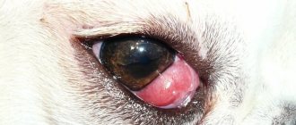
An adenoma can form in one or both eyes at the same time. A characteristic symptom and the first sign of the disease is the appearance of a protrusion from the inner corner of the eye, resembling a miniature head of cauliflower.
As the tumor grows, it begins to irritate the cornea and prevent the eyelids from closing. As a result, the following symptoms quickly develop:
- redness of the conjunctiva;
- lacrimation;
- photophobia.
Since the dog is experiencing discomfort, it begins to hit its eyes with its paws, injuring the eyelids and conjunctiva. The process is often complicated by the development of pathogenic microflora, leading to purulent inflammation.
Surgical intervention
The only effective treatment for the third eyelid is surgery. After a properly performed operation, the disease does not recur, and the lacrimal glands continue to perform their function.
Operation technique
Removal of a third eyelid adenoma is not a complex or lengthy operation and can therefore be performed under local anesthesia. However, problems with restraining the animal in most cases require the use of general anesthesia. An exception is made for pets with pathologies of the cardiovascular system.
The surgical technique is as follows:
- using general anesthesia or instilling 2-3 drops of 5-10% novocaine into the eye;
- injection under the conjunctiva of 1-2 ml of a mixture of 0.5% novocaine and 0.1% adrenaline in a ratio of 10:1;
- grasping the tumor with surgical tweezers;
- removal of the tumor with eye scissors.
Prolapse of the third eyelid gland
Prolapse of the lacrimal gland of the third eyelid is a pathological condition in which the lacrimal gland of the third eyelid is displaced from its normal anatomical position and becomes visible in the medial corner of the eye.
Normally, the lacrimal gland of the 3rd eyelid is located at the base of the third eyelid under the conjunctiva (Figure 1).
The lacrimal gland of the third eyelid is accessory and secretes 10-30% of all tears.
Lacrimal gland prolapse is common in certain breeds of dogs: Cane Corso, Mastino, English Bulldog, Spaniel, Chihuahua. Dog breeds with a loose constitution and pathologies of the lower eyelids – inversion (ectropion) – are predisposed. Most often, prolapse of the lacrimal gland of the third eyelid occurs in young animals 5-9 months old.
In cats, the lacrimal gland prolapses less frequently, but this condition also occurs, more often in cats of brachycephalic breeds (Persian, British).
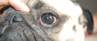
The condition can be unilateral or bilateral, with dogs of predisposed breeds likely to experience sequential loss first in one eye and then in the other eye (Figure 2).
Clinical signs : with prolapse, the displaced gland is pinched in an abnormal position, swells, increases in volume, the mucous membrane becomes hyperemic, the animal may experience discomfort, scratch the eye, and purulent conjunctivitis often occurs.
If the gland remains in the wrong anatomical position, then its nutrition is disrupted, its function decreases, which can lead to the development of dry keratoconjunctivitis (lack of tear fluid), curvature (crease) of the cartilage of the third eyelid may develop (this disease can also be independent and occur regardless of position of the lacrimal gland).
Sometimes, with prolapse of the lacrimal gland of the third eyelid, it spontaneously reduces and falls out again, the same thing happens with mechanical reduction (massage, tweezers), since this method does not lead to fixation of the gland, and it falls out again.
Previously, prolapse was mistakenly called adenoma of the lacrimal gland of the third eyelid and they resorted to surgical removal of the gland, which entailed irreversible loss of the organ of tear production and, accordingly, a high risk of developing keratoconjunctivitis sicca.
At the moment, for an animal with prolapse of the lacrimal gland, there is a reliable surgical treatment method - reduction of the gland , an operation is performed using general anesthesia, which takes about 10 minutes.
The operation involves immersing the lacrimal gland into a pocket from the conjunctiva and suturing this pocket to prevent recurrent prolapse of the gland (pocket method).
A submerged seam with a thin thread (6-0) is used, so it does not cause discomfort to the animal. The suture material is absorbable and does not require removal from the tissue.
Video of pocket method operation:
Also, one of the new methods has proven itself - the method of fixation with a non-absorbable thread such as a purse-string suture. This thread also does not need to be removed, since the seam is submersible and does not injure the tissue.
Video of the operation using the purse string suture method:
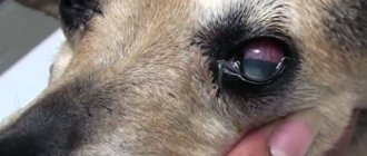
Immediately after surgery, the lacrimal gland is in its normal position, but the third eyelid may be swollen and protruding (Figure 3).
The postoperative period requires the use of antibiotic drops in the conjunctival sac and short-term (5-7 days) systemic antimicrobial therapy, wearing a protective collar if the patient scratches his eyes (most often not required).
Prevention and care for dog eyes
When purchasing a puppy of a bracheocephalic or hunting breed, you should pay attention to the prevention and care of the dog’s eyes from the first days. The set of measures should include the following points:
- constant maintenance of visual hygiene;
- prevention of injuries to the conjunctiva and third eyelid;
- avoiding contact with eyes with detergents while swimming;
- preventing exposure to chemicals on the eyes;
- timely vaccination of your pet against canine distemper and adenovirus, which can cause conjunctivitis;
- immediate contact with a veterinarian if ophthalmic pathologies occur;
- cutting off the hair hanging over the eyes of representatives of long-haired breeds.
Particular attention should be paid to the place where the pet sleeps. The room should be free of drafts, accumulation of dust and irritating substances, which include household aerosols, varnishes and paints.