4.2 / 5 ( 6 votes)
When a dog's eyelids are turned up, the planes of the free edges of the eyelids, which are usually adjacent to the eyeball, are completely or partially turned inward. Sometimes not only the free edge, but also the skin surface of the eyelid is turned towards the eye in such a way that eyelashes and hair injure the cornea. The palpebral fissure narrows sharply, lacrimation, inflammation and even perforation of the cornea are observed. While not a life-threatening condition, the disease can lead to undesirable consequences, including loss of an eye, if left untreated.
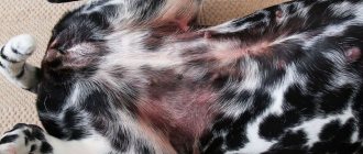
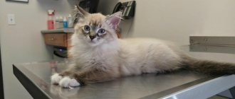
Dog on day 14 after surgery.
Causes of eyelid enversion
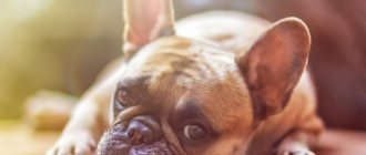
The appearance of bloat is typical in breeds that have an excess of skin folds on the head: Shar-Pei, Bulldogs, Mastino and others.
Acquired causes include:
- Damage to the cornea, causing the dog to squint its eye for relief. This leads to overstrain of the eyelid muscles, which causes spastic-type volvulus. Self-healing is possible when corneal pathology is cured.
- Reduced eye size, which may be congenital or acquired. In such cases, the eyelid support provided by the eyeball is lost, which leads to entropion.
- Injuries, eye diseases, some systemic diseases. These factors can cause deformation of the eyelids, as a result of which the dog develops entropion.
Separately, there is senile weakening of the ligamentous apparatus, in which volvulus develops in older animals.
As the disease develops, contact of fur with the mucous membrane leads to irritation of the conjunctiva and cornea. Erosion forms on the affected cornea, which subsequently leads to an ulcerative defect and loss of the internal structures of the eye. In addition, the development of keratitis is possible. In this case, the cornea becomes cloudy over time, becomes vascularized, and the dog’s vision decreases and ultimately goes blind.
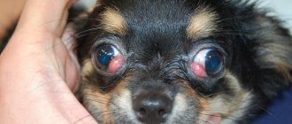
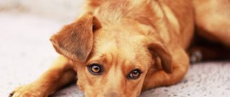
Medial inversion of the eyelids in a dog.
Diagnosis and prognosis
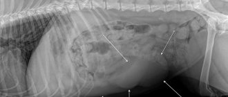
Puppies of both of the above-mentioned breeds are often diagnosed (before 16 weeks of age) with idiopathic Horner's syndrome, and as they grow older, its symptoms spontaneously “resolve.” If the lesion appears in a cat at an older age, a medical examination is necessary to identify the true cause of what is happening. In many cases, when suspicion falls on a tumor or bacterial cause of the disease, it is necessary to resort to chest radiology, ultrasound, and also take blood and urine tests - in a word, they try to determine the pathogen that caused all this.
When the nerve damage is in the sympathetic ganglion (behind the eye), or outside it, we are talking about the postganglionic variety of Horner's syndrome. It is to this type that cases of an idiopathic nature belong, for which the prognosis is favorable. If the sympathetic nerve injury is located somewhere between the brain and the sympathetic ganglion, then it is a preganglionic type, in which the prognosis may be questionable. In the second case, a “thoughtful” diagnosis is vitally important, allowing one to identify exactly where the damage to the sympathetic fibers is localized and what exactly caused it.
Symptoms of the disease
Initial manifestations include increased lacrimation and photophobia. As the disease progresses, the secreted secretion thickens and becomes mucous in nature. Photophobia is detected when a dog begins to rub its eyes with its paw after looking at light sources. This causes discomfort, so the animal becomes restless and tries to avoid light sources. Increasing pain syndrome forces the dog to look askance.
Without treatment, eye tics develop and signs of keratitis appear. Due to the weakness of the ligamentous apparatus, the skin surrounding the eye sockets sags. Sometimes corneal ruptures develop, turning into ulcers.
In animals of brachycephalic breeds (breeds of dogs with a relatively short and wide head shape, approaching round), owners often miss the onset of the development of bloat due to the characteristic constant lacrimation.
The main causes of entropion in dogs
- Genetics. It is very difficult to identify a clear cause of entropion in a dog. Sometimes we can say that such a phenomenon is genetically provoked. But this is just a guess. Basically, entropion can be found in purebred dogs, more often when crossing genetic relatives. But the gene that is responsible for the inversion of the eyelid in a dog cannot be identified, so it is also impossible to say that it is genetically programmed.
- Shape of a dog's skull.
- Location of the eyeballs.
- Length of the eyelid, their elasticity.
- Animal eyelid injuries.
- If your dog constantly squints his eyes. This feature is extremely rare, but sometimes it also causes entropion of the eyelids.
Volvulus also occurs as a result of scarring of the eyelid or severe conjunctivitis.
Entropion of the third eyelid in dogs
This form of the disease often affects shepherd dogs, Great Danes, and Pinschers. It is rarely diagnosed in other breeds.
Entropion of the third eyelid in dogs develops as a result of degeneration of the cartilaginous part of the eyelid or as a complication of follicular conjunctivitis.
It manifests itself as deformation of the third eyelid, hyperemia of the conjunctiva. The nature of the exudate during lacrimation is serous-mucosal. Some individuals may develop blepharospasm, which disappears with treatment for bloat.
Adenoma of the third century.
Entropion (turning of the eyelid) in dogs and cats
Inversion of the eyelids is when the eyelid is turned over the entire gap or only a small part of it into the eye.
CAUSE of entropion of the eyelids in animals of various etiologies:
- primary (common) - breed predisposition, which has congenital and acquired etiology;
- secondary - as a result of other diseases (conjunctivitis, blepharitis, trauma, burns (thermal and chemical), eyelid tumors, foreign body and others.
I would like to pay more attention to primary inversions, because... They are the ones that are ignored by owners at the beginning of development. It would seem no big deal, such a small trifle. At first, no one can imagine that this could result in a big problem, including the loss of an eyeball.
As a result of breed predisposition, some dogs and cats have congenital bloat as a result of abnormal development and acquired in connection with breed characteristics of the structure of the skull, skin structure and habitat. The pathology is characterized by the fact that the folded edge of the eyelid, having cilia and hair, when in contact with the cornea of the eye, forms a mechanical irritation, causing it every minute and daily injury. Before the first visit to an ophthalmologist, many owners have to observe the development of symptoms of the disease in stages.
Stage 1. EPIPHOR (lacrimation) Traumatizing the cornea of the eye, a protective reaction occurs: a large amount of tears is produced, as a result of inversion, the tear lake decreases and the tear begins to be released out. The pet has constantly wet fur around the eye, which additionally contributes to bacterial contamination of the conjunctiva of the eye.
Stage 2. BLEPHAROSPASMS and PHOTOphobia. This is a squinting of the eye due to involuntary contraction of the orbicularis oculi muscle. The reason for this phenomenon is soreness of the cornea due to damage to the layers and swelling.
Stage 3. CORNEAAL EDEMA
Stage 4. CORNEAAL ULCER
Layer by layer the cornea of the eye is damaged, forming an ulcer (superficial, stromal, descemetacele). The deeper the damage, the more eye pain the animal experiences. Some lose their appetite, hide in dark corners, and ignore their owners.
The most terrible end result of the disease that one has encountered is a perforated ulcer of the cornea of the eye, resulting in the loss of the eyeball.
TREATMENT.
- Blepharoplasty. (at the initial stage).
- Keratoplasty. Keratoconjunctivalplasty. Tarsorrhaphy (in subsequent stages)
- Removal (enucleation) or prosthetic replacement of the eyeball in cases of perforated corneal ulcer and loss of intraocular pressure.
Contacting an ophthalmologist in the first stages will save your pet’s eyes!
Diagnosis and treatment of entropion
The main diagnostic method is a general examination. To reduce discomfort during the procedure, drops with an anesthetic are first instilled into the eyes. Fluorescent solutions are used to detect erosions or ulcers on the cornea. After treatment, the affected areas glow under ultraviolet light.
In mild cases, the veterinarian prescribes antiseptic drops to relieve inflammation and destroy pathogenic microflora. Treatment for entropion in dogs involves treating the skin around the eyes with gels that contain antiseptics. Additionally, anti-inflammatory drugs are prescribed for secondary infections.
Treatment of entropion in animals
The method of treating entropion is selected by the veterinarian individually in each case, since it is necessary first of all to identify the cause and classify the entropion.
If volvulus is diagnosed due to spasm of the orbicularis oculi muscle, drug treatment can help reduce the degree of volvulus or eliminate it completely. In other cases, it is necessary to resort to surgery.
The author of the article is veterinarian Ksenia Vladimirovna Tarasova.
Eyelid inversion surgery
The surgical method plays a leading role in treatment. Even in case of serious complications (keratitis, conjunctivitis), the operation will alleviate the general condition and allow for high-quality sanitation of infectious foci.
During the operation, entropion of the eyelids in dogs is stopped by straightening and cutting off the folded part. After this, supporting sutures are applied to fix the ligaments in a certain position. Sutures are often used, which dissolve over time and do not require removal. In adult dogs, several operations may be required to fully strengthen the ligamentous apparatus.
After the operation, regular antiseptic treatment of the orbital area is carried out. Additionally, a plastic collar is put on for 7-14 days, which prevents the paw from touching the seams. The decision to prescribe systemic antibiotics is made by the doctor.
The main signs of entropion
- Pain in the eyes, “sand” in the eyeballs.
- Squinting of eyes.
- Tearing.
- Mucous secretions from the eyes of an animal.
- Tear tracks under the dog's eyes.
With all this, it is almost impossible to examine the cornea of an animal’s eyes. Eyes are constantly squinted. Sometimes the dog does not open his eyes at all.
Diagnosing entropion in a dog is quite simple. The pet will look sideways, and liquid will begin to ooze from the eyes, which will change in consistency over time. The dog also exhibits photophobia, a reaction to light and the sun.
Degrees of severity of entropion in a dog
The disease manifests itself in several degrees. The main ones can be identified:
- Extremely tight fit of the dog's eyelid;
- Turning of the eyelid touching the cornea at an angle of 90 degrees;
- Inversion with the skin and fur of the eyelid touching the cornea at an angle of 180 degrees.
In each case, the dog rubs its eyes very hard and experiences discomfort. Pekingese, bulldogs, pugs, Labradors, toy terriers and boxers are predisposed to the most common eyelid entropions.
Inversion of the eyelids in dogs can be central or lateral . With central entropion, the central part of the eyelid is everted and droops. With lateral – the eyelid droops from the middle to the outer corner of the eye.
Sometimes the problem of entropion in a dog can go away on its own as the puppy grows. In this case, it does not harm the pet. This happens when the bones of the skull do not grow in accordance with the growth of the puppy's skin. In any case, if one of the symptoms of entropion occurs, a quick consultation with a doctor and further observation or treatment of the dog is required.
Entropion (turning of the eyelids)
This is an incorrect position of the upper, lower or both eyelids, in which the free edge of the eyelid is shifted inward towards the eyeball.
There are 2 types of entropion:
- Congenital:
- hereditary etiology: the genetic basis for the development of entropion has not been established. There is a breed predisposition: Chow Chow, Shar Pei, St. Bernard, Leonberger, Bernese Mountain Dog, Cocker Spaniel, English Bulldog, Labrador Retriever, Mastiffs;
- the presence of skin folds, lack of tone of the skin of the eyelids, the size of the eyeball, the length of the palpebral fissure - all these are predisposing factors to the development of entropion;
- Acquired:
- Spastic (resulting in soreness in the eye area)
- Scarring (from injury or chronic inflammation in the area)
The clinical manifestations of this pathology are very diverse - from asymptomatic disease to more severe clinical manifestations (blepharospasm, epiphora, corneal diseases)
Diagnosis: complete ophthalmological examination + fluorescein staining of the cornea.
Treatment: surgical eyelid surgery