Third eyelid
In addition to fish, reptiles, amphibians, birds and sharks, some mammals such as camels, polar bears and seals have a fully functional membrane known as the "third eyelid". It protects the eyes of crocodiles and beavers from water.
In humans it is present only as a small fold known as the plica semilunaris, connected to the tissue covering the whites of the eyes and the insides of the eyelids, which no longer serves its original purpose but to aid in the drainage of tears. In addition, without it, the conjunctiva would be attached directly to the eyeball, limiting the mobility of the latter.
What it is
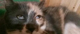
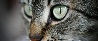
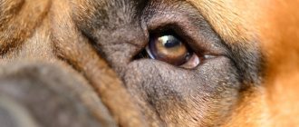
The third eyelid is located in the inner corner of the eyes. In humans this is a poorly developed rudimentary organ, but in some animals this fold is well developed. In cats and chickens, you can see a translucent nictitating membrane that performs a protective function. It protects the birds' eyes from small debris in flight. A crocodile needs such a film to see clearly in the water.
This film protects the eyes well and helps moisturize the mucous membrane. At the same time, the quality of vision is maintained.
It is noteworthy that the rudiments of the nictitating membrane are present in the human embryo, but as the fetus develops, this part of the eye atrophies.
Pointed part of the ear
It was first described by Charles Darwin in his book The Descent of Man and Sexual Selection. The scientist named it "Wolner's tip" after the British sculptor. The tubercle occurs in 58% of Swedish children, 40% of adults in India and 10.4% of adults in Spain. It is usually present on both ears, but may be present on only one.
“I will give more freedom”: 6 vows that will help you establish a connection with your teenage child
Portable air purifier and other new products for a post-pandemic world
Roskoshestvo warned of three dangerous cases for charging a phone
Ear movement
Macaques and other monkeys have more developed auricular muscles, used to move their ears, than humans, chimpanzees, or orangutans. In our country, these muscles, although quite large, do not function.
However, some people can move their ears at will, and anyone can learn to do this. The inability to move the ears in humans and other animals is compensated by the ability to turn the head horizontally, which most monkeys cannot.
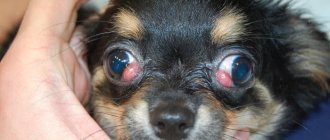
What is the third eyelid in cats?
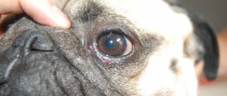
A cat's third eyelid, or nictitating membrane, is a thin bluish fold hiding in the inner corner of the eye. Normally, it is invisible, and only when the cat is dozing, falling asleep or bowing its head can you pay attention to it.
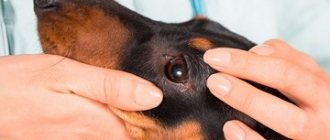
The third eyelid is also present in dogs and many other animals; Its rudiment in humans is the semilunar fold of the eye.
It should be noted that in brachycephalic cats (British, Himalayan, Persian) the third eyelid is more pronounced than in cats with a normal skull structure.
The third eyelid is best visible when the cat's eyes are half-closed.
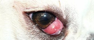
The nictitating membrane is part of the conjunctival sac, which forms the epithelium of the mucous membrane of the eye. Its dimensions are very large and comparable to the area of the anterior surface of the eyeball. The structure of the nictitating membrane contains cartilage, T-shaped and small in size; it also contains smooth and striated muscle fibers, the latter allowing for voluntary movements. Small accumulations of lymphoid tissue lie near the surfaces of the third eyelid.
The inner side of the nictitating membrane contains the lacrimal gland, whose secretion serves to wash the cornea of the eye. This gland is additional and secretes 10–30% of the tear fluid from the total volume.
The nictitating membrane performs the functions:
- protective - together with the upper and lower eyelids, protects the eye from possible external damage;
- moisturizing - prevents the cornea from drying out;
- cleansing - rids the cornea of small particles that have entered the cat's eye;
- immune - lymphoid tissue is a zone of production of secretory immunoglobulins that protect the surface of the eye from the development of various infections.
When the eyelids close, the nictitating membrane expands from the inner corner of the eye, distributes tears along its front surface, and also removes small debris.
The third eyelid contains small cartilage, muscle fibers, and lymphoid tissue; the lacrimal gland is adjacent to it
Grasp reflex
This ability is formed in the 11-15th week of pregnancy. This is one of the most important reflexes that a newborn must exhibit in order to be considered neurologically healthy.
The reflex can be tested by placing a finger on the child's palm. When trying to remove the finger, the child will grab it tightly enough to support his own weight. The latch can be unpredictable as the baby may let go without warning.
So that Saint Gregory does not punish. Sadness and work: what not to do on January 21
Vinegar for cleaning a washing machine: a non-standard use of a well-known product
Prigozhin changed his image with a beard and hair (photo). Everyone is wondering who's on the left
The reflex disappears after 2-3 months, maximum after six months, when the child gains control over his body. If this does not happen, you may have difficulty holding objects. The reflex can also be observed on the legs.
Jacobson organ
The vomeronasal organ was first discovered by the Danish anatomist Ludwig Levin Jacobson in 1811. It is located at the base of the nasal cavity just above the palate. The organ contains receptors that help detect certain types of non-volatile or liquid, organic compounds that may come from prey, predators or potential mates.
Unlike the olfactory bulb, which sends signals directly to the olfactory cortex, the Jacobson's organ sends signals to the hypothalamus, which is responsible for regulating hormone production and body temperature.
Jacobson's organ is present in all snakes and lizards, as well as in many mammals.
In humans, it develops in the embryo, but after that it regresses and the nerve connections disappear. The cavities persist in some adults, but without a functional nervous system the organ simply does not function.
Goose pimples
Skin bumps appear when tiny muscles at the base of the hair contract in an emergency or due to irritants such as a gust of cold wind. In animals with fur, this helps trap air between the hairs, which creates a layer of insulation and prevents hypothermia.
A girl found a stray cat by the river: after a month of living in the family, she was unrecognizable
Singer Biser Kirov loved one woman all his life: the story of dating and relationships
The guy became popular on TikTok: the Scottish postman sings about the sea and sailors
People experience goose bumps from screeching, metal scraping, music, feelings or strong emotions, watching horror movies, or experiencing exciting events such as winning a sporting event. Some are able to deliberately cause goose bumps in themselves.
Functions in animals
The third eyelid in animals plays a protective role. It promotes high-quality hydration of the mucous membrane and helps remove small debris that gets into the eyes during the day.
For waterfowl and birds, such a membrane helps them see well under water, acting like swimming goggles. In birds, the nictitating membrane protects the eyes from debris during flight. You can also see the third eyelid in cats; in these animals it is especially well developed. To see how the membrane functions, it is enough to slightly open the cat’s eyes while sleeping. The eyeball will seem to be covered with a thick film.
It is unknown why the human body lost this function during the evolutionary process. Scientists suggest that a few centuries ago such a membrane was very well developed in people.
Any inflammatory diseases in the inner corner of the eyes lead to disruption of the process of tear fluid removal.
Hiccups
According to the hypothesis, this reflex is necessary for tadpoles before their lungs are fully formed. It forces you to swallow air and water through your gills and is similar to the hiccups of mammals.
Hiccups are normal for babies in the last three months of pregnancy and are part of the fetal development process. It is also seen in premature babies, and researchers believe it is "an evolutionary precursor to modern pulmonary breathing."
Useful video
The appearance of a feature does not always signal congenital pathologies or genetic abnormalities in children. The formation can be provoked by the negative influence of various factors on the intrauterine formation of the fetus. The development of such a feature can be expressed through Mongoloid roots.
Author's rating
Author of the article
Alexandrova O.M.
Articles written
2029
about the author
Was the article helpful?
Rate the material on a five-point scale!
( 1 ratings, average: 5.00 out of 5)
If you have any questions or want to share your opinion or experience, write a comment below.
Wisdom teeth
Our ancestors compensated for their lack of ability to properly digest plant foods by chewing them thoroughly. But with the advent of agriculture, the human diet changed, and as a result, the jaws began to move forward less, leaving less space for wisdom teeth. Because of this, in some people they do not fully grow in because they are blocked by other teeth and must be removed.
Treatment Options
If there are no problems with vision or the functions of the visual apparatus, there is no need to treat the fold. Educational therapy is necessary if there are additional disorders:
- strabismus;
- active lacrimation;
- ptosis of the upper eyelid;
- incomplete closure of the organs of vision;
- pressure on the eyeball due to development;
- reduction in picture quality;
- narrowing of the viewing angle;
- overstrain of the visual organs.
Until the age of seven, the fold is not removed. The operation is carried out according to the stages:
- marking the area for removal;
- excision according to established lines;
- removal of fatty tissue that fills the feature;
- formation of an identical area, but with a smaller volume;
- tightening the muscle fibers of the organ of vision.
Rehabilitation is about 2 weeks. At this time, it is recommended to observe the following restrictions:
- stop watching TV;
- eliminate computer games;
- when going outside, use special optics;
- Avoid getting dust or small particles into the visual apparatus.
Surgeries are common among teenage children. It is recommended to postpone the operation to remove the third fold until the child reaches adulthood, since the formation of muscle fibers and fat deposits in this area stops.