What is sarcoptic mange
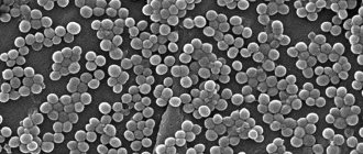
The causative agents of sarcoptic mange are mites Sarcoptes scabiei var. canis or scabies. These are small arthropods measuring 0.2 - 0.5 mm in size that live and reproduce in the thickness of the epidermis. Parasites feed on lymph and skin particles, gnawing out tunnels with ventilation outlets to the surface with their mouth appendages.
The biological cycle of Sarcoptes canis lasts from 15 to 19 days and goes through 5 stages of development: egg, larva, protonymph, teleonymph, imago.
Sexually mature females live 1.5 months. Moving through the tunnel, they lay 2–6 eggs at a time, up to 60 throughout the entire period. Males live in the epidermis for up to 1 month, and die after mating.
On the surface of the skin, Sarcoptes canis moves at a speed of 3 cm/min, in the epidermis - 2.5 mm per day. Over the course of its entire life, the itch gnaws a canal about 6 cm long.
In the external environment, the scabies mite lives for 6 days, at a favorable temperature of 10 - 15℃ and high humidity - up to 3 weeks. The eggs remain viable for up to a month. At temperatures below 0℃ and above 21℃, parasites quickly die. The video shows a magnified scabies mite.
Dogs often suffer from scabies, regardless of season, gender, age and breed. Animals are affected only by teleonymphs and sexually mature females. The source of infection is sick dogs and common care items - bedding, combs, towels. It is less common to catch a tick in a room where an infected dog has been.
Is sarcoptic mange contagious to the owner?
A person is attacked by another type of scabies mite. However, if there is close contact with an infected pet, canine itch can spread to the owner and cause pseudosarcoptosis.
Only the larvae spread to humans. When they get on the skin, they sink under the keratinized scales of the epidermis, into the hair follicles and cause an inflammatory reaction. In places where there is frequent contact with the pet, bright red or pink itchy spots, rashes, and blisters appear.
Itchy rash on the skin with the development of pseudosarcoptosis.
However, human skin is not suitable for life and reproduction of dog itch. They do not penetrate the epidermis, do not dig tunnels, and do not lay eggs. Before reaching the stage of sexual maturity, the larvae are rejected and die.
Pseudosarcoptosis is not transmitted to another person and does not require treatment. Redness and itching disappear spontaneously when the dog recovers.
Sometimes a dog suffers from sarcoptic mange without obvious symptoms, and the owner develops pseudoscabies. In this case, the appearance of a rash, spots, and itching in areas of contact with the dog indirectly signal the development of sarcoptic mange.
Symptoms of sarcoptic mange in dogs
The clinical picture of the disease depends on the individual immuno-allergic reaction to saliva and parasite excrement. Therefore, symptoms do not appear from the moment of infection, but after sensitization - the development of sensitivity to allergens and irritants.
The first symptoms of sarcoptic mange appear after 1 to 3 weeks. With repeated infection, the disease manifests itself much faster – within 1–3 days.
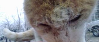
Scabies mites love to live in areas with short hair. The first outbreaks most often occur:
- on the back of the nose;
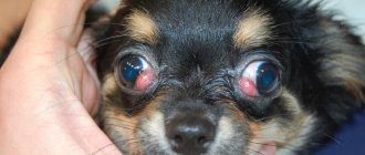
- around eyes;
- eyebrows;
- along the edges of the ears;
- on the elbow and hock joints;
- on the stomach;
- in the groin;
- armpits.
Damage to the ear.
Here are the main signs of infection:
- The main symptom of sarcoptic mange is increasing itching. It occurs in response to parasite bites and as an allergic reaction to excrement, eggs, and saliva. The dog chews, licks places where parasites accumulate, and begins to itch even where there are no ticks. The itching intensifies in the evening and at night, in warm rooms, near heating devices.
- The animal develops an ear-pedal reflex. If you rub the tip of the ear between your fingers, the dog involuntarily jerks its back paw, as if trying to scratch its ear.
- In places where itching accumulates, the skin first turns red and then becomes covered with nodules. The fur falls out and thins.
Scabies on the abdomen: nodules, redness.
- The nodules turn into blisters with clear inflammatory exudate. Due to severe itching, the dog leaves scratches and abrasions on the skin, and injures blisters.
Consequences of scratching with severe itching.
- The released liquid sticks the hair together, then dries and turns into crusts and scabs.
Redness, crusts, baldness on the elbow.
- In places where severe scratching occurs, the skin becomes rough and thick, hair falls out, up to complete baldness.
Damage to paws with scabies.
- Sometimes the skin peels over the entire surface, and dandruff appears.
- In advanced cases, a bacterial infection spreads in places where scratching occurs, and ulcers with an unpleasant odor appear.
- Against the background of general intoxication, the temperature sometimes rises and the lymph nodes become inflamed.
- Behavior changes due to exhausting itching. The dog becomes irritated or apathetic, loses appetite, and loses weight.
Animals with a weak immuno-allergic reaction and older dogs often tolerate sarcoptic mange without itching.
Sarcoptic lesion on the bridge of the nose without itching.
Diagnosis, treatment and prevention of psoroptosis
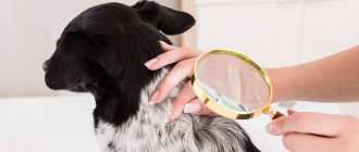
Diagnostics. To make a diagnosis, it is necessary to take into account the epizootic situation, the clinical manifestation and be sure to examine scrapings taken from damaged areas of the skin.
Laboratory methods for examining skin scrapings for diagnosis are divided into biotic (detection of living mites) and abiotic (detection of dead ones).
Biotic methods.
A scraping taken from the site of the lesion is placed on a watch glass and slightly heated to body temperature. The ticks begin to move and are clearly visible. You can examine scrapings on black paper, which is heated (placed on a warm object or held in the palm of your hand) to make it easier to identify ticks.
Abiotic methods.
Most often, scrapings are examined after treating the crusts with an alkali solution. To do this, the taken material is placed in a bacteriological dish or test tube, filled with a 10% solution of sodium or potassium hydroxide and heated for 15-20 minutes until steam appears. Then the liquid is drained, and the sediment is examined under a microscope or magnifying glass. You can add kerosene to the material, which clarifies the contents of the scraping, dissolves small crusts, and the mites become clearly visible.
Treatment of animals with psoroptosis is carried out with many drugs in the form of ointments, liniments, solutions, emulsions, suspensions, powders, aerosols.
The most effective method of treating animals is bathing them in acaricidal baths. You can use various washing devices, both stationary and portable. If the number of animals is small, they are washed with acaricides manually using a brush.
For bathing or washing animals, solutions, emulsions and suspensions of various drugs are used: aqueous emulsions of neocidol
(0.05% according to ADV): vetionol (0.25% according to ADV);
cyodrine
(0.25% ADV);
emulsion K
(6%);
emulsion SK-9
(4%);
emulsion of creolin
(2-5%),
lysol
(2-5%),
butox-50
(1:1000),
pyrene
(1:500),
tanacetos liniment.
In winter, animals are treated with dusts, ointments, liniments; When it gets warm, they must be treated with liquid acaricides.
You can treat the ears of rabbits with a 2% emulsion of chlorophos in petroleum jelly,
liniment, which contains 6 hecachlorane, 50 sulfur, 44 talc and 250 vaseline oil;
an emulsion of kerosene with vaseline oil
in equal parts;
emulsions of butox-50
(1:1000),
pyrene
(1:500),
tanacetos
or
polysulfide liniments.
Good results are obtained by using aerosols “Acrodex-sa”, “Aerosol-dicresyl”, “Aerosol-cyodrin”, “Dermatosol”, “Psoroptol”
etc.
Ivomec
at 1 ml per 50 kg of animal weight,
closantel
at 2 mg/kg live weight (ADW).
Preventive actions. When importing animals to a farm, it is necessary to keep them in quarantine, examining them at this time for psoroptosis and other scabies diseases. When carriers of scabies mites are identified, all imported livestock are treated with acaricides. Periodically, all farm animals are examined, paying attention to the condition of the skin. When animals with skin changes are identified, they must be examined for the presence of scabies mites.
In farms unaffected by psoroptosis, all animals are divided into three groups: first - animals sick and suspected of the disease; the second - animals suspected of infection (without signs of disease, but having had contact with patients); the third is healthy animals. Each group is kept separately and should not have contact either through care items or through staff. Animals of the first group are treated, and the second group is treated for preventive purposes. Animals of the third group are examined. If sick or suspicious animals are identified, they are immediately isolated, and the premises where they were kept are cleaned mechanically. Manure is stored for biothermal disinfection.
For decontamination of premises and care items, use a 5% hot mixture of creolin, a 10% solution of bleach, a 20% solution of freshly slaked lime, a 3% solution of Lysol or naphthalizol, a 30% solution of hot ash alkali, HB-1 solution.
Chorioptoses
Chorioptoses -
invasive diseases of horses, small and large ruminants and other species, which are caused by carpet beetle ticks of the genus
Chorioptes
(family
Psoroptidae).
Occurs with skin damage,
Pathogens. The skin of horses is parasitized by Ch.
food/, cattle -
Ch.
bovis, sheep -
Ch.
ovis, goats -
Ch.
caprae, Biology of pathogens. The development of Chorioptes is similar to that of mites of the genus Psoroptes.
On the skin of an animal, the causative agents of chorioptosis are most often localized in certain areas: in horses - on the skin of the limbs, in the area of the fetlock joint and tail; in cattle - in the area of the root of the tail, the flexor surface of the fetlock joint; in sheep - on the scalp and legs.
Equine chorioptosis
It is characterized by damage to the skin of the legs and is accompanied by itching, which is especially noticeable at night and after work. The most characteristic manifestation of the disease is hair loss in the area of the brushes and inner thighs, and frequent non-stepping while resting. Diagnosis is the same as for psoroposis, but a scraping is taken in the middle of the lesion. During treatment, the entire body of the animal is treated with acaricides (see “Psoroptosis”). At the same time, decontamination of harness, premises and care items is carried out.
Chorioptosis in cattle
The skin of the legs, tail root and udder is affected. The process usually does not apply to the entire body. The disease often occurs simultaneously with psoroptosis. Diagnosis, treatment and preventive measures are the same as for psoroptosis.
Chorioptosis of small ruminants
Carpet beetles first affect the legs in the fetlock area. In some cases, the process spreads to all parts of the body. In damaged areas, animals feel severe itching, sloughing of the epidermis and hair loss occur, then the skin thickens, wrinkles and becomes covered with scabs.
Diagnosis, treatment and prevention of chorioptosis
The diagnosis of chorioptosis is based on the clinical manifestation of the disease, epidemiological data and the detection of mites during acarological examination of scrapings from the affected areas.
Chorioptosis, unlike other sarcoptic diseases, is relatively easy to treat. This is due to the superficial parasitism of ticks, which contributes to the direct effect of acaricidal drugs on them. When treating animals with chorioptosis, the same means and methods are used as for psoroptosis. To ensure therapeutic effectiveness, one cannot limit oneself to local treatment of lesions; it must be total.
When carrying out therapeutic and preventive measures, it is necessary to take into account that goats (especially thin-haired ones) do not respond well to bathing in acaricidal solutions. Therefore, before
When using acaricides, it is necessary to carry out a biological test on several animals. Usually goats are treated with acaricides after shearing.
Activities to eliminate the disease should also include decontamination of premises, machines, equipment and care items using chemical and biological methods of neutralization. In farms that are unfavorable for chorioptosis, it is necessary to carry out planned preventive treatment of animals with acaricides in the fall, before placing them in stalls.
Otodectosis carnivores
Otodectosis -
a chronic invasive disease of dogs, cats, foxes, arctic foxes and other animals, which is caused by skin beetle mites of the genus
Otodectes
(family
Psoroptidae)
and is accompanied by itching and dermatitis of the ears.
The causative agent of otodectosis is the mite Otodectes cynotis,
Biology of the pathogen. The development cycle of Otodectes is similar to the development of skin mites. Under optimal conditions, their development from egg to sexually mature individuals lasts 9-12 days. Mites are localized on the inner surface of the ears, in the external auditory canal and on the eardrum.
Epizootology. Mostly young dogs with long ears and foxes are affected. The source of infection is sick animals. On fur farms, otodectosis can occur as an enzootic. Infection occurs through contact with infested animals or through care items. Puppies usually become infected from sick bitches.
Pathogenesis. Mites have a mechanical and toxic effect on the skin of the auricle, resulting in irritation of nerve endings, destruction of the epidermis, and atrophy of the sebaceous glands. Conditions are created for the development of secondary microflora.
Clinical manifestation. Patients experience hyperemia, itching, peeling of the skin of the auricle and ear canal. Animals are worried, whining, shaking their heads, scratching their ears with their paws. Serous and then purulent exudate forms in the auricle. Hearing decreases; when you press your fingers on the base of the ear, a splash is heard. If left untreated, the eardrum perforates and the inflammatory process spreads to the middle ear, then to the inner ear, and sometimes to the membranes of the brain. The head of sick animals is often turned 90°C towards the affected ear. When the processes are complicated by a secondary infection, the body temperature rises.
Diagnostics. The final diagnosis is made by microscopy of a scraping from the auricle dissolved in a 10% solution of sodium hydroxide or kerosene. In this case, different stages of mites are identified.
Treatment and prevention. hydrogen peroxide solution is poured into the auricle
and use a cotton swab to clean the inflammatory exudate from it.
After this, acaricidal preparations are introduced into the sink: mixtures of 3% chlorophos in camphor oil,
5%
hexachlorane in liquid mineral oil,
hexatalpa
oil emulsions containing 0.03% gamma isomer of hexachlorane.
Also effective are a 5% oil suspension of gardan
a 40% oil suspension of phenothiazine
aerosol acaricides suspensions of butox
:1000), pyrene
:500), etc. Ivomec
which is administered subcutaneously in a dose of 1 ml per 50 kg of animal weight, or
pharmacy (aversect-2).
The preparations are also applied to areas of the skin that the ears touch. The treatment is repeated after 5-6 days. If the middle and inner ear are affected, antibiotics are prescribed (streptomycin, tetracycline subcutaneously).
For preventive purposes, it is necessary to isolate animals entering the farm, periodically examine their ears, and promptly treat patients; carry out decontamination of premises where sick animals were located and items for their care.
Previous26Next
Diagnostics
Sarcoptic mange is diagnosed based on clinical presentation, microscopic analysis of skin scrapings, and trial treatment.
It is not always possible to make an accurate diagnosis based on external signs, since the symptoms of scabies resemble other skin pathologies:
- atopic, flea, contact dermatitis;
- food allergies;
- demodicosis;
- superficial pyoderma;
- cheyleliosis;
- dermatophytosis;
- notoedrosis.
To clarify the diagnosis, a microscopic analysis is prescribed. Numerous superficial scrapings are taken from the dog from typical areas of scabies. The diagnosis is confirmed when mites, larvae, eggs, and excrement are found under a microscope. However, traces of parasites can be detected in only half of the dogs; according to other sources, in 20%.
An adult scabies itch under a microscope.
If the tests show a negative result, and the clinical picture of sarcoptic mange is obvious, then a trial treatment is prescribed. The diagnosis is confirmed when anti-tick therapy helps alleviate the symptoms and the dog is on the mend.
Before visiting the veterinarian and taking scrapings, the pet is not washed, treated with anything, or given any medications.
Diagnosis of the disease
Trying to diagnose mange in dogs can be difficult. The standard method is to scrape the skin and then identify the mite under a microscope. Unfortunately, on average, only twenty percent of infested dogs will have mites on any given scraping. Therefore, if a dog has a positive skin scrape, the diagnosis is confirmed, but a negative skin scrape does not rule out sarcoptic mange in the dog. Most cases are diagnosed based on the patient's medical history and the animal's response to mange treatment.
How to treat sarcoptic mange in dogs and puppies
The treatment regimen for scabies includes:
- Shampoos with a keratolytic, antiseptic effect for disinfection, softening and cleansing of the skin from crusts, scales, scabs.
- Acaricidal agents for the destruction of itches. Solutions for local treatments and drip application to the withers, ointments, and concentrates are produced against scabies mites. In difficult cases, systemic medications are prescribed - tablets, injection solutions.
Additionally you may need:
- Antipruritic for severe scratching.
- Antibiotics for the appearance of suppuration.
If several animals live in the house, the sick dog is isolated. The rest also undergo treatment, even if there are no visible symptoms of infection.
Shampoos
Veterinary shampoos for removing crusts.
Before applying acaricidal preparations, the animal is completely bathed with a keratolytic detergent, for example:
- Doctor cleansing. Shampoo with benzoyl peroxide. This synthetic substance softens rough skin, exfoliates keratinized scales and crusts, and inhibits pathogenic microflora.
- DermaBenSs. Therapeutic shampoo for bathing pets with bacterial, parasitic skin diseases, scratching. The composition is enriched with benzoyl peroxide, 1% sulfuric acid, salicylic acid, which disinfect and dry the skin.
- Peroxiderm. Antibacterial agent with benzoyl peroxide.
Lather the shampoo on wet wool and leave for 5 – 7 minutes. The crusts are first soaked, then those that come off easily are removed.
At the end, the shampoo is washed off with running water, and the pet is rinsed with a decoction of medicinal plants: chamomile, celandine or calendula. After bathing, the fur is carefully blotted with a towel so as not to transfer parasites to healthy parts of the body.
Acaricidal ointments, solutions
Before treatment, the hair from the affected area and around the lesion is cut off. The dog is put on a veterinary collar or muzzle. The preparations are rubbed from the borders of the affected area to the center, covering 2–4 cm of healthy skin.
Here are examples of effective external remedies against scabies mites:
- Aversectin ointment. The main component is the acaricide aversectin C. Treatments are repeated 2–5 times every 5–7 days. The ointment is contraindicated in puppies under 8 weeks of age, during pregnancy, and while nursing offspring. A similar drug is Amidel gel. It is allowed for puppies from 2 weeks of age.
- Decta Forte. Combined solution based on 3 components. Fipronil destroys mites, chloramphenicol blocks the spread of pathogenic bacteria, lidocaine reduces sensitivity. The animal is treated 2–5 times at intervals of 5 days. Decta is not recommended for treating puppies under 4 weeks of age, pregnant or lactating females. Similar solutions: Amitrazine, Amit Forte.
- Simple sulfur, sulfur-tar ointment. The drugs stop the spread of ticks, accelerate tissue regeneration, and stimulate hair growth. Treatments are carried out daily 1 - 2 times a day until the epidermis is restored. There are no contraindications based on age. Both ointments can be combined with other drugs.
Emulsion concentrates
Emulsion concentrates against scabies mites.
Concentrates based on synthetic insectoacaricides work well against arthropod parasites:
- Butox;
- Neostomozan;
- Delcid.
For dogs and cats, the liquid is poured into glass ampoules in a volume of 1 to 5 ml. Concentrates are diluted with water according to the instructions immediately before processing. Rub the prepared solution with a sponge into the skin and fur over the entire surface.
After treatment, the liquid is not washed off and the animal is not wiped. The muzzle can be removed when the coat is dry. The solution is applied 2 - 3 times every 7 - 10 days. The prepared aqueous emulsion is stored for no longer than 2 hours.
Drops on the withers
Spot-on solutions are effective against scabies mites. Drops are applied pointwise to intact dry skin in places where the dog cannot lick them off - at the base of the neck, between the shoulder blades.
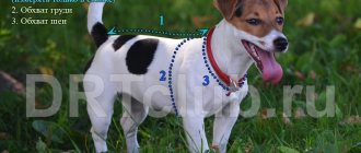
Solutions are produced in pipettes with a dose of the active substance already calculated based on body weight, so the pet is weighed before choosing drops.
Here are examples of effective solutions for the withers to destroy itch mites:
- Stronghold. Drops with selamectin. To treat scabies, the dog is treated twice with a 1 month interval. The drug is allowed from 6 weeks of age.
- Advocate. The drops combine 2 potent insectoacaricides: moxidectin and imidacloprid. When treating scabies, the solution is applied 2 times with a time interval of 30 days. Puppies can be treated from 7 weeks, miniature dogs weighing less than 1 kg - only with the permission of the treating veterinarian.
- Bravecto Spot On. Drops based on fluralaner. To destroy sarcoptic mites, one treatment is enough. Drops are not used for animals younger than 8 weeks and lighter than 2 kg.
Pills
Tablets for the treatment and prevention of sarcoptic mange.
Relatively recently, tablets began to be produced to combat ticks. They are relatively safe for mammals, and they are good at treating parasitic diseases: sarcoptic mange, demodectic mange, and otodectic mange.
Preparations are selected strictly according to the weight of the animal after weighing; they cannot be crushed or broken. Tablets are given by hand or with food. For picky pets they hide it in a piece of meat.
- Nexguard Spectra. The active ingredient is afoxolaner. For sarcoptic mange, feed 1 tablet, after 30 days another one. Nexgard Spectra is contraindicated in small dogs lighter than 2 kg. A similar drug is Frontline Nexgard.
- Simparica. A drug based on sarolaner. When treating scabies, 2 doses of 1 tablet are prescribed with a break of 1 month. The permissible body weight limit is lower than that of other tablets - 1.3 kg.
- Bravecto. To eliminate scabies, 1 tablet of the required dosage is enough. The next one can be given for prevention after 12 weeks.
Anti-mite tablets should not be used on puppies under 8 weeks of age.
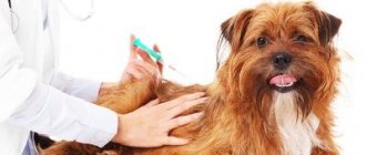
Injections
For extensive lesions, injectable drugs of the avermectin series are prescribed:
- Aversect K&S. They produce 2 types with different concentrations of the active component aversectin C: 0.2% for cats and dogs weighing up to 10 kg, 0.5% for dogs weighing more than 10 kg. For sarcoptic mange, injections are given 2 times with a week's break, following the dosage according to the instructions.
- Otodectin. Ivermectin based solution. It is administered subcutaneously twice with an interval of 8–10 days at the rate of 0.2 ml per 1 kg of body weight.
Both drugs are not approved for puppies under 2 months, animals with kidney or liver diseases. Due to breed sensitivity, avermectins are poorly tolerated by bobtails, collies, and shelties.
Preparations for itching
The itching can be so severe that the dog scratches itself until it bleeds, and bacteria and fungi quickly spread to open wounds. Therefore, in case of unbearable itching, drugs that suppress the immuno-allergic reaction are prescribed.
Apoquel. They produce 3 types with different contents of the active component oclacitinib for small, medium, and large dogs. The tablets are fed with food or by hand twice a day. Single dose 0.4 – 0.6 mg/1 kg. The instructions contain a detailed dosage table by body weight.
The duration of admission is determined individually according to the condition of the animal, but no longer than 14 days. The medicine is safe when taken for a short time. Apoquel is prohibited for puppies under 1 year of age, mini dogs lighter than 3 kg, during pregnancy, and for malignant neoplasms.
Prednisolone or veterinary analogue Prednivet. The medicine is administered subcutaneously for 3 to 5 days during the period of exacerbation of itching. The average daily dose of prednisolone for dogs is 0.5 – 1 mg per kg of weight. According to reviews from owners, prednisolone does little to relieve itching caused by scabies mites.
Antibacterial drugs
Antibiotics are prescribed if suppuration begins at the scratch site. Skin bacterial infections are treated with penicillin drugs:
- Amoxicillin 15%, veterinary. Suspension for injection. The medicine is administered subcutaneously or intramuscularly 1 time. If there is no noticeable improvement, a second injection is given after 48 hours. According to the instructions, a single dose of amoxicillin is 15 mg/1 kg, which is 1 ml of suspension per 10 kg of body weight.
Amoxicillin veterinary.
- Sinulox. Injectable suspension and tablets containing a combination of amoxicillin and clavulanic acid. Injections are given into the muscle or under the skin for 3–5 days, once a day. 1 ml of suspension is designed for 20 kg of body weight, so for injections it is better to take an insulin syringe. Sinulox tablets are given twice a day at the rate of 12.5 mg/1 kg. The course of treatment for skin infections lasts from 1 to 4 weeks. The “human” analogue of Sinolox is Amoxiclav tablets.
Sinulox suspension and tablets.
The veterinarian can adjust the dose of the antibiotic and the duration of the course depending on the dog’s condition.
Treatment
Therapeutic measures are carried out by the dog's owner under the supervision of a veterinarian. It should be borne in mind that some recommended medications act only on the imago, others on the larval stages. Tick eggs are practically invulnerable to acaricides. Therefore, the instructions for use indicate the frequency of treatments and the timing between them. You should not use the medications more often than recommended because they contain substances that are toxic to ticks and dogs. If treatment is missed, the therapeutic effect is reduced.
Therapy begins with preparing the affected area - cutting the hair, soaking the crusts with antiseptic solutions - Furacilin, Perhydrol and others.
Anti-tick action in most situations is ensured by the use of the following acaricidal drugs:
- Liniments, ointments, drops for external use.
- Insecticidal emulsions.
- Parenteral agents.
- Anti-mite tablets.
The choice of certain drugs is determined by the scale of the lesion, the presence of concomitant diseases, as well as the size of the dog.
Liniments, ointments, drops for external use.
The following drugs are in demand:
- Amitrazine.
- Amit - Forte.
- Demos.
- Ivermek-gel.
- Drops on the withers.
Used for small areas of lesions or small animals.
Insecticidal emulsions
For large affected areas and large dogs, they use preparations for bathing animals - Butox, Diazinon, Ectomin, etc. This method is convenient and does not involve high financial costs. Up to six weekly treatments may be required for complete cure. If there are no containers for bathing, or it is cold outside, use similar medications in aerosol form.
Parenteral agents
A relatively convenient method of treatment when organizing bathing is difficult. Subcutaneous injections of Ivermectin or Dectomax are in demand. However, some dog breeds are hypersensitive to the drugs and may die.
Anti-mite tablets
Milbemycin or Sayfly are non-toxic for dogs, but the effect is achieved with a long course of treatment.
If the disease is advanced, the veterinarian prescribes antibiotics and antifungal drugs.
Prevention
It is necessary to maintain the dog’s strong immunity with timely disinsections, deworming, vaccinations and adequate nutrition with ready-made food. Contacts with the yard and relatives must be limited.
We invite you to join our Zen channel and group on VKontakte or Odnoklassniki, where new articles for pet owners are published.
Similar articles:
- What is separation anxiety in dogs and how to deal with it?
- Tuberculosis in dogs
- Umbilical hernia in dogs
Forecast
Sarcoptic mange is debilitating for a dog and does not go away spontaneously, but is highly treatable. The prognosis is favorable.
It will take 4 to 8 weeks to completely eliminate itching and restore skin and fur. However, noticeable improvement occurs within the first 14 days. The rate of recovery depends on the size of the lesion, the severity of the lesion, and the animal’s immunity.
If after 6 weeks from the start of treatment there are no noticeable improvements, the itching does not go away, additional studies are prescribed to determine the real cause of the disease.
Infection with scabies can be avoided if you keep your pet away from dogs with obvious signs of sarcoptic mange and use anti-mite means: drops on the withers, collars or tablets.