Owners do not always notice the appearance of neoplasms on the body of pets. The similarity of such seals to bumps diverts the attention of owners of four-legged friends from the problem. Lack of knowledge in this area is fraught with the development of minor pathologies into dangerous diseases. The article will discuss one of the skin pathologies - histiocytoma in dogs, its inherent symptoms and treatment methods.
Histiocytoma in dogs: symptoms, treatment
Histiocytic skin diseases in dogs
This group includes rare skin disorders caused by excessive cell proliferation.
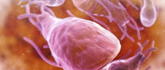
Histiocytes are cells of hematopoietic stem origin. During development, they turn into macrophages or dendritic cells, to which IDCs and Langerhans cells belong.
This pathology is classified into several types:
- cutaneous histiocytoma;
- Langerhans cell histiocytoma;
- histiocytoma from IDC;
- systemic and malignant histiocytosis.
This tumor is not always found in a benign form. There are known cases of a neoplasm transforming into a cancerous form.
Histiocytes are connective tissue cells that have a protective function
The disease manifests itself in the form of a tumor that forms in tissue cells during immune failures. Neoplasms form mainly on the head and hind limbs. The favorite place for pathology is the ears.
Histiocytoma in dogs is the most common skin pathology, occupying a leading position in the list of sarcomas.
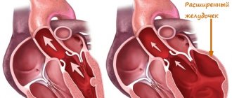
This type of tumor is characterized by spasmodic growth
Disease susceptibility
Regardless of gender, the disease is most often observed in pets during the juvenile period. In 80% of cases, the disease attacks dogs under 2 years of age. Representatives of smooth-haired breeds are most prone to the disease. Genetic predisposition is observed in Bernese Mountain Dogs , Rottweilers, Golden Retrievers, and Flat-Coated Retrievers.
Rottweiler is at risk
Causes of the disease
The factors that provoke the development of this disease are not fully understood. Scientists have found that mutations occur in the cell genome, leading to rapid tissue growth. Also among the possible reasons are the following:
- Cellular inflammation.
- Exposure to carcinogens.
- Non-cancerous pathologies.
- The influence of ionizing rays.
- Poor immune response.
- Soft tissue injury
Among existing breeds, up to 25% of neoplasms are observed in the Bernese Mountain Dog
General information about what is histiocytoma in dogs
Histiocytoma is a group of tumor diseases of the skin. Most of them are benign. But benign formations can become malignant. The results of treatment depend on the moment of contacting the veterinary center. The sooner an attentive owner notices changes in the pet, the better the effect will be. Some veterinarians do not consider histiocytoma a classic tumor, since in its development and current symptoms it is more similar to reactive hyperplasia of certain types of cells. Despite popular opinions, there are cases of histiocytoma degeneration into malignant formations.
It is better to perceive the disease as potentially dangerous. Cases of morbidity are more often recorded in 3-year-old animals
. In older animals, histiocytoma is less common. Scientists are working on the causes of the disease. There is no data confirming any factors for tumor formation.
Symptoms of the disease
The manifestations of the disease depend on the location of the tumor development:
- If a tumor forms in the tissues, the animal suffers from abdominal pain and fever. The disease is accompanied by increased body temperature, loss of appetite, decreased body weight and severe malaise.
- If a histiocytoma appears on the skin, the pet loses mobility of the joints located in the area of tumor growth. The seal on the dermis is colored red, and when pressed, discomfort occurs. As the formation grows, it puts pressure on neighboring tissues, causing peeling and ulcers.
Symptoms depend on the location of the tumor
If a malignant tumor develops on the extremities, there is a risk of fractures. In pets with cutaneous and systemic histiocytosis, the listed symptoms may be absent.
Cutaneous histiocytoma
Belongs to the category of fast-growing tumors. The surface of the skin often becomes bald, but the new growth does not cause any discomfort.
Main symptoms:
- lesions are concentrated in the dermis and subcutaneous layer;
- the skin is dotted with numerous seals on the trunk, muzzle, limbs and neck.
The pathology is characterized by a chronic nature, in which periodic relief of symptoms is noted. In the advanced form of the disease, there is an overgrowth of secondary microflora, leading to severe itching.
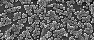
Cutaneous histiocytoma is the most common type of disease
Cutaneous Langerhans cell histiocytosis
Langerhans cells contribute to the development of the disease. Normally, these structural units are part of the mucous membranes and dermis. When the disease occurs, they grow rapidly and provoke multiple histiocytoma of the skin. The size of the tumor ranges from small nodules to large red formations. Compared to the previous variety, the pathology has a worse prognosis. If in normal histiocytosis systemic organs are not involved, then in this form penetration into them is possible. The tumor also affects the lymph nodes.
Shar-Peis are susceptible to Langerhans cell histiocytoma
Cutaneous histiocytosis from IDC
Interstitial dendritic cells contribute to the formation of compactions in the subcutaneous tissue and dermis. The formation of numerous neoplasms, reaching a diameter of up to 4 cm, is characteristic. Localization is the neck, head, limbs and torso. Damage to the lymph nodes is sometimes observed. The average age of pets most susceptible to the disease is 4 years. Unlike the pathology caused by the proliferation of Langerhans cells, this disease has an inherent tendency to penetrate into the deep layers of the epidermis.
Langerhans cells are concentrated exclusively in the epithelium
Systemic histiocytoma
It is not as common as the cutaneous type. The disease is characterized by generalized damage to the mucous membranes, skin and lymph nodes.
Main symptoms:
- multiple formations in the form of nodules, covered with scabs;
- alopecia of the dermis;
- conjunctivitis;
- weight loss;
- noises when listening to the lungs.
Symptoms of conjunctivitis in dogs
The disease most often attacks Bernese mountain herding dogs between 2 and 8 years of age.
The Bernese Mountain Shepherd is susceptible to a systemic type of pathology
Malignant histiocytoma
Histiocytic sarcoma originates predominantly from IDCs. Main symptoms:
- paleness of the skin;
- lethargy;
- dyspnea;
- the presence of noise in the lungs;
- temporary paralysis of the hind legs;
- nervous attacks;
- enlargement of the liver, lymph nodes and spleen.
This type of pathology practically does not affect the skin, progressing mainly on the internal organs. The average age of infected animals is 7 years. The disease develops rapidly and ends in death.
With malignant histiocytoma, compactions form in the liver and spleen
How to distinguish a lump from a histiocytoma?
Despite the fact that these formations have much in common, there are a number of significant differences.
A lump is a compaction that appears on the dermis as a result of a blow or insect bite. It varies in diameter from a few millimeters to 2-3 centimeters.
The lump may be accompanied by a slight increase in temperature. By its nature, this is a small hematoma that resolves after a few days. Histiocytoma remains on the skin for a long time and does not disappear without appropriate treatment. This formation has a more defined shape and brighter color.
If a lump on a pet’s body tends to grow, is colored red and moves when pressed, in most cases a histiocytoma is diagnosed.
The lump can easily be confused with a histiocytoma
Diagnosis of pathology
Since there is a risk of the neoplasm becoming malignant, timely detection of the disease becomes important. Due to the fact that it is not always possible to obtain accurate test results, diagnosing the disease is not easy. During the examination of the pet, the veterinarian performs the following tests and examinations:
- Blood and urine analysis.
- Biopsy of affected tissue.
- Cytology of bone marrow cells.
- Immunohistochemical examination. It is this diagnosis that allows us to identify the pathological origin of cells.
- MRI. Makes it possible to identify hidden tumors and their locations.
- Ultrasound. Shows the size of the tumor located on the internal organs.
Depending on the type of pathology, the specialist uses the appropriate diagnostic method.
Diseases are determined using immunohistochemical studies
Histiocytoma in dogs: methods for diagnosing a tumor
Carrying out a cytological examination makes it possible to make a more accurate diagnosis. This method involves examining the material under a microscope.
.
For this, a biopsy is used . A puncture is made in the tumor area and the cellular material is removed. But this method does not provide complete confidence
in the diagnosis, as with histological examination. A histological examination provides a complete and accurate picture. A thin section of the tumor is prepared, fixed, stained using a certain technique, and examined under a microscope. To exclude the presence of metastases, X-ray and ultrasound examinations are performed.
Treatment of the disease
The severity of symptoms and the stage of development of the disease determine the choice of therapeutic method. When making a decision in favor of surgical or drug treatment, the doctor takes into account the extent of skin lesions.
Surgery
It is used primarily in the development of fibrous histiocytoma. The main methods of influencing the tumor are cutting off or cryosurgery. Removal of the seal occurs with cutting out adjacent tissue within a radius of 2 cm. This avoids relapse. Surgical intervention is used for the following symptoms:
- severe itching;
- acute inflammatory process;
- multiple neoplasms.
Depending on the clinical picture, the animal may require infusion therapy or blood transfusion.
In parallel with surgical measures, in severe cases, a course of radiation and chemotherapy is prescribed. These procedures will stop the growth of new seals and destroy existing ones.
Cryosurgery is used to treat histiocytoma.
Drug treatment
This type of therapy is used less frequently. It is used in the presence of inoperable tumors or minor skin lesions:
- Taking hormonal drugs. It is administered mainly in the form of blockades. An injection is made into the seal with a substance containing hormones in large quantities. They successfully neutralize the process of tumor formation and help reduce it.
- Local treatment with dimethyl sulfoxide and corticosteroids.
For systemic histiocytosis, antitumor anthracycline antibiotics are prescribed.
"Dimethyl sulfoxide" is used in the conservative treatment of histiocytoma
Disease prognosis
With timely treatment, a complete cure for the pathology is possible, but the cure rate is not too high. Doctors associate this with a high probability of metastases. A specific feature of this type of tumor is its resistance to radiation therapy or chemotherapy.
In 15% of cases, histiocytoma in dogs develops into a malignant tumor of bone tissue.
To prevent negative consequences, it is important not only to regularly examine the animal for the presence of tumors, but also to reduce the risk of their occurrence. To do this, you should not expose your pet to vaccines that contain aluminum hydroxide. If there is a need for an injection, it is advisable to give the injection in the tail area - below the third vertebra. If a sarcoma forms after administration of the medication, cutting off the posterior appendage will not have a negative impact on the pet's health.
Histiocytoma in dogs: diagnosis, symptoms, manifestations and treatment
How does histiocytoma occur in dogs ? Oncology occupies one of the first places in terms of incidence. The reasons for such high growth are unknown. Scientists around the world are working on a cure for this terrible disease. Animals are also not spared from this problem. Pets cannot tell in human language what hurts or bothers them, so early diagnosis is difficult. Doctors say: “you need to contact us on time, since treatment of the disease in the early stages is easier and there are more chances for a full recovery.” But what about animals? Histiocytoma in dogs would not be so dangerous if the owner knew its symptoms and immediately contacted the specialists of the veterinary center . Let's talk about the manifestation of the disease in a dog.