Lymphoma, or lymphosarcoma in dogs, is a group of cancers that affect the lymphatic system. Malignant cell degeneration detected at a late stage cannot be treated.
It is possible to save an animal from sudden death only at the very beginning of the development of pathology. To do this, you need to know its main signs that appear as the tumor grows.
What diseases fall under the definition of “lymphoma”
This definition includes 2 types of tumors: lymphogranulomatosis and non-Hodgkin lymphosarcoma. The former are found only in humans, and the latter are further divided into several varieties depending on the location of the lesion:
- Nutritional.
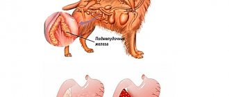
Lymphoma develops in the gastrointestinal tract, causing problems with stool and vomiting. Despite weight loss, abdominal volume is steadily increasing. This occurs due to excess accumulation of free fluid in the abdominal cavity.
- Multicentric.
Develops in the prescapular, popliteal, mandibular and inguinal lymph nodes while maintaining their mobility. It is painless even in advanced forms.
- Mediastinal.
Affects the chest cavity, causing breathing problems. The dog develops shortness of breath and a severe cough. Her pulse slows and her mucous membranes turn blue due to lack of oxygen.
- Cutaneous.
Accompanied by the growth of lymph nodes located close to the skin, as well as the formation of ulcers and eczema. Pain syndrome manifests itself only at a late stage of the pathology.
- Extranodal.
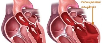
This type of tumor grows in organs outside the lymphatic system (kidneys, heart, eyes, bones, brain). Lymph nodes become involved only as it grows.
In all of these cases, malignant degeneration of lymphocytes, the most important cells responsible for the production of immunity, is observed. Degeneration leads to the gradual failure of the affected organ, and the transfer of diseased lymphocytes through the bloodstream and the appearance of metastases aggravate the picture, capturing more and more new tissues.
The disease can be confused with bacterial infection, parasitosis, allergies, intoxication and autoimmune pathologies. They are united by the same symptom - lymphadenitis, or lymphadenopathy - inflammation of the lymph nodes, accompanied by an increase in their size. This change is most easily detected in short-haired animals in the neck and lower jaw area.
Diagnostics in a veterinary clinic
Lymphoma can only be diagnosed in a veterinary clinic. When making a diagnosis, laboratory blood tests (general clinical and biochemical) and ultrasound examination of organs located in the abdominal cavity are taken into account. X-ray examination and magnetic resonance imaging are also carried out.
Histological and cytological studies can confirm the final diagnosis. Biological material is taken from accessible, enlarged superficial lymph nodes using a medical syringe.
Histological examination is necessary to diagnose the affected lymph node. After making a diagnosis, the veterinarian prescribes an adequate treatment regimen that suits the individual characteristics of the sick dog’s body.
Reasons for the development of pathology
The exact causes of lymphoma in dogs are not known. The theory about the influence of viruses, radiation exposure and prolonged use of glucocorticosteroids has not been proven.
Animals with low immunity are at risk, therefore there is a relationship with a number of unfavorable external factors. Under their influence, the body's strength weakens, facilitating the development of the tumor.
External factors
Various carcinogens have a negative effect on your pet. These include:
- herbicides used to fertilize a summer cottage;
- artificial additives included in low quality dry food;
- paint and varnish materials used during repair work;
- volatile fumes emitted by factories.
Being kept in such conditions not only reduces immunity, but also leads to disruptions within the DNA. Compounds that are dangerous to the body are integrated into its genetic code, provoking the degeneration of healthy cells into tumor cells.
Breed and genetic predisposition
Some breeds are more likely to get cancer than others. These include:
- boxers;
- terriers;
- Pekingese;
- retrievers;
- Dobermans;
- Great Danes;
- dachshunds;
- bulldogs;
- mastiffs;
- Rottweilers.
There is also a high tendency to develop oncology in offspring obtained from one or two sick parents. It is recommended to sterilize all babies from such a litter to prevent the transmission of the pathogenic gene to new generations.
Before buying a puppy, be sure to check all the information about its father and mother. All inherited diseases are indicated in the pedigree.
Diagnosis of the disease
Diagnosing lymphoma is a very meticulous and multi-step process. The initial step is to examine the animal. The doctor determines how enlarged the lymph nodes are, determines their tenderness and mobility upon palpation.
The next stage is an ultrasound scan of the lymph nodes and abdominal cavity, checking how enlarged the liver and spleen are. An x-ray of the chest cavity is taken to check for changes in the lungs. After this, a blood test is performed, including markers for cancer activity. Sometimes magnetic resonance imaging and computed tomography are performed. It is performed under general anesthesia. The animal's blood and urine are also subject to mandatory testing.
If an enlarged lymph node is found, a knife biopsy is performed. Next, the material is sent for research to confirm or refute the malignancy of the tumor. Only after receiving the results of all tests can conclusions be drawn about the tumor itself, the rate of its development, and the development of a treatment regimen.
Signs of lymphoma in a dog
The most severe symptoms are observed at the age of 5 years. Before this, the tumor process proceeds very secretly, accompanied by minor behavioral changes.
Lymphosarcoma has no specific symptoms, so it cannot be detected without laboratory diagnostics. Symptoms that appear depend on the type and may include:
- ascites, vomiting, bloody diarrhea, or constipation;
- the appearance of neoplasms under the conjunctiva;
- change in the color and smell of urine, as well as disturbances in urination (reduction in volume, pain);
- shortness of breath, cough, slow pulse and blue mucous membranes;
- ulceration and hardening of the skin;
- nosebleeds and frequent sneezing.
A sick dog becomes apathetic. His lymph nodes become inflamed, and his weight decreases even though he maintains his appetite. Due to weakened immunity, pathogenic microorganisms begin to enter the body more and more often.
As a result of extensive inflammation and secondary infections, body temperature can rise greatly without responding to antipyretic drugs for a very long time.
Varieties and symptoms
The clinical manifestations of lymphoma in dogs depend on which organs and systems are affected and to what extent. Enlargement of the subcutaneous lymph nodes, which can be easily detected by palpation, is a characteristic, but not obligatory, symptom of this disease. Among the signs common to any serious disease, lymphosarcoma may include:
- general weakness, fatigue;
- decreased appetite;
- disorders of the digestive system;
- periodic causeless vomiting;
- weight loss;
- increased body temperature, often only to low-grade fever;
- When examining the dog, ascites (fluid in the abdominal cavity), enlargement of the liver or spleen is detected;
- in the mediastinal form - cough, shortness of breath, difficulty swallowing;
- with damage to the nervous system - loss of coordination of movements, convulsions, paresis;
- in case of skin damage - multiple, poorly healing ulcers.
Symptoms vary at different stages of lymphoma in dogs. However, the early stages are characterized by the following general symptoms:
- lack of appetite;
- weight loss;
- regular indigestion or vomiting;
- increased body temperature;
- lethargy and depression;
- hair loss or thinning;
- an increase in the amount of water consumed and, as a result, frequent urination;
- an increase in the size of the lymph nodes, which causes pain.
There are five main types of this disease: multipolar, mediastinal, gastrointestinal, cutaneous, and spinal lymphoma (a disease of the spinal cord). To a certain extent, lymphoid lesions of the central nervous system can be considered a variation of the latter type. The most common type is multi-pole. It mainly affects the external lymph nodes.
Symptoms depend on which system was affected by the cancerous degeneration of cells. There are no two completely identical cases of this disease. First, cutaneous lymphoma in dogs often presents as large, swollen, painful lumps under the skin (swollen lymph nodes).
In many cases, there are no specific signs of the disease for a long time. Anorexia, weight loss, ascites (pathological accumulation of fluid in the abdominal cavity), shortness of breath (difficulty breathing), polydipsia (pathological thirst), polyuria (excessive urine production), fever, anemia, bleeding from the nasal passages, sepsis are sometimes observed.
Stages of the disease
Treatment methods for lymphoma in a dog and the prognosis for recovery depend on the stage of its development. Until the animal dies, the tumor goes through 5 stages:
- The lesion does not extend beyond one lymph node. The size of the organ increases, but pain does not always occur. The animal feels satisfactory, but you can suspect something is wrong by palpating its neck.
- Neighboring lymph nodes are involved in the tumor process. The first alarming symptoms appear.
- Changes affect lymph nodes not only within one system, but throughout the body. This condition is called generalized lymphadenopathy.
- Not only the lymphatic system, but also the internal organs are affected. The liver and spleen are primarily affected. Both changes are clearly visible on ultrasound.
- The tumor reaches the bone marrow and central nervous system. As a result of extensive growth of metastases, death occurs.
The most favorable prognosis is typical for the first two stages. In all other cases, it is almost impossible to save the animal.
Possible complications
Lymphoma is divided into five stages. On the first and second, one or more lymph nodes are affected, the animal’s condition is normal. There may be no signs. The third is characterized by spread to all lymph nodes. The fourth involves damage to the spleen and enlargement of the liver. On the fifth, the bone marrow and nervous system are affected.
Depending on the stage, complications are mentioned. At the third stage, in addition to the lymph nodes, nearby organs are already affected. Kidneys, bladder, lungs, heart, etc. Then the functions of these organs weaken and stop working. This condition most often leads to death.
What tests need to be done at the veterinary clinic?
Symptoms alone are not enough to treat lymphoma in a dog, but this is a serious reason to undergo an unscheduled examination. Enlarged lymph nodes are characteristic of many diseases, so it is very important to confirm the preliminary diagnosis with the results of laboratory and instrumental studies. These include:
- General and biochemical blood tests. They demonstrate an increase in protein and white blood cells, an anemic state and a decrease in platelet count.
- Ultrasound of the abdominal cavity. Evaluates the condition of lymph nodes that are not amenable to conventional palpation, and pathological changes in the liver and spleen.
- X-ray, MRI and endoscopy. Detect disorders of the heart, lungs, central nervous system and gastrointestinal tract.
- Cytology of the lymph sample and biopsy of the affected organ. The degree of malignancy of the tumor and the stage of its development are determined.
Please note that after taking a sample of the biomaterial, the four-legged patient is left in the hospital for another 8 hours. The puncture may cause spontaneous hemorrhage or bleeding, which can be fatal. For this reason, veterinarians need to make sure there are no complications before releasing the pet to its owner.
How do symptoms appear?
The symptoms of lymphoma in dogs are as varied as the different types of tumor itself. And this makes diagnosis difficult.
| Name of lymphoma | Symptoms |
| Multicenter | The first symptom that usually appears is swollen lymph nodes. They are 3–10 times larger than usual. But the nodes themselves do not hurt at all. They are simply large and feel like an elastic, moving lump under the skin. Over time, dogs diagnosed with this condition may develop lethargy, fever, anorexia, weakness, and dehydration. |
| Food | Alimentary lymphoma is accompanied by vomiting, abdominal pain, anorexia, diarrhea and weight loss. |
| Mediastinal | Dogs with this diagnosis have difficulty breathing. This is due to the presence of a tumor inside the chest or due to a build-up of fluid (pleural effusion). Animals may experience swelling of the face and front legs, as well as increased thirst and urination. |
| Extranodal | This lymphoma affects the skin. Its signs include isolated raised nodules or more widespread scaly lesions. They can appear in the mouth, affecting the gums, lips, and upper mouth. If the tumor is in the lungs, symptoms of respiratory distress are likely. And if located in the kidneys, it causes kidney failure. A tumor of the central nervous system is accompanied by seizures, and a tumor in the bones can cause pain and cause a sudden fracture. |
The lymphatic system helps fight infection. Therefore, fever is often one of the first signs of illness. Additionally, because lymphoma weakens the immune system, dogs may become more susceptible to other diseases. And it’s hard not to notice if your pet was previously absolutely healthy and active. Lymphoma itself, however, is not considered painful in animals.
Important! Because cutaneous lymphoma causes itching and flaking, it is often mistaken for a fungal infection.
Treatment at the veterinarian and at home
Lymphosarcoma is incurable. The types of therapy used are aimed at slowing down the rate of its development and improving the patient’s quality of life.
Types of therapy
Treatment in the early stages allows you to achieve a fairly long and stable remission. For this purpose use:
- Chemotherapy.
Antitumor drugs are very aggressive. They affect not only the tumor, but also healthy cells. Most often, several medications are used at once, which results in a whole list of side effects. To relieve them, the patient is prescribed antisymptomatic drugs.
- Radiation therapy, or radiotherapy.
Effective only in the first two stages. Unlike chemotherapy, it does not destroy tumor cells, but stops their division.
- Operation.
Used to remove small tumors in limited areas and for conditions whose danger exceeds the risks of anesthesia. These include excessive blood loss, intestinal obstruction and compression of the heart.
A sick pet will require constant care and more gentle nutrition. His condition will have to be constantly monitored by an oncologist, since remission will certainly, albeit not soon, be followed by a relapse.
Care and nutrition
The animal is provided with comfortable living conditions. It is prohibited to leave it outside in a booth or enclosure, as even a small temperature change can worsen the condition. The sleeping area is placed in a warm and quiet room, protected from drafts and excess noise.
Depending on the affected organs, an individual therapeutic diet is selected for the pet. The amount of fat is reduced to reduce the load on the gastrointestinal tract. For the same purpose, the portion size is also reduced, increasing the frequency of feedings.
To restore intestinal microflora and maintain liver function, the patient is prescribed probiotics and gastroprotectors. He may also need vitamins or immunomodulators that restore micronutrient deficiencies and the effects of anemia. The length of time you take these drugs depends on the severity of side effects from chemotherapy.
The tumor itself must be monitored through regular blood tests, ultrasound or x-rays (for mediastinal pathology). The frequency of such examination should be at least once a month.
Treatment
Treatment of lymphoma is effective only at the early stage of the disease. The disease cannot be completely cured, but with the help of medications, remission of the disease can be achieved for a period of several months to a year.
Without treatment, lymphoma develops rapidly and death occurs within 3-4 weeks. Death occurs due to the fact that the affected organ completely ceases to function.
The main method of treatment is chemotherapy. There are many drugs for this purpose and none of them are perfect. Which one to use, what chemotherapy regimen to use, how to avoid complications as much as possible - all this is determined by the veterinarian. His decision depends on the stage of the disease, the condition of the dog and many other related factors.
If the lymphoma has entered one of the last stages, and the dog’s general condition has deteriorated significantly, then there is no point in carrying out therapy. Medicines stop the development of the disease, but at the same time they negatively affect other organs and systems. In this case, the doctor prescribes painkillers and corticosteroids.
Lymphoma cannot be defeated. Even after remission, the disease will again begin its destructive effect after some time. The goal of any treatment is to improve the dog’s well-being and prolong its life as much as possible with this diagnosis.
Forecasts and statistics
A disappointing prognosis is given starting from the third stage. Even with a response to chemotherapy, the tumor returns after some time, becoming more aggressive due to metastases. Serious complications are also observed in internal organs that lose their functionality.
The life expectancy of sick animals varies from 1 month to 2 years. The minimum period is typical for the advanced stage, and the maximum for the initial stage, when the longest remission is achieved.
In addition to the stage of the pathology, other factors also influence the prognosis: the location of the tumor, the age of the patient, the presence of concomitant infections and the strength of the immune system.
Canine cutaneous epitheliotropic lymphoma
Svetlana Belova, Estonian University of Life Sciences Photos of the author are used in the article
Introduction
Epitheliotropic lymphoma (EL) is the most common cutaneous lymphoma diagnosed in dogs, although it accounts for only 5% of all types of canine lymphoma. It is characterized by tumor proliferation of cytotoxic (CD8+) T-lymphocytes with tropism for the epithelium of the skin (epidermis), its adjacent structures (hair follicles, sebaceous and sweat glands) and the epithelium of the mucous membranes, especially the epithelium of the oral cavity. In addition to cutaneous EL, dogs can also be diagnosed with primary EL of other locations (for example, bladder, intestines). In humane medicine, cutaneous EL, or rather, one of its forms, is called mycosis fungoides, and this term is sometimes used in veterinary medicine, although it is considered controversial and outdated. The average age of onset of cutaneous EL is 10 years. Boxers, English Cocker Spaniels and Bichon Frize are considered predisposed breeds. There is no gender predisposition.
In addition to dogs, cutaneous EL is less common but is also diagnosed in cats, rabbits, ferrets, guinea pigs, rats, mice, hamsters and cows. The cause of EL is not clear. Some, but not all, studies have noted a relationship between atopic dermatitis and subsequent detection of EL.
| Photo 1. Alopecia, crusts and depigmentation of the eyelid area |
| Photo 2. Erythema, crusts and exfoliation |
| Photo 3. Plaque |
| Photo 4. Extensive erosion in the area of the auricle |
Clinical picture
It is extremely varied, and although it is common to distinguish four clinical syndromes of EL (exfoliative erythroderma, mucocutaneous form, single or multiple plaques and nodules, and peptic ulcer disease of the oral mucosa), they usually do not occur alone, and signs can often be seen in one dog all of the above syndromes simultaneously. Signs of EL, in descending order: diffuse erythema, plaques, erosions, desquamation (before exfoliation), nodules, hypopigmentation, crusts, and alopecia (Figures 1–6). Lesions are usually diffusely distributed throughout the body, less often localized only on the head. Damage to the limbs and even paw pads is possible (crusts, depigmentation, ulcers, photo 7). Localized EL limited to a single lesion is extremely rare. The mucous membranes and mucocutaneous border (erosions, ulcers, depigmentation) are affected in approximately 50% of cases and almost always together with the skin (photos 8–9). In the oral cavity, both the gums and the tongue and palate may be affected (photos 10–11). The most common mucocutaneous area affected will be the area between the nose and upper lip (Figure 12).
Itching is present in approximately 40% of cases and can be severe.
In addition, cases of EL with the formation of numerous vesicles and bullae, which turn into erosions when ruptured, have been described.
Most often, cutaneous EL is limited to the skin and mucous membranes, but systemic dissemination is also possible with lesions of the epithelium of lymph nodes and internal organs (spleen, liver, kidneys, heart, trachea and lungs, gastrointestinal tract, bladder, prostate, eyes, etc.). d.). In addition, the presence of tumor lymphocytes in the peripheral blood is possible (leukemic stage, Sezary syndrome).
| Photo 5. Generalized alopecia |
| Photo 6. Depigmentation and crusts on the nose, reminiscent of pemphigus foliaceus |
| Photo 7. Depigmentation, crusting and erosion in the area of the pads and interdigital spaces |
| Photo 8. Alopecia, depigmentation and erosions on the mucocutaneous border of the prepuce |
Diagnostics
Due to the extremely variable clinical signs, EL can mimic a large number of dermatoses of various etiologies - from parasitic to autoimmune, so the list of differential diagnoses is very long.
A presumptive diagnosis is made on the basis of anamnesis, clinical picture and cytological examination (the most informative will be material taken by fine-needle aspiration from plaques and nodes), and the final diagnosis is made only on the basis of histology results. The average time between the onset of clinical signs and the final diagnosis is usually five months.
| Photo 9. Depigmentation of eyelids |
| Photo 10. Erosion on the gums |
| Photo 11. Erosion and depigmentation of the oral mucosa |
| Photo 12. Alopecia, depigmentation and erosion |
Treatment
For localized EL, surgical excision is recommended. In other cases, many drugs have proven to be potentially effective: retinoids, lomustine, glucocorticosteroids, masitinib, human interferon (alpha-2a), L-asparaginase, vincristine, cyclophosphamide.
In addition, photo- and photodynamic therapy and radiation (including electron therapy) can be used.
The prognosis is poor, and in most cases survival is 2 to 6 months after diagnosis, regardless of the treatment method chosen. If the diagnosis was made at an early stage and treatment was started immediately, the prognosis is better. Rarely, but a more favorable course of EL with survival of up to 2 years is possible.
Literature
1. Gross TL, Ihrke PJ, Walder EL, et al. Discoid lupus erythematosus. In: Gross TL, Ihrke PJ, eds. Skin diseases of the dog and cat. 2nd ed. Blackwell Science 2005, pp. 52–55.
2. Muller and Kirk's Small Animal Dermatology, 7th Ed. Miller W., Griffin C., Campbell C. WB Saunders, 2012.
3. Holtermann N., Kiupel M., Kessler M., Teske E., Betz D., Hirschberger J. Masitinib monotherapy in canine epitheliotropic lymphoma. Vet Comp Oncol. 2016;14 Suppl 1:127–35.
4. Fontaine J., Heimann M., Day MJ Canine cutaneous epitheliotropic T-cell lymphoma: a review of 30 cases. Vet Dermatol. 2010;21:267–75.
5. Santoro D. et al. Total skin electron therapy as treatment for epitheliotropic lymphoma in a dog. Vet Dermatol. 2017 Apr;28(2):246-e65.
Doctors interested in veterinary dermatology are welcome to Svetlana Belova’s website: www.vetderm.eu.
Here you will find information about the School of Veterinary Dermatology in Tartu, about webinars, upcoming interesting events, and you can also look through the dermatological atlas and subscribe to the blog.
SVM No. 3/2017
Rate material
Like Like Congratulations Sympathy Outrageous Funny Thoughtful No words
Main symptoms
Symptoms of lymphoma are completely different, depending on the type of disease. No two sick animals have the same signs.
Based on the type of progression, lymphosarcoma is divided into:
- multi-pole;
- gastrointestinal (alimentary);
- mediastinal;
- cutaneous;
- spinal (spinal cord is affected).
The most common is multipolar, when all lymph nodes are affected. Superficial ones are more susceptible. Cancer spreads to neighboring organs. Dysfunction occurs, which is why the pet dies.
If the kidneys are affected, the blood stops being cleansed as it should. Death occurs from intoxication of the body.
The cutaneous type of lymphoma is characterized by the formation of compactions. They are located under the skin. They grow and become painful. These are subcutaneous lymph nodes affected by cancer. Weeping ulcers form and do not heal for a long time.
Signs do not appear for a long time; this is a hidden form. They also appear blurry. There are no specific symptoms. Anorexia, shortness of breath, and ascites (accumulation of fluid in the abdominal cavity) occur. There is constant thirst, frequent urination, anemia, and fever.
With the mediastinal type, cyanosis of the mucous membranes, cough, and shortness of breath are observed. This occurs due to the release of fluid in the pleural cavity.
With the gastrointestinal type, weight loss, vomiting, and diarrhea occur. Palpating the abdomen reveals intestinal compaction and an enlarged spleen.
The spinal form is characterized by damage to the bone marrow. Then the hematopoietic system produces cancer cells instead of normal ones. This type occurs at stages 4-5.