Preparing for the examination
For more accurate results during a routine examination, special preparation of the animal is required. On the eve of the test, you should stop feeding the dog 10-12 hours in advance.
You need to make sure that the dog does not eat its usual food, does not beg for treats, does not steal anything from the table, and does not find anything edible during a walk. It is important to provide the animal with a sufficient amount of clean drinking water.
In some cases, before conducting an extended biochemical analysis, the doctor may advise limiting the animal’s physical activity. In this case, you should avoid long walks, jogging with the dog, active games and training lessons.
Self-prescription of any medications, vitamins and dietary supplements for animals is prohibited. If your pet is already taking any medications, you should consult with your veterinarian about the advisability of taking or stopping them on the day of the examination.
Before taking blood for analysis, any physiotherapeutic procedures, ultrasound, or massage are prohibited. It is also recommended to avoid stressful situations.
Causes
Various reasons can provoke deviations in the composition of the blood. An increased number of lymphocytes in the blood is called lymphocytosis, and a decreased number is called lymphopenia.
An increased number of lymphocytes appears in the following cases:
- Lymphocytic leukemia. This serious disease is also called lymphocytic leukemia. This is a commonly found variant of leukemia, or blood cancer. It is characterized by a predominance of neoplastic clonal cells of malignant origin in the dog’s blood. The same effect is provoked by other malignant diseases of the hematopoietic and lymphatic systems: lymphoma, lymphogranulomatosis, lymphosarcoma, myeloma.
- Inflammatory processes and infectious diseases. The immune system produces many white blood cells that fight infection and inflammation. Not all infections are accompanied by an increase in the number of lymphocytes, since some pathogens can be destroyed by other types of leukocytes.
- Allergic reactions. An increase in the number of lymphocytes can occur as a response of the body to the penetration of allergens into it.
- An increase in the number of lymphocytes may be a consequence of long-term use of certain medications.
- Poisoning with heavy metals and other highly toxic substances.
- Endocrine problems.
- Lack of vitamin B12.
- High physical activity.
- Stress.
- Injuries.
- Starvation.
- Predominance of protein foods.
- Hyperthyroidism.
In some cases, the use of certain vaccines may be the cause of a high lymphocyte count. Also, this condition can be temporary (after illness, injury, surgery) or permanent.
A reduced number of cells is observed in the following cases:
- bone marrow lesions;
- diseases of the lymphatic system;
- long-term debilitating infections and inflammations;
- severe renal and heart failure;
- immunodeficiencies;
- treatment with certain types of drugs (cytostatics, corticosteroids, antipsychotics);
- pregnancy (slight reduction in the number of lymphocytes).
How does the procedure work?
During the study, a certain amount (several milliliters) of venous blood is taken from the animal. The procedure is carried out by an experienced specialist in a veterinary hospital. By agreement, blood is taken from the animal at home.
During the procedure, the presence of the owner is necessary, as well as the presence of a muzzle for large, aggressive or agitated animals due to illness. The manipulation is painless and easily tolerated by the animal. But sometimes it may be necessary to restrain the dog on a table and administer local anesthesia.
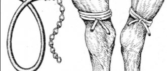
Blood is taken from a large vessel on the paw (no matter the front or back). To do this, a small area of fur is shaved or cut short, the skin at the puncture site is treated with an antiseptic solution, and a tourniquet is applied above this area.
Blood is drawn using a disposable syringe inserted into a vein or using a needle and a vacuum tube with an anticoagulant.
After the procedure, the dog’s paw is re-treated with a disinfectant solution, and a sterile gauze bandage is applied to it.
Possible complications
Depending on what the dog is sick with, complications of varying severity may occur. Untreated infections, acute or chronic inflammatory processes, diseases of the blood and hematopoietic organs have a sharply negative impact on the animal’s immune system, which can cause the appearance of protracted and difficult to treat colds - bronchitis and pneumonia.
Since the dog is very weak, such diseases can be fatal.
Biochemical analysis
No less important in diagnostic terms is a biochemical study. Using its parameters, you can evaluate metabolism, study the function of the liver, pancreas, kidneys and other internal organs.
Biochemical analysis makes it possible to assess the acid-base balance (pH). The normal limits of which are from 7.35 to 7.45. It is considered one of the main parameters of the analysis, since deviations from the standard indicate serious problems that can cause severe dysfunction of internal organs.
An increase in pH is a shift to the alkaline side, alkalosis. Its causes are frequent uncontrollable vomiting, overdose of diuretics, kidney disease, and improper infusion therapy (excess bicarbonates).
A decrease in pH value indicates a shift in the blood reaction to the acidic side, acidosis. It can be caused by prolonged diarrhea, diabetes, poisoning with certain poisons (ethylene glycol, methanol), and certain medications.
Biochemical blood test in cats
A biochemical blood test is a laboratory research method used in veterinary medicine, which reflects the functional state of the organs and systems of the animal’s body.
A biochemical blood test in cats requires certain preparation of the animal for the procedure. Blood is taken from the pet on an empty stomach before diagnostic and therapeutic procedures. A needle is inserted into the vein, through which blood is drawn. The resulting material is collected into a test tube and sent along with a referral to the laboratory.
Blood biochemistry
in cats can help with:
- making a final diagnosis,
- determining the prognosis of the disease - its course and further development,
- disease monitoring - monitoring the course and results of treatment,
- screening—detection of a disease at a preclinical stage.
The range of biochemical indicators is quite large.
The main indicators for the study are:
enzymes
(molecules or their complexes that accelerate (catalyze) chemical reactions in living systems) and
substrates
(the initial product converted by the enzyme as a result of a specific enzyme-substrate interaction into one or more final products). The interpretation of the biochemical blood test in cats is based on the data of the studied enzymes and substrates.
The main indicators characterizing the enzymatic activity of the body are:
1. Alanine aminotransferase (ALT)
– found mainly in the liver cells of cats and when they are damaged enters the bloodstream. Therefore, when ALT increases, they speak of acute or chronic hepatitis, liver tumors, and fatty liver. This enzyme is also found in the kidneys, cardiac and skeletal muscles.
2. Aspartate aminotransferase (AST)
— high activity of this enzyme is characteristic of many tissues.
Determination of AST activity is used to identify disorders of the liver and striated muscles (skeletal and cardiac).
When the cells of the above tissues are damaged, they are destroyed, which may indicate necrosis of liver cells of any etiology (hepatitis), necrosis of the heart muscle, necrosis or injury of skeletal muscles.
3. Alkaline phosphatase (ALP)
– The activity of this enzyme is found mainly in the liver, intestines and bones.
The total activity of alkaline phosphatase in the circulating blood of healthy animals consists of the activity of liver and bone isoenzymes. Therefore, in growing animals, the bone ALP isoenzyme is elevated.
But in adult animals, this increase indicates bone tumors, osteomalacia or active healing of fractures.
An increase in the level of alkaline phosphatase in the blood is also the result of a delay in the secretion of bile (cholestasis and, as a consequence, cholangitis). However, in cats, the half-life of circulating ALP is only a few hours, limiting the value of ALP testing as a marker of cholestatic disease.
The ALP isoenzyme, which is responsible for the activity of the latter in the intestine, is found mainly in the small intestine. At the moment, it has not been sufficiently studied in cats, therefore, when the activity of intestinal alkaline phosphatase changes, one can indirectly judge the pathological processes of the gastrointestinal tract.
In cats, an increase in the activity of alkaline phosphatase and other liver enzymes is often found in hyperthyroidism, and a decrease in the latter in hypothyroidism.
4. Amylase -
refers to digestive enzymes. Serum alpha amylase primarily originates from the pancreas and salivary glands.
Enzyme activity increases with inflammation or obstruction of pancreatic tissue, which may indicate pancreatitis or acute hepatitis.
However, in cats, traditional amylase tests for pancreatitis do not have sufficient diagnostic value. Also, an increase in amylase activity is observed in acute and chronic renal failure.
Other organs, such as the small and large intestines and skeletal muscles, also have some amylase activity. Therefore, an increase in blood amylase may indicate intussusception, peritonitis.
The following substrates are of primary importance for clinical research:
1. Total protein.
Proteins are essential components of all living organisms; they are involved in most of the life processes of cells. Proteins carry out metabolism and energy transformations. They are part of cellular structures - organelles, secreted into the extracellular space for the exchange of signals between cells, hydrolysis of food and the formation of intercellular substance.
The diagnostic value of this indicator is quite broad and can indicate complex processes occurring in the body.
An increase in total protein is observed with general dehydration of the body, infectious and inflammatory processes.
Loss (decrease) occurs in diseases of the liver, gastrointestinal tract, kidneys, which result in impaired protein absorption, as well as in the depletion of animals, nutritional dystrophy.
2. Albumin.
Serum albumin is synthesized in the liver and makes up the largest portion of all whey proteins. Since albumin makes up the majority of the total blood protein, they have a close relationship with each other. Thus, an increase or decrease in total protein occurs due to the albumin fraction. Therefore, these indicators have similar diagnostic value.
3. Glucose
. In the animal body, glucose is the main and most universal source of energy for metabolic processes. Glucose is involved in the formation of glycogen, nutrition of brain tissue and working muscles.
Glucose serves as the main indicator for diagnosing diabetes mellitus in animals, which develops as a result of absolute or relative deficiency of the hormone insulin. This, in turn, provokes the development of hyperglycemia - a persistent increase in blood glucose. A significant increase in blood glucose levels is also observed in chronic kidney disease.
An increase in glucose can also be observed under various physiological conditions: stress, shock, physical activity.
Hypoglycemia (low glucose levels) can occur as a result of acute necrosis of the liver or pancreas.
4. Urea -
the end product of protein metabolism in animals. Found in blood, muscles, saliva, lymph.
In clinical diagnostics, the determination of urea in the blood is usually used to assess the excretory function of the kidneys.
Thus, a significant increase in urea levels is observed in cases of impaired renal function (acute or chronic renal failure).
Shock or severe stress can also contribute to an increase in urea levels. Low values are observed with insufficient protein intake in the body and severe liver diseases.
5. Creatinine -
end product of protein metabolism. Most of the creatinine is synthesized in the liver and transported to skeletal muscles and then released into the blood, participating in the energy metabolism of muscle and nervous tissue. Creatinine is excreted from the body by the kidneys in the urine, so creatinine (its amount in the blood) is an important indicator of kidney activity.
High creatinine is an indicator of a rich meat diet (if there is an increase in the blood and urine), kidney failure (if there is an increase only in the blood). Creatinine levels also increase with dehydration and muscle damage. A low level is observed with reduced meat consumption and fasting.
About the reasons for fluctuations in hematocrit
What does hematocrit show? How well does blood transport oxygen to cells and tissues? For most dogs, the norm is 35-55%. Above, we looked at which dog breeds the indicator can be elevated, and it fits within the norm.
If the hematocrit increases or decreases, the specialist is alarmed. Such fluctuations in the indicator indicate that some kind of pathology is occurring in the pet’s body.
It could be:
- heart disease;
- gastrointestinal disease;
- anemia;
- oncology;
- anemia.
For puppies or bitches expecting offspring, NST rates are lower. The veterinarian considers it normal when their hematocrit is from 23 to 52%.
There are factors that can provoke a hematocrit increase from 55 to 65%. Let's look at them:
- primary erythrocytosis or erythremia;
- secondary erythrocytosis, due to: hypoxia for various reasons; hydronephrosis; polycystic disease; tumors in the kidneys, when erythropoietin is intensively produced - this is the hormone due to which red blood cells are released;
- burns, peritonitis and other troubles leading to a decrease in the volume of plasma that circulates in the pet’s body;
- cardiac, with pulmonary failure;
- upset stomach, with vomiting and consequences - the body becomes dehydrated.
Most often, a pet's Ht value increases due to dehydration. In dogs, the hematocrit increases after large physical exercises. loads The pet will rest and the indicator will return to normal.
Clinical blood test (basic studies and indicators) in dogs and cats
Material for research. Venous blood is taken into a special tube with a stabilizer (preferably K₃EDTA, Trilon - B. We strongly ask you not to use heparin!) in an amount of 0.5 ml. Deliver to the laboratory on the day of the test.
HEMATOCRIT (Ht, HCT) - the ratio of the volumes of erythrocytes and plasma (volume fraction of erythrocytes in the blood).
Average for:
- dogs - 37 - 55%;
- cats - 26 - 48%.
Increased:
- Primary and secondary erythrocytosis (increased number of red blood cells);
- Dehydration (gastrointestinal diseases accompanied by profuse diarrhea, vomiting; diabetes);
- Decrease in circulating plasma volume (peritonitis, burn disease);
Downgraded:
- Anemia;
- Increased circulating plasma volume (heart and kidney failure, hyperproteinemia);
- Chronic inflammatory process, trauma, fasting, chronic hyperazotemia, cancer;
- Hemodilution (intravenous administration of fluids, especially with reduced renal function).
HEMOGLOBIN (Hb, HGB) is a blood pigment (complex protein) contained in red blood cells, the main function of which is the transport of oxygen and carbon dioxide, regulation of the acid-base state.
Average for:
- dogs - 120 - 180 g/l;
- cats - 80-150 g/l;
Increased:
- Primary and secondary erythrocytosis;
- Relative erythrocytosis during dehydration;
Downgraded:
- Anemia (iron deficiency, hemolytic, hypoplastic, B12-folate deficiency);
- Acute blood loss (on the first day of blood loss due to blood thickening caused by large loss of fluid, the hemoglobin concentration does not correspond to the picture of true anemia);
- Hidden bleeding;
- Endogenous intoxication (malignant tumors and their metastases);
- Damage to the bone marrow, kidneys and some other organs;
- Hemodilution (intravenous fluids, false anemia).
ERYTHROCYTES (RBC) are nuclear-free blood cells containing hemoglobin. They make up the bulk of the formed elements of blood.
Average for:
- dogs - 5.6 - 8.0 x 10⁶ /l;
- cats - 5.3 - 10.0 x 10⁶/l;
Increased:
- Erythremia - absolute primary erythrocytosis (increased production of red blood cells);
- Reactive erythrocytosis caused by hypoxia (ventilation failure in bronchopulmonary pathology, heart defects);
- Secondary erythrocytosis caused by increased production of erythropoietin (hydronephrosis and polycystic kidney disease, kidney and liver tumors);
- Relative erythrocytosis during dehydration.
Downgraded:
- Anemia (iron deficiency, hemolytic, hypoplastic, B12 deficiency);
- Acute blood loss;
- Late pregnancy;
- Chronic inflammatory process;
- Overhydration.
COLOR INDICATOR - characterizes the average hemoglobin content in one red blood cell. Reflects the average color intensity of erythrocytes. Used to divide anemia into hypochromic, normochromic and hyperchromic.
Average for:
- dogs - 0.75 - 1.05;
- cats - 0.65 - 0.90;
MEAN ERYTHROCYTE VOLUME (MCV) is a measure used to characterize the type of anemia.
Average for:
- dogs - 60 -75 µm3;
- cats - 43 - 53 µm3;
Increased:
- Macrocytic and megaloblastic anemia (B12-folate deficiency);
- Anemia that may be accompanied by macrocytosis (hemolytic);
Norm:
- Normocytic anemia (aplastic, hemolytic, blood loss, hemoglobinopathies);
- Anemia that may be accompanied by normocytosis (regenerative phase of iron deficiency anemia, myelodysplastic syndromes;
Downgraded:
- Microcytic anemia (iron deficiency, sideroblastic, thalassemia);
- Anemia that may be accompanied by microcytosis (hemolytic, hemoglobinopathies).
AVERAGE CONCENTRATION OF HEMOGLOBIN IN ERYTHROCYTE (MCHC) is an indicator that determines the saturation of red blood cells with hemoglobin.
Average for:
- dogs - 33 - 38%;
- cats - 31 - 36%
Increased:
- Hyperchromic anemia (spherocytosis, ovalocytosis);
Downgraded:
- Hypochromic anemia (iron deficiency, spheroblastic, thalassemia).
MEAN ERYTHROCYTE HEMOGLOBIN CONTENT (MCH) - rarely used to characterize anemia.
Average for:
- dogs - 21 - 27 pg;
- cats - 14 - 19 pg,
Increased:
- Hyperchromic anemia (megaloblastic, liver cirrhosis); Downgraded:
- Hypochromic anemia (iron deficiency);
- Anemia in malignant tumors.
RED CYTOSIS INDICATOR (RDW) is a condition in which erythrocytes of various sizes (normocytes, microcytes, macrocytes) are simultaneously detected.
Average for:
- dogs - 11.9 - 16.0%;
- cats - 14.0 - 18.0%;
Increased:
- Macrocytic anemia;
- Myelodysplastic syndromes;
- Metastases of neoplasms to the bone marrow;
- Iron deficiency anemia.
RETICULOCYTES - immature red blood cells containing RNA residues in ribosomes. They circulate in the blood for 2 days, after which, as the RNA decreases, they turn into mature red blood cells.
Average for:
- dogs 0.5 -1.2% of RBC;
- cats 0.5 - 1.5% of RBC;
Increased:
- Stimulation of erythropoiesis (blood loss, hemolysis, acute lack of oxygen);
Decreased (absent):
- Inhibition of erythropoiesis (aplastic and hypoplastic anemia, B12-folate deficiency anemia).
Erythrocyte sedimentation rate (response) (ESR, ROE, ESR) is a nonspecific indicator of dysproteinemia accompanying the disease process. Average for:
- dogs - 0 - 22 mm/h;
- cats - 0 - 13 mm/h;
Enhanced (accelerated):
- Any inflammatory processes and infections accompanied by the accumulation of fibrinogen, a- and B-globulins in the blood;
- Diseases accompanied by tissue decay (necrosis) (heart attacks, malignant neoplasms, etc.);
- Intoxication, poisoning;
- Metabolic diseases (diabetes mellitus, etc.);
- Kidney diseases accompanied by nephrotic syndrome (hyperalbuminemia);
- Diseases of the liver parenchyma leading to severe dysproteinemia;
- Pregnancy;
- Shock, trauma, surgery.
!!! The most significant increases in ESR (more than 50 - 80 mm/h) are observed with: 1. paraproteinemic hemoblastoses (myeloma); 2. connective tissue diseases and systemic vasculitis. !!!
cells (WBC) are blood cells whose main function is to protect the body from foreign agents.
Average for:
- dogs - 6.0 - 16.0 x 10³ /l;
- cats -5.5-18.5 x 10³/l;
Increased (leukocytosis):
- Bacterial infections;
- Inflammation and tissue necrosis;
- Intoxication;
- Malignant neoplasms;
- Leukemia;
- Allergies;
- The result of the action of corticosteroids, adrenaline, histamine, acetylcholine, insect poisons, endotoxins, digitalis preparations.
!!! A relatively long-term increase in the number of leukocytes is observed in pregnant women and with a long course of corticosteroids. !!! !!! The most pronounced leukocytosis is observed in: 1. chronic, acute leukemia; 2. purulent diseases of internal organs (pyometra, abscesses, etc.) !!!
Decreased (leukopenia):
- Viral and some bacterial infections;
- Aplasia and hypoplasia of the bone marrow, metastases of neoplasms in the bone marrow;
- Ionizing radiation;
- Hypersplenism (splenomegaly);
- Aleukemic forms of leukemia;
- Anaphylactic shock;
- The use of sulfonamides, analgesics, anticonvulsants, antithyroid and other drugs.
!!! The most pronounced (so-called organic) leukopenia is observed with aplastic anemia; agranulocytosis; feline viral panleukopenia. !!!
Neutrophils are granulocytic leukocytes whose main function is to protect the body from infections. In the blood there are band neutrophils - younger cells, and segmented neutrophils - mature cells.
Average for:
- dogs - stab - 0-3% of WBC; segmented - 60 - 70% of WBC;
- cats - stab - 0-3% of WBC; segmented - 35 - 75% of WBC;
Increased (neutrophilia):
- Bacterial infections (sepsis, pyometra, peritonitis, abscesses, pneumonia, etc.),
- Inflammation or necrosis of tissue (rheumatoid attack, heart attacks, gangrene, burns);
- Progressive tumor with decay;
- Acute and chronic leukemia;
- Intoxication (uremia, ketoacidosis, eclampsia, etc.);
- The result of the action of corticosteroids, adrenaline, histamine, acetylcholine, insect poisons, endotoxins, digitalis preparations.
- Increased carbon dioxide concentration.
Decreased (neutropenia):
- Viral (canine distemper, feline panleukopenia, parvovirus gastroenteritis, etc.)
- Some bacterial infections (salmonellosis, brucellosis, tuberculosis, bacterial endocarditis, other chronic infections);
- Infections caused by protozoa, fungi, rickettsia;
- Aplasia and hypoplasia of the bone marrow, metastases of neoplasms in the bone marrow;
- Ionizing radiation;
- Hypersplenism (splenomegaly);
- Aleukemic forms of leukemia;
- Anaphylactic shock;
- Collagenoses;
- The use of sulfonamides, analgesics, anticonvulsants, antithyroid and other drugs.
!!! Neutropenia, accompanied by a neutrophilic shift to the left against the background of purulent-inflammatory processes, indicates a significant decrease in the body's resistance and an unfavorable prognosis of the disease. !!!
Shift to the left - an increase in the proportion of young forms of neutrophils - stab, metamyelocytes (young, myelocytes, promyelocytes). Reflects the severity of the pathological process. Occurs during infections, poisoning, blood diseases, blood loss, after surgical interventions). Shift to the right - an increase in the proportion of segmented neutrophils. May be normal. The constant absence of band neutrophils is usually regarded as a violation of DNA synthesis in the body. Occurs with hereditary hypersegmentation, megaloblastic anemia, liver and kidney diseases. Signs of neutrophil degeneration - toxic granularity, vacuolization of the cytoplasm and nucleus, pyknosis of nuclei, cytolysis, Deli bodies in the cytoplasm - occur in severe intoxications. The severity of these changes depends on the severity of intoxication.
Elevated leukocytes and lymphocytes in the blood of a dog
Table of norms for indicators determined during a dog’s blood test:
Unfortunately, our pets sometimes get sick and we have to turn to specialists to help us cure our four-legged friend.
General blood test of a dog interpretation
It is not uncommon for pet dogs to be given a blood test. But having received the result of a dog’s blood test, owners cannot always figure out what’s what and what is written on the piece of paper. Our site wants to explain to you, dear readers, what a blood test for dogs includes.
Blood test parameters in dogs.
Hemoglobin is the blood pigment of red blood cells that carries oxygen and carbon dioxide. An increase in hemoglobin levels can occur due to an increase in the number of red blood cells (polycythemia), or may be a consequence of excessive physical activity. Also, an increase in hemoglobin levels is characteristic of dehydration and blood thickening. A decrease in hemoglobin levels indicates anemia.
Red blood cells are nuclear-free blood elements containing hemoglobin. They make up the bulk of the formed elements of blood. An increased number of red blood cells (erythrocytosis) may be due to bronchopulmonary pathology, heart defects, polycystic disease or neoplasms of the kidneys or liver, as well as dehydration. A decrease in the number of red blood cells can be caused by anemia, large blood loss, chronic inflammatory processes and overhydration. The rate of erythrocyte sedimentation (ESR) in the form of a column when blood settles depends on their number, “weight” and shape, as well as on the properties of the plasma - the amount of proteins in it and viscosity. An increased ESR value is characteristic of various infectious diseases, inflammatory processes, and tumors. An increased ESR value is also observed during pregnancy.
Platelets are platelets of blood formed from bone marrow cells. They are responsible for blood clotting. An increased level of platelets in the blood can be caused by diseases such as polycythemia, myeloid leukemia, and inflammatory processes. Platelet counts may also increase after some surgical procedures. A decrease in the number of platelets in the blood is characteristic of systemic autoimmune diseases (lupus erythematosus), aplastic and hemolytic anemia.
Leukocytes are white blood cells formed in the red bone marrow. They perform a very important immune function: they protect the body from foreign substances and microbes. There are different types of leukocytes. Each species is characterized by some specific function. The change in the number of individual types of leukocytes, and not all leukocytes in total, is of diagnostic importance. An increase in the number of leukocytes (leukocytosis) can be caused by leukemia, infectious and inflammatory processes, allergic reactions, and long-term use of certain medications. A decrease in the number of leukocytes (leukopenia) can be caused by infectious pathologies of the bone marrow, hyperfunction of the spleen, genetic abnormalities, and anaphylactic shock.
The leukocyte formula is the percentage of different types of leukocytes in the blood.
Types of leukocytes in dog blood
1. Neutrophils are white blood cells responsible for fighting inflammatory and infectious processes in the body, as well as for removing their own dead and dead cells. Young neutrophils have a rod-shaped nucleus, while the nucleus of mature neutrophils is segmented. When diagnosing inflammation, it is the increase in the number of band neutrophils (band shift) that is important. Normally, they make up 60-75% of the total number of leukocytes, band cells - up to 6%. An increase in the content of neutrophils in the blood (neutrophilia) indicates the presence of an infectious or inflammatory process in the body, intoxication of the body or psycho-emotional agitation. A decrease in the number of neutrophils (neutropenia) can be caused by certain infectious diseases (most often viral or chronic), bone marrow pathology, and genetic disorders.
2. Eosinophils are leukocytes involved in the fight against parasitic infestations and allergies. Normally, their content in the blood is 1-5% of the total number of leukocytes. An increase in the number of eosinophils (eosinophilia) is characteristic of allergic conditions, parasitic infestations, malignant tumors, and myeloid leukemia.
3. Basophils are leukocytes that participate in immediate hypersensitivity reactions. Normally, their number is no more than 1% of the total number of leukocytes. An increase in the number of basophils (basophilia) may indicate the presence of an allergic reaction to the introduction of a foreign protein (including an allergy to food), chronic inflammatory processes in the gastrointestinal tract, and blood diseases.
4. Lymphocytes are the main cells of the immune system that fight viral infections. They destroy foreign cells and altered body cells. Lymphocytes provide so-called specific immunity: they recognize foreign proteins - antigens, and selectively destroy cells containing them. Lymphocytes secrete antibodies (immunoglobulins) into the blood - these are substances that can block antigen molecules and remove them from the body. Lymphocytes make up 18-25% of the total number of leukocytes. Lymphocytosis (increased levels of lymphocytes) can be caused by viral infections or lymphocytic leukemia. A decrease in the level of lymphocytes (lymphopenia) can be caused by the use of corticosteroids, immunosuppressants, as well as malignancies, or renal failure, or chronic liver diseases, or immunodeficiency conditions.
5. Monocytes are the largest leukocytes, the so-called tissue macrophages. Their function is the final destruction of foreign cells and proteins, foci of inflammation, and destroyed tissues. Monocytes are the most important cells of the immune system that are the first to encounter antigen. Monocytes present antigen to lymphocytes to develop a full immune response. Their number is 0-2% of the total number of leukocytes. An increase in the level of monocytes occurs as a result of an infectious lesion of the body, either with blood-parasitic diseases, or with the presence of inflammatory processes in the tissues.


