What is mastocytoma?
Mastocytoma is a mast cell tumor, a collection of mast cells. The precursors of these cells live in the bone marrow. They enter the blood and spread to tissues, where they turn into mastocytes. Most of them are found in mucous membranes and skin. The cytoplasm of the cells is filled with granules containing histamine, heparin, and enzymes. These components allow mast cells to participate in inflammatory processes and the formation of allergic reactions. In healthy animals, these cells are not found in the blood.
In dogs, there are two main forms:
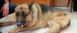
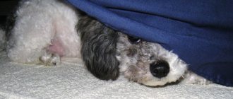
- Skin. The lesions are located on the surface of the skin and grow slowly. The formations are often solitary and hairless. Animal owners do not have any concerns when detecting such sores, since they are similar to dermatitis. Therefore, they consult a doctor after long-term home observation.
- Subcutaneous. The neoplasms look like lipomas, the hair does not fall out. Can appear on any part of the body. The growth rate of such a tumor depends on the degree of malignancy.
Diagnosis of mast tumors
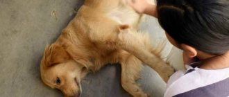
Cancer is diagnosed using fine needle aspiration. The essence of such diagnostics is to take a certain number of cells from the tumor and examine them under a microscope. The behavior of cancer is complex and depends on many factors. Therefore, the doctor must assess how aggressive the neoplasm is and correlate it with grades 1, 2, 3.
To clarify the diagnosis, they sometimes do:
- biopsy - tissue examination;
- blood test for tumor markers;
- Ultrasound of an organ that could presumably be affected by the disease.
Did you know? About 25% of all neoplasms in dogs and cats are mast cell tumors (mastocytomas).
Dog owners need to remember that mastocytomas can mimic anything. They may look like a wart, a small subcutaneous formation. Therefore, any skin abnormalities should be checked by a veterinarian.
Causes of occurrence in dogs
The exact reasons why mastocytomas occur have not yet been identified. The process is believed to be associated with a genetic mutation in some cells. Such atypical structures begin to divide uncontrollably, resulting in an increase in the volume of tissue where they are located.
During the study of the disease, it was revealed that:
- Mastocytoma in dogs can develop at any age, but the average age is about 9 years.
- There is no gender predisposition; both males and females are affected.
- The disease is not inherited.
- Breed predisposition: Boston terriers, pit bull terriers, and retrievers are more prone to developing neoplasms.
- Highly malignant tumors occur in Shar Peis, especially at a young age.
- In boxers and pugs, in most cases the formations are benign.
Treatment methods
Actions in the treatment of mastocytoma in a dog will depend on the overall clinical picture and the individual characteristics of the animal. Taking these factors into account, the most suitable method is selected:
- Surgical removal is especially effective for type 1 or type 2 tumors, but is contraindicated for multiple or poorly differentiated mastocytomas. Before the operation, a so-called diagnostic resection is performed - the collection of pathological material to determine the boundaries of the formation. During the operation, some healthy tissue is necessarily removed to minimize the risk of relapse. Subsequently, the animal requires regular examinations by a veterinarian (every 2.5-3 months).
- Chemotherapy – can be used after surgery or instead of it (if surgery is contraindicated). For this purpose, drugs are selected to prevent or delay the growth of the tumor, and sometimes to reduce its size. The most commonly used drug is prednisolone.
Important! Success in the treatment of mastocytoma can only be counted on if it is detected in a timely manner and the type and stage of development are determined. In this regard, if you discover any suspicious formations on your pet’s body, it is important to promptly contact a veterinarian.
Symptoms of Mastocytoma in Dogs
In the initial stages, the symptoms of mastocytoma can be difficult to recognize, especially if your pet has had skin problems before.
Main signs of the disease:
- Single nodes appear on your pet's skin. Less common are multiple tumors.
- The lesions have any shape (round, shapeless, etc.).
- The boundaries of the neoplasm are clear or unclear.
- The size of the tumor is any (from a pea to several centimeters).
- Blisters and redness appear on the mastocytoma.
- Swelling of nearby tissues develops.
- Wound healing is slow.
- They can appear anywhere, including on mucous membranes. Most often, localization is noted on the torso and paws, less often on the neck and muzzle.
Signs that the tumor is malignant:
- fast growth;
- an increase in the number of tumors themselves;
- has no clear boundaries;
- the affected area of the skin is inflamed and there are ulcerations;
- the pet is itching;
- nearby lymph nodes are enlarged;
- enlarged liver and spleen (determined by the doctor during the examination).
Classification of mastocytoma
Mastocytomas can be of several types:
- Solitary single - characterized by slow growth.
- Single neoplasms that metastasize to the lymph nodes.
- Multiple tumors.
- Mast cell leukemia or mastocytosis.
The peculiarity of mastocytes is that they can occur either with severe symptoms or asymptomatically.
It is customary to distinguish three stages of mast cell tumors in dogs:
- In the first degree, neoplasms form in the skin and are benign in nature. They may be large and difficult to operate on, but there is no risk of spreading to other organs or areas of the body.
- In the second stage, tumors form in the subcutaneous layer. There are some signs of malignancy here, and therefore it is unknown how the cells will respond to treatment.
- In the third degree, deep subcutaneous areas are affected. This type of mastocytoma is quite aggressive and requires a special approach to treatment.
Diagnostic methods
Diagnosis of mastocytoma is possible only in a veterinary clinic and includes a number of studies:
- General examination of the pet, auscultation, palpation, history taking (how long ago the tumor appeared, whether any drugs were used at home, data on feeding and keeping the animal, information on vaccination and deworming).
- A smear is taken from the affected area for cytological examination. The resulting material is stained in the laboratory using the Romanovsky-Giemsa method, or using the metachromatic method. In tumor cells, granules in the cytoplasm are poorly stained and have an unusual appearance.
- Patients referred for surgical removal of the tumor undergo a full examination, which includes echocardiography, blood tests (general and biochemical) and urine tests.
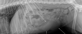
- To detect metastases, the condition of the liver and spleen, the animal undergoes an ultrasound, MRI or x-ray examination.
- The most reliable study of the nature of the tumor is a fine-needle aspiration biopsy. The material obtained by this method is sent for histology.
The biopsy must be performed by a qualified specialist, because The procedure has some nuances:
- The puncture site is excised at the end of the procedure.
- Samples for testing are taken from the periphery of the tumor, not from the center.
- In some cases, the biopsy is performed under ultrasound guidance, since the tumor cannot be pierced through. Such an error contributes to the fact that dangerous cells will be introduced into other tissues.
How is mastocytoma diagnosed in cats and dogs?
If any neoplasms appear on your animal's skin, take your pet to the veterinary clinic as soon as possible for an examination by an oncologist. During the appointment, the veterinarian will prescribe:
- comprehensive tests - urine and blood are taken from the pet for a complete clinical and biochemical analysis, which will help establish the general level of health of the animal and the spread of mastocytoma to internal organs;
- X-ray of the abdominal cavity and chest - allows you to most accurately determine how the tumor spreads throughout the body;
- Ultrasound - provides detailed visualization of mastocytoma and helps in detecting metastases.
The doctor can also perform a fine-needle biopsy - removing a fragment of the tumor for the most detailed examination. Remember that the sooner treatment for mastocytoma begins, the less time it will take for treatment, and the more successful the result will be.
Treatment of mastocytoma in dogs
► The main method of treatment for cutaneous mastocytoma in dogs is surgical. Surgery is scheduled if the dog's condition is satisfactory and the anesthesiologist approves the administration of anesthesia. The location of the tumor, its size and the number of tumors are also important. When removing a mastocytoma, the surgeon captures 1-2 cm of nearby tissue.
If there are many tumors, all that are possible are removed. Sometimes, in one operation, only part of the tumors are eliminated, and after a few months the rest are removed. Repeat surgery will be required if the lesion cannot be completely removed. Then, after some time, the formed scar and 1-2 cm of tissue are removed. If the tumor occupies a large area, experienced surgeons resort to skin grafts. Tissue flaps are taken from the patient from other areas of the body (as a rule, places where the skin can be folded - the area of the shoulder blade, neck).
After the operation, the dog is put on a special blanket or protective collar (depending on the location of the wound). It is important to use these devices constantly, even if the pet is experiencing discomfort. Otherwise, the animal will scratch or lick the seam area. The blanket can only be removed while the wound is being treated. To do this, use a solution of Chlorhexidine or hydrogen peroxide. Next, apply a thin layer of Levomekol ointment. The condition of the seam should be monitored daily: it should not be wet, red, swollen or hot. Skin healing lasts 10-14 days. The stitches are removed and the doctor prescribes further therapy or monitoring of the condition.
Observation scheme for dogs after removal of highly differentiated mastocytoma:
- one month after surgery;
- every 3 months throughout the year;
- once every 6 months during the second year;
- then once a year for life.
Such observation is necessary in order to detect the occurrence of possible relapses in time.
The doctor will examine the skin and do an ultrasound to check the internal organs. ► Drug therapy is prescribed to reduce the volume of the tumor, and also if the tumor is located in places difficult to reach for surgery (eyelid, armpit area, on the face). For this method of treatment, glucocorticosteroids are used - long-term prednisolone at a dosage of 2 mg/kg. This drug is also indicated if mastocytoma has affected internal organs (stomach, intestines).
► Tumors are sensitive to radiation therapy. This method is used if it is impossible to completely remove the tumor surgically. The procedure is expensive and not available in all regions.
► Chemotherapy is indicated if there are signs of metastasis or the patient will not undergo elective surgery. For this method, the following drugs are used: Vincristine (the course consists of 4 intravenous injections over 4 weeks), Vinblastine (the regimen includes concomitant oral prednisolone).
Chemotherapy is used for:
- reducing tumor volume;
- reduction of metastases;
- control the spread of disease;
- if it is impossible to remove surgically;
- if surgically it was impossible to remove the tumor completely.
► There is also targeted therapy - the use of a new class of drugs - tyrosine kinase inhibitors (masitinib, imatinib). Treatment is lifelong, effectiveness is achieved only in 30-40% of cases. This type of therapy is used in the following cases:
- impossibility of surgical removal of a mast cell tumor;
- if the tumor is highly malignant or recurrent;
- if traditional chemotherapy does not help, is not available or is contraindicated.
Expert opinion
Kuzmenko Olga Olegovna
Information about the expert
Ask a Question
Traditional medicine, the use of the ASD fraction in the treatment of oncological processes is inappropriate. Such methods have no proven effectiveness and only delay the start of effective methods.
Mastocytoma and histiocytoma of dogs in the city of Ulan-Ude
Khankhasykov S.P. Federal State Budgetary Educational Institution of Higher Professional Education "Buryat State Agricultural Academy named after. V.R. Filippova", Ulan-Ude
Considering the problem of skin tumors in small domestic animals, N. Gorman and D. Dobson noted that they are diagnosed much more often than neoplasms of other organs [1, 3]. In dogs they account for 30% of all tumors. Accurate data on the dependence of incidence on sex, age and breed are very scarce. Available sources indicate that the average age of dogs suffering from skin tumors is 10.5 years. Available observations suggest that among dogs the most predisposed to these tumors are the following breeds: boxers, Boston terriers, Scottish terriers, schnauzers, cocker spaniels, bullmastiffs, Labrador retrievers and basset hounds [3, 5, 6]. It has been noted that the most common skin tumors are:
— mastocytoma is a mast cell tumor, accounting for 9 to 12% of all skin neoplasms; can develop in the dermis or subcutaneous tissue, observed in animals of all age groups (the average age of patients is 8.5 years); the highest incidence is observed in brachycephalic dogs, especially boxers;
— cutaneous histiocytoma is a tumor unique to dogs and accounting for 10% of all skin neoplasms; in 50% of cases it develops in dogs under 2 years of age; Most often, tumors are found on the head (especially on the ears), on the feet, pelvic limbs and torso.
The high frequency of occurrence of these tumors noted in literary sources determined the purpose of the research - to determine the place in the structure of skin neoplasms of dogs in the conditions of Ulan-Ude, to determine the age and breed predisposition of animals to these diseases, to determine the typical localization sites of these neoplasms.
Material and research methods. The research material was dogs of various breeds and age and gender groups with tumor-like skin formations. For histological examination, biopsy or surgical material was used, which was fixed in 10% neutral formalin, followed by histological processing and preparation of histological sections 5-7 μm thick [2]. Verification of tumors was carried out using pathological diagnostic methods [2, 4].
Results and discussions. Histiocytoma was diagnosed in 12.10% of the total number of skin tumors in dogs. The age of sick animals ranges from 2 to 6 years and 1 month, on average being 4 years; mongrel dogs are more susceptible to the disease - 60%. 20% of the total number of histiocytes were diagnosed in Boxer and Giant Schnauzer dogs.
The tumor was detected in various areas of the scalp. It looked like a round-oval knot with a dense consistency, the size of a walnut. The skin is hairless, hyperemic, with slight ulceration on the surface.
Microscopic examination of the cells reveals round and oval nuclei containing one or more nucleoli. Lipofuscin granules are found in the cytoplasm; the edge is fuzzy, merging with the background of the preparation. Mitoses practically do not occur (Figure 1). Small cells with a heterochromatic, unevenly cut nucleus are also observed. Lipofuscin inclusions are located in the cytoplasm, collagen fibers are numerous (Figure 2).
Fig.1. Histiocytoma: 1 - cells with a large round nucleus; arrows indicate lipofuscin vacuoles (stained with methylene blue with azure II; magnification 400)
Fig.2. Histiocytoma: 1 - cells with a large round nucleus; 2 - small cells with an unevenly cut nucleus; 3 - collagen fibers; m - mitosis; arrows indicate lipofuscin vacuoles, lipid droplets (stained with methylene blue with azure II; magnification 1000).
Mastocytoma was diagnosed in 7.31% of cases of the total number of skin tumors. Moreover, 66.67% are Boxer dogs and 33.33% are Bullmastiffs. The average age of sick animals was 7 years and 9 months.
The tumor is represented by a single node of dense consistency. The node is mobile, hairless, reddish-violet in color, the size of a walnut. The main location of the tumor is the thigh area.
Microscopic examination reveals round cells with a large nucleus filled with euchromatin. Also, there are smaller cells with an often unevenly cut nucleus filled with heterochromatin. The nucleolus is clearly visible in the nucleus. The cytoplasm of both types of cells contains granules - lipofuscin, glycogen, but in round cells with a large nucleus they are more numerous. There are no mitoses. The cords formed by tumor cells are oriented along the collagen fibers (Figure 3).
Fig.3. Mastocytoma: 1 - cells with a large nucleus; 2 - small cells with an uneven nucleus; 3 - collagen fibers (stained with methylene blue with azure II; magnification 400).
As a result of the research, the following conclusions can be drawn:
1. In the structure of skin tumors in the city of Ulan-Ude, histiocytoma is represented in 12.10% of cases. The average age of sick animals is 4 years; mongrel dogs are more often affected. The main localization of the tumor is various areas of the scalp.
2. Mastocytoma in the structure of the named pathology is 7.31%. Brachycephalic dogs (boxers and bullmastiffs) are affected. The average age of sick animals is 7 years and 9 months, the tumor is localized in the hip area.
3. The macroscopic picture of these neoplasms is significantly similar, so their diagnosis must be carried out based on the results of histological examination. Also, the age of sick animals should be taken into account.
Bibliography:
- Kutsina O.A. Skin neoplasms in dogs and cats / O.A. Kutsina // United Scientific Journal, M., 2006, N° 5. P. 69-72.
- Merkulov G.A. Course of pathohistological techniques /G.A. Merkulov. L.: Medicine, 1969. 87 p.
- Oncological diseases of small domestic animals /ed. Richard A.S. White. M.: AQUARIUM LTD LLC, 2003. P.88-97,177-189.
- Guide to pathological diagnosis of human tumors / ed. ON THE. Kraevsky, A.V. Smolyannikova, D.S. Sarkisova. 3rd ed. M.: Medicine, 1982. 511 p.
- Selina V.M. Skin tumors of dogs / V.M. Selina. Irkutsk: PC “RIEL”, 2012. pp. 36-27.
- Stephen J., Withrow E., MacEwen G. Small animal clinical oncology - 3rd ed, 2001. P. 311 - 317.
Summary. Skin tumors are diagnosed much more often than neoplasms of other organs, accounting for 30% of all tumors in dogs. Accurate data on the dependence of incidence on age and breed are very scarce. It has been noted that the most common skin tumors are: mastocytoma (mast cell tumor), accounting for 9 to 12% of all skin tumors, and cutaneous histiocytoma, a tumor unique to dogs and accounting for 10% of all skin tumors.
The article presents the results of a histological study of dog skin tumors. The authors noted that in the structure of tumors in the city of Ulan-Ude, histiocytoma is represented in 12.10% of cases. The average age of sick animals is 4 years. Outbred dogs are more often affected. The main location of the tumor is various areas of the scalp. Mastocytoma in the structure of this pathology makes up 7.31%. Brachycephalic dogs (boxers and bullmastiffs) are affected. The average age of sick animals is 7 years and 9 months. The tumor is localized in the thigh area.
The macroscopic picture of these neoplasms has significant similarities, so their diagnosis must be carried out based on the results of histological examination. Also, the age of sick animals should be taken into account.
Key words: skin, neoplasms, dogs, mastocytoma, histiocytoma, structure, age, breed, macroscopic and microscopic characteristics, diagnosis.
Information about the author: Sergey Pavlovich Khankhasykov, Candidate of Veterinary Sciences, senior lecturer at the Department of Anatomy, Histology and Pathomorphology, Buryat State Agricultural Academy named after. V.R. Filippova"; 670034, Ulan-Ude, st. Pushkina, 8; tel.: 8(3012)230793, 8 (914) 8424580; e-mail responsible for correspondence with the editors
UDS 619:616-006:636.7
CANINE MASTOCYTIS AND HISTIOCYTOMA IN CONDITIONS OF ULAN-UDE
Khanhasykov SP
Summary. Boxers, Boston terriers, Scottish terriers, Schnauzers, Cocker spaniels, Bull mastiffs, Labrador Retrievers and Basset Hounds are the most susceptible breeds of dogs to skin tumors.
Mastocytis may develop in the dermis or subcutaneous tissue and is noted in animals of all age groups. More common in boxers.
Cutaneous histiocytoma develops in dogs between 2 and 6 years old. Tumors occur on the head, especially on the ears, on the feet of pelvic limbs and torso.
Authors have determined its place in the structure of canine skin tumors in Ulan-Ude, age and breed predisposition of animals, as well as localization of tumors.
Authors diagnosed histiocytes in 12.10% of the total number of canine skin tumors. Age of dogs was in an average of 4 years. Mongrel dogs were more susceptible to disease (60%). 20% of the total number of histiocytes was observed in dogs of Boxer and the Giant Schnauzer breed. Authors detected tumors of rounded-oval form with dense consistency and the size of a walnut, on hyperemic areas on the scalp. Microscopic examination of the cells revealed round and oval nuclei containing one or more nucleoli. Collagen fibers are plentiful.
Mastocytoma was diagnosed in 7.31% of the total number of skin tumors. In this case, 66.67% is accounted for Boxers and 33.33% - Bull mastiffs. Average age of dogs was 7 years and 9 months. Tumor is represented by a single node of dense consistency. Node is movable, hairless, reddish-purple in color, as a walnut in a size. Main location of the tumor is the femur area. Microscopic examination revealed rounded cells with large nucleus filled with euchromatin. Cytoplasm of both types of cells contain lipofuscin, glycogen granules.
Key words: skin tumors, dog mastocytoma, histiocytoma, structure, age, breed, macroscopic and microscopic characteristics, diagnostics.
References:
- Kutsina OA Novoobrazovaniya kozhi u sobak i koshek Canine and feline skin tumors]. - Moscow, 2006. - pp. 69-72.
- Merkulov GA Kurs pathogistologicheskoy tekhniki
Clinical cases of mastocytomas
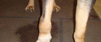
Labrador, male, not neutered, 5 years old. The owners noticed a lump in the area of the right hind limb about 3 weeks ago. Further, the dynamics of compaction growth was noted.
Fig.1. Labrador, 5 years old, male, neoplasm on the right hind limb in the area of the knee joint.
A cytological examination was performed using a fine-needle puncture method, which revealed the presence of mast cells. Next, a general examination was carried out, including an x-ray. Due to the absence of signs of metastasis, as well as due to the presence of a single lesion, a decision was made to surgically excise the tumor with further histological examination.
Fig.2 Labrador, 5 years old, male, preoperative condition.
Fig.3 Labrador, 5 years old, male, postoperative wound condition.
In this and the next case, it is important to note the planning of the operation. Preoperative planning includes determining the goals of therapy, and most importantly, working on the surgical defect. In this case, radical resection was performed. This option was used because the tumor was localized without metastasis. It is important to know that the first operation of this type has the highest chance of success.
Based on the results of histological examination, a well-differentiated mastocytoma was diagnosed. Prescribed treatment: prednisolone, at the rate of 1 mg/kg per day for 4 months. Next, reduce the dosage. The general course of treatment was 7 months.
Labrador, 7 years old, female, not neutered. The owner noticed a spot on the right hip area about 7 days ago. About 1 cm in diameter, no growth dynamics.
A cytological examination was carried out using a surface smear-imprint, which revealed the presence of cocci, dead epidermis, and blood cells. It was decided to send this material to a cytologist. The pathologist identified the presence of mast cells, and resection of the neoplasm with further histological examination was recommended. The examination, including x-ray, did not reveal any signs of metastasis. The tumor was removed.
Rice. 4. Labrador, female, 7 years old, neoplasm on the outer surface of the thigh of the right hind limb.
Fig.5 Labrador, female, 7 years old, neoplasm on the outer surface of the thigh of the right hind limb.
Fig.6 Labrador, female, 7 years old, neoplasm on the outer surface of the thigh of the right hind limb, progress of the operation.
In this case, prednisolone was also used at a rate of 1 mg/kg per day. The timing of taking the drug is the same as in the previous case.
In cases where two or more lesions occur, chemotherapy must be used in combination with surgery!
There are a number of chemotherapy protocols. Veterinarians can choose protocols based on the conclusion of pathologists, the financial capabilities of the animal owner, and most importantly, based on the patient’s health condition.
Among foreign authors one can find vinblastine/prednisolone protocols 2,3. In Russia, the cyclophosphamide/prednisolone regimen is also applicable. Both schemes are applicable to animals. When using regimens, it is important to monitor the blood picture, liver and kidney function. On average, the course of treatment can take about 15 weeks. Dosage of vinblastine 2 mg/m2. Dosage of cyclophosphamide 50 mg/m2.
In the two described cases, relapse was not observed for more than 1 year. In the literature, relapses during excision are possible in 30-50% of cases, and during excision with a wide sweep, only in 3-11%.
Mastocytoma in dogs is one of the malignant tumors. This is a very serious disease that can be fatal. In our review we will describe the signs of this scourge, its diagnosis, symptoms and treatment.
If at least one of the signs is present, you should immediately contact a veterinarian. To receive high-quality and effective assistance, we recommend contacting the Emergency Veterinary Care Center, where experienced specialists using modern equipment will conduct diagnostics, make a clinically based diagnosis and, of course, prescribe adequate treatment for your pet. Trust your pets only to professionals!