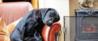
Urolithiasis (UKD) in dogs is a fairly common disease that can cause significant harm to the health of your pet. It is difficult to diagnose it at an early stage, especially for a layman. The processes that take place in the animal’s body at the beginning of the formation of urolithiasis appear imperceptibly on the outside. That is why by the time the owner notices changes in the dog’s behavior, the disease has time to progress significantly. But there is good news: if you immediately contact a veterinary clinic, KSD can be treated!
General information about urolithiasis in dogs
Urolithiasis is the process of formation of sand and stones in the kidneys and bladder. Just like in humans, urolithiasis in dogs is accompanied by very painful sensations. The animal whines, takes strange poses and appears frightened during simple urination. If you suddenly notice changes in your pet’s behavior described above, then under no circumstances delay visiting the veterinarian. The dog is in a lot of pain, and it will get even worse later!
There are quite a few types of stones formed in the animal’s body. All of them consist of various microelements. There are also many reasons for the occurrence of ICD. Without understanding the causes of the disease and what type of stone we are dealing with at the moment, it is impossible to prescribe the correct treatment.
Stones in the urethra and bladder in a male dog
Bladder stones in a dog
Content
- Causes
- Diagnostics
- Treatment
Almost every day, patients with various problems of the urinary system are seen by veterinarians.
Among them there are many animals that are diagnosed with pathologies caused by urolithiasis. This is a disease in which stones form in the organs of the urinary system. Urolithiasis occurs in both males and females. But it is detected more often in males and female cats, due to the anatomical features of the structure of the urethra. Along its length, it has one fairly pronounced bend (S-shaped bend), and this is where, most often, problems arise associated with blockage of the urethra by uroliths.
But first things first.
Causes
Without going into details and focusing on key points, we can say that the urinary system is formed by the kidneys, ureters, bladder and urethra (urethra). The kidneys are a paired organ whose main task is to filter electrolytes and products of nitrogen metabolism. Normally, these compounds are eliminated from the body through urine, however, sometimes, some of the chemical compounds found in the urine can form crystals and be deposited in both the kidneys and bladder, gradually forming stones.
Among the most likely causes predisposing to the formation of stones are the following:
- Urinary tract infections;
- Metabolic disorders (including in animals after castration and sterilization);
- Errors in the diet leading to disruption of the acid-base balance.
Stones in the bladder cavity cannot in any way be considered normal, even if their presence is discovered by chance, during routine examinations, and even if they do not cause any visible concern to the animal. By the way, a very small percentage of animals with stones in the bladder cavity do not experience any discomfort at all. Not in one hundred percent of cases are clinical symptoms of the disease present - the presence of blood in the urine, frequent and/or painful urination, licking of the prepuce area.
Diagnostics
It is possible to recognize urolithiasis, which leads, among other things, to the formation of stones in the bladder cavity, at an early stage if you undergo an annual medical examination.
The diagnosis is made comprehensively based on the clinical picture of the disease, the results of laboratory tests (blood and urine tests), and the results of visual diagnostic methods (x-rays and ultrasound).
Treatment
If the diagnosis is suspected and confirmed, emergency or planned surgery is recommended, depending on the stage at which the disease is recognized. A cystotomy is performed - access to the abdominal cavity, and then into the bladder cavity, followed by extraction of uroliths.
In the future, a long-term or lifelong therapeutic diet is prescribed, the purpose of which is to prevent further formation of new stones.
The article was prepared by A.V. Goryacheva,
veterinary therapist at MEDVET © 2021 SEC MEDVET
Causes of urolithiasis in dogs
In most cases, stones form directly in the animal's bladder. Much less often - in the kidneys. All the causes of the formation of urolithiasis, or, as it is also called, urolithiasis, are not fully known. But the main ones have long been proven:
- genetic predisposition. If your pet's parents had urolithiasis, there is a high probability that he himself will develop this disease;
- breed. Unfortunately, small breed dogs (dachshunds, pugs, hounds, bulldogs, etc.) are much more likely to be diagnosed with urolithiasis;
- congenital pathologies. Many factors in a dog’s body influence the formation of urolithiasis. Impaired metabolic processes, kidney disease, liver disease and even vascular disease can lead to the formation of urolithiasis;
- Almost any infection can lead to the formation of urolithiasis. Especially urinary tract infections;
From natural causes we move on to causes that arise as a result of incorrect content.
The first of them is an unbalanced diet. Very often, owners want to do what’s best: give their pet, who is used to eating dry food, a tasty morsel from their table. Or, conversely, due to lack of time, feed a dog that is accustomed to natural food with crackers from a bag. All this, as well as an excess of proteins and carbohydrates (you should not feed the dog only meat or cereals) are some of the main reasons for the formation of urolithiasis.
Besides:
- Don't make your dog endure it. Walk with her as often as possible! Urine that has been in the animal’s body for a long time begins to crystallize. That is, to turn into those same stones;
- Insufficient activity leads to obesity. And obesity leads to stagnation of fluid in the body, including urine;
- drink. Intermittent access to water, or drinking untreated tap water contributes to the formation of sand in the dog’s body. Keep track of what your pet drinks!
Causes of urolithiasis
Before treating an animal, it is necessary to find out the cause of the disease. Stones in urine may appear based on patterns. Infectious diseases can change the structure of the blood and the chemical composition of urine. When the balance of the elements begins to become unbalanced, some components begin to harden. Particular attention should be paid to sexual diseases. Most often, they leave fossils in the bladder.
Poor nutrition also affects the body. A dog needs a certain balance of vitamins and microelements. And if, for example, dog food is combined with regular food, this can lead to the deposition of salts and very high pressure on the digestive organs. Therefore, it is necessary for a dog to balance his diet.
Dirty or poor quality water can cause increased salt deposits. For example, it is undesirable for a dog, like a person, to drink tap water. It is better to drink settled water or purified (purchased) water. Although, if the owner knows that tap water contains a normal amount of salts, then it can be consumed. And it is strictly forbidden to let your pet near puddles. Since dirty water can cause not only stone deposits, but also infection with various kinds of parasites.
Irregular urination. This reason appears when the owners do not follow the regime and walk the dog irregularly. The dog develops a reflex (even to defecation). And when urine accumulates, the body begins to defend itself and crystallizes the liquid on its own. Obesity due to lack of mobility leads to the fact that the excretory system ceases to cope with all loads. This causes stagnation of urine, which affects the functioning of the heart.
Genetic predisposition is associated with various deviations from the norm in the body. For example, deformation of the canals can cause urine retention. Improper structure of organs that affect the genitourinary system can lead to the deposition of fossils.
Although some breeds are more prone to kidney stones, other dogs may also develop these symptoms. But, the risk of contracting this particular disease increases significantly if you do not follow the dog’s diet, feed it anything and allow it to drink water from dirty sources.
Symptoms of urolithiasis in dogs
Urolithiasis in dogs, the symptoms and treatment of which can vary dramatically even between two puppies from the same litter, is further complicated by the fact that it is very difficult to detect its signs at an early stage. The urine becomes slightly cloudy and its quantity decreases slightly. By the time the dog begins to show obvious signs of discomfort and pain, the disease has progressed greatly. Remember: urolithiasis does not happen overnight! If your pet is examined and tested at a veterinary clinic at least once a year, you will prevent the development of the following symptoms:
- frequent urination. The dog pees a little at a time and cannot always wait until the next walk;
- the pet begins to lick the genitals frequently;
- the urine becomes very cloudy or takes on a pinkish or even dark red color. The presence of blood and sometimes pus in it is noticeable;
- lethargy, apathy, the dog begins to refuse food.
If the disease is completely neglected, blockage (obstruction) of the urinary tract occurs. This makes the symptoms even worse. The dog begins to pee a few drops at a time, experiencing very painful sensations. There is more and more blood in the urine, appetite worsens, and signs of anorexia appear.
As a result, the pet begins to vomit frequently, convulsions appear, the temperature rises, and the dog completely stops going to the toilet. At this stage of the disease, death is possible; the clock literally counts. You should consult a doctor immediately!
Symptoms of the disease
The problem with this disease is that you can visually notice a change in your pet’s behavior only when stones have already formed in the urinary system. But the problem is alleviated if the dog is regularly taken to the veterinarian for examination. Ultrasound, for example, can detect signs of urolithiasis in dogs at an early stage.
If your dog is at risk, then regular screening will reduce the likelihood of your pet becoming ill. If the dog begins to develop stone deposits in the genitourinary system, then the following symptoms can be observed:
- the dog begins to go to the toilet very often, but sometimes he cannot bear to go outside and does it right in the house;
- each time the amount of urine during emptying changes;
- the color of the urine becomes darker, there are droplets of blood in the puddle made by the pet;
- during urination, the animal experiences unbearable pain, whines and can take strange positions;
- when the canal is blocked, the dog may develop a fever, the pain becomes simply unbearable, and the dog tries to avoid any touch.
Your pet's urine may have an unpleasant odor and its volume may decrease. But even with such problems and acute pain, a pet can suffer for years. Therefore, at the slightest sign, you should contact your veterinarian for a diagnosis.
Diagnosis of urolithiasis in dogs
To diagnose urolithiasis in your pet, the doctor will first take fresh urine for analysis. It is very important that the waste product is collected immediately before the study. If the urine has time to sit and cool, crystals will begin to form. Essentially the same sand. This will lead to an incorrect diagnosis. A urine test will help determine not only the presence of the disease, but also the type of stones. Let us remind you that the treatment tactics for urolithiasis in dogs depend on the type of stones. Different stones are treated differently. You cannot prescribe a course of tablets or injections without making sure which stone you are treating. A panacea for one can accelerate the growth of another urolith.
Next, in order to understand exactly where the stone is located, what size it is, and also to assess the general condition of the body, the doctor will do an ultrasound and possibly an x-ray. In addition, a biochemical blood test may be necessary to make a diagnosis.
Stones in a dog's bladder
Remember: only after the doctor has collected a complete history, asked about the symptoms, and conducted the necessary research, can he correctly prescribe a course of treatment. Otherwise, if the disease has not been studied thoroughly, treatment will turn into Russian roulette. Whether you're lucky or unlucky.
Dog Bladder Stones
Treatment and prevention of urolithiasis in cats and dogs
One of the aspects of pharmacological therapy for urolithiasis is the removal of uroliths, creating conditions leading to their dissolution. Such treatment is required much more often and more important than surgical removal of stones. In addition, drug therapy is always indicated to prevent recurrence of urolithiasis, either after surgical removal of stones or after their pharmacological dissolution. Drug therapy is also indicated to control stone-related urinary tract infections.
Pharmacological dissolution of stones
Dissolution is based on the principle of undersaturation of urine with the mineral components of the crystals that we plan to dissolve. In other words, the urine should be undersaturated with the chemical components that make up the stone, which will lead to the opposite tendency - the transition of crystals from the solid substance of sand (or stone) into urine. And vice versa, the more concentrated the urine, the faster the calculus will form. Thus, using certain drugs, we must change the chemical composition of urine. In order for the stone to dissolve, it must be constantly immersed in this less concentrated urine, so the ideal place for dissolution would be the bladder. Kidney stones can also be dissolved if organ function is adequate in the affected kidney, but kidney stones take longer to dissolve. Stones located in the ureter or urethra cannot be dissolved unless they are moved into the bladder naturally or by medical intervention. It takes 2 to 4 months for stones to dissolve in the bladder. If stones are found in the urethra in male dogs, if possible they should be moved back into the bladder so that they can be treated, but in such cases there is a risk of re-displacement into the urethra before it decreases or disappears. There are developed methods for dissolving sand and stones of struvite origin in dogs and cats, as well as for resolving urate and cystine stones in dogs, but there are no such effective methods for calcium-containing stones. The therapy required for dissolution depends on the type of urinary stone. Thus, it is very important to know the type of stone based on urinalysis along with clinical data.
Treatment cannot be carried out at random, assuming one reason or another. If the treatment decision is based on a guess, the calculus will not dissolve as expected, since the guess may be incorrect or the stone may contain more than one type of urolith. Surgical removal of the stone is indicated if the stone does not dissolve after 2 months of drug therapy or if it causes acute pain, bleeding, or blockage of the urethra or ureter.
Animals treated in this way should be examined regularly. This examination should include a complete blood count, biochemical profile, urinalysis, urine culture, and abdominal x-ray (ultrasound). Assessments are carried out at 3-week intervals unless the animal's condition requires more frequent examination.
The decision to use conservative or surgical therapy for urolithiasis must be individualized for each animal. The decision must take into account the location and type of stone, the age and sex of the animal and the severity of its clinical signs, the presence of other diseases, the risk of anesthesia, the wishes of the owner and the willingness to comply with the strict requirements of pharmacotherapy for stone dissolution. The size and number of stones do not affect the effectiveness of drug therapy. In some cases, surgery is the preferred treatment.
Urolithiasis caused by infection-induced struvite
Dissolution of struvite uroliths, which form secondary to urea-splitting infection, first of all requires control of this infection, as well as a decrease in the concentration of ammonium, magnesium and phosphate in the urine.
By eliminating the infection, the urine pH shifts to the acidic side, thereby increasing the solubility of struvite. The infection is cleared by administering appropriate antimicrobials. It is best to choose an antibiotic based on urine culture, isolation of the pathogen, and testing its sensitivity to various antibiotics. Most staphylococci and Proteus Spp, which are involved in the induction of struvite, are sensitive to ampicillin, amoxicillin, penicillin, unless they have become resistant due to frequent use of the above antibiotics. Antibiotics should be prescribed at full dose for 2 weeks until the uroliths become radiologically invisible. As noted, urinalysis and urine culture should be performed at each examination to ensure that the infection is under control.
Special diets are usually used by doctors to reduce the concentration of the mineral constituents of struvite. If it is dry food, then it is S/D food (Prescription Diets, Hill's Pet Products). These foods are formulated with reduced amounts of protein (to reduce urinary urea concentrations), magnesium, phosphorus, and calcium (to reduce the concentrations of these minerals and maintain Ca/P balance). Diets often contain extra salt to stimulate thirst and produce less concentrated urine. Contraindications to this diet include any conditions in which salt should not be increased, including edema, hypertension, cardiac failure, liver failure, and animals with hypoalbuminemia. For animals with kidney disease, the diet should be introduced slowly over several weeks to ensure that hypertension does not develop.
If the owner feeds only S/D food, a decrease in urea concentration should occur, urine specific gravity becomes less than 1.016 and urine pH less than 6.5. If such changes do not occur, this means that the owner is not following dietary recommendations. Blood alkaline phosphatase concentrations increase. These changes were associated primarily with a decrease in dietary protein concentration.
For cats, there is an appropriate S/D diet that is limited in magnesium, phosphorus, and calcium with added salt, but with normal protein content. It is also designed to bring urine pH to around 6.0 acidity. It has the same contraindications as those listed for dogs when fed S/D food.
When using such diets, no additives are used to acidify urine. The diet should be applied for 2 to 4 weeks after the stones disappear. Due to the different nutritional needs of cats and dogs, each species needs to be fed the appropriate food. Let us note again that these diets should not be fed to growing dogs or cats (up to one year). Surgery is more appropriate for young animals with struvite stones.
If the animal eats a natural diet, then the food should be made as moist as possible, the diet should be limited (without overfeeding), softened water should be given to the animal and the diet should be balanced in terms of mineral composition, in particular calcium and phosphorus, by including the mineral supplement previously recommended article.
The most important target of dietary changes is acidic urine (pH-6.0), which creates unfavorable conditions for bacteria and stones.
In dogs with struvite-induced infection, stones dissolve after 3 months of therapy (2 to 7 months). This type of struvite is rare in cats and dissolves much later: from 9 to 13 weeks.
Urolithiasis caused by “sterile” struvite
For sterile struvite uroliths, treatment consists solely of the use of diet or urine acidifying drugs. The same food with the abbreviation S/D is used for dogs and cats. The same precautions and contraindications as mentioned above. Sterile struvite uroliths dissolve more quickly than those associated with infection. In dogs, the average dissolution time is 6 weeks, with a range of 1 to 3 months. In cats, sterile struvite uroliths dissolve on average within 5 weeks, with a range of 2 to 20 weeks.
Urolithiasis caused by urate stones
Pharmacological dissolution of urates involves the use of drugs that reduce the formation of uric acid and alkalinize the urine. It takes 2 to 4 months to dissolve. Due to the fact that kidney stones consisting of urates are difficult to visualize radiographically, therapy is continued for another 1 month after they have disappeared during visual diagnosis. The time required for dissolution varies from 8 to 11 weeks.
These recommendations are for dogs only. They have not been evaluated for safety and effectiveness in cats. Surgery remains the recommended treatment for urate uroliths in cats, which is rare in cats.
Allopurinol reduces urate formation by inhibiting the fermentation of xanthine oxidase, which normally converts xanthine to uric acid. The dosage corresponds to 30 mg/kg/day, divided into two or three doses. In dogs, no side effects are observed with its use, in contrast to medical practice, when kidney failure increases in humans.
Alkalinization is usually achieved using sodium bicarbonate. The dosage of this product should maintain urine pH around 7.0. The initial dose is 2 tablets/kg/day (approximately 125 mg/kg/day) in two to three doses. The owner can monitor the urine pH using special test strips so that appropriate dosage adjustments can be made. Doses even higher than 600 mg/kg/day may be necessary. As a guide, a quarter teaspoon of sodium bicarbonate powder contains about 1 g.
The goal of dietary changes is to reduce the intake of uric acid, which is primarily found in purine-containing organs (eg, kidneys, liver). For dry foods, the U/D diet has been recommended by some scientists, while others recommend S/D. Both diets are limited in protein. The advantage of U/D is that it tends to produce more alkaline urine, while S/D acidifies the urine. The advantage of the S/D diet is that it contains a large amount of salt to increase urination. There is no ideal dry diet that combines all the advantages and disadvantages.
Sodium bicarbonate can also be added to S/D feed. Salt (13 tsp/10 kg/day, or 200 mg/kg/day) or water can be added to the U/D feed to increase diuresis. We must not forget about the above-mentioned contraindications to adding salt.
Urolithiasis caused by cystine stones
Therapeutic resolution of cystine stones involves the use of drugs to reduce cystine formation, alkalinization of urine, and dietary changes. It takes 2 to 4 months to dissolve. Drug therapy is considered more important than diet or urine alkalization.
N-(2-mercaptopropionyl)-glycine reduces urinary cystine concentrations by forming cysteine disulfide, which is much more soluble than cystine. This drug is less toxic than D-penicillamine (Cuprenil), which acts similarly. The dosage of both drugs is 30 mg/kg/day once a day or divided into 2 equal doses. The optimal dosage for dogs has not yet been established and may vary from patient to patient. These drugs may cause skin lesions, vomiting, fever, hemolytic anemia, and lymphadenopathy.
Using sodium bicarbonate or potassium citrate, it is possible to easily alkalize urine to a pH of 7.0. Reducing dietary protein has been recommended through the U/D diet. Some scientists note that low protein intake has little effect on the formation of cystine in urine, at least in human practice. Currently, the effect of a low protein diet on cystine excretion in dogs remains unknown.
Urine volume can be increased by adding water to any diet or by adding salt to food (14 tsp/10 kg/day) to increase water intake, but salting should be avoided if possible. The goal of these recommendations is to maintain a urine specific gravity of less than 1.025, which also requires monitoring.
Prevention of urolithiasis with natural feeding and factors contributing to the occurrence of the disease
To understand the issues of preventing urolithiasis, you should understand the factors and causes that contribute to their occurrence. They are as follows (listed in order of importance):
1. Food quality and quantity. Improper natural feeding contributes to the formation of stones. Overeating, and especially frequent feeding, as mentioned above, contributes to the constant alkalization of urine, since immediately after eating, the urine shifts to the alkaline side for 2-4 hours. When feeding 5-7 times a day, the urine becomes constantly alkaline. This is a common practice when the food bowl is always full.
Lack of calcium in food (lack of whole bone or in the form of a calcium supplement) in a natural diet contributes to disruption of mineral metabolism and the formation of phosphate stones.
Incorrect natural food (with cereals or carbohydrates) or feeding dry food can lead to a decrease in natural resistance (in particular, lysozyme activity of the mucous membranes), which contributes to the disruption of the barrier functions of the mucous membranes, including the bladder, which in turn leads to the development of ascending infections and the appearance of infection-induced struvite stones.
2. Water quality and quantity. Raw tap water is rich in soluble salts, which further aggravate mineral imbalances. It is recommended to drink soft, standardized purchased water when living in areas with hard water.
The disadvantage of dry food is that when feeding it, the animal consumes less water than with natural nutrition, which contributes to the development of urolithiasis. It is advisable to moisten dry food or avoid feeding it, and add additional water to natural food (especially for cats).
3. Sedentary lifestyle of cats (mostly) and dogs. During the summer season in cats, all urological disorders often go away. This is due to changes in lifestyle, quality of food and water consumed.
4. Stagnation of urine with rare urination (often associated with low water consumption), which creates conditions for crystallization and the formation of stones and sand.
5. Genetic predisposition. It is difficult to deal with this, but all recommendations will be relevant in this case.
Some points are more relevant for cats, others for dogs. Compliance with the above recommendations will prevent most types of crystals, especially those whose appearance is not associated with genetic predisposition.
| Doses of drugs used to dissolve and prevent stones in the urinary system of dogs and cats | |
| A drug | Dose |
| Bicarbonate of soda | 120-200 mg/kg/day. (1g = 1/4 tsp) |
| Salt | 200 mg/kg/day (2 g=1/3 tsp) |
| Allopurinol | 30 mg/kg/day in 3 divided doses for dissolution and 10 mg/kg/day for prophylaxis |
| Penicillamine | 30 mg/kg/day for 2 doses |
| 2-Methylproprionyl-glycine | 30 mg/kg/day at a time or in 2 doses |
| Hydrochlorothiazide | 4-8 mg/kg/day for 2 doses |
| Potassium citrate | 100-150 mg/kg/day for 2 doses |
| Ammonium chloride | 200 mg/kg/day for 2 doses with food |
| DL-methionine | 600 mg/day for all feedings |
Indications, contraindications, performance monitoring, see text.
Misconceptions about the causes of urolithiasis
1. “Feeding fish leads to the development of urolithiasis.” The use of fish as food for cats has nothing to do with the above factors that contribute to the development of stones and urolithiasis.
2. “Castration of a male cat affects the development of urolithiasis.” Castration has nothing to do with the above factors and does not in any way affect the occurrence of urolithiasis. Except that castrati move even less than non-castrati.
Prevention of stone recurrence
Once they occur, stones tend to form again if preventive measures are not taken. Factors that predispose to stone formation cannot be cured by surgery. Although the principles of prophylaxis are the same as for medicinal dissolution, prophylactic measures should be safer for continuous and long-term use.
In cases where the doctor does not give recommendations for prevention, stones in most cases recur.
Prevention of infection-induced struvite formation
The most important factor in preventing recurrence is to eliminate the infection and then monitor for possible recurrence of the infection. We need to make sure that the infection has been eliminated and not just suppressed. A urine culture test should be performed at each re-examination before and after the dissolution process. Antibacterial therapy should be continuous for 2 weeks during treatment. If stones were removed surgically, antibiotic therapy should be continued for 3 weeks after surgery. Antibiotic therapy continues for 1 week after stone resorption. Urine testing and culture can detect recurrent infections, which may require a repeat course of antibiotics. In addition, the owner should monitor the urine pH several times a week, using urine collected in the morning before feeding (to avoid “after-prandial” alkalinization of the urine). If the owner notes a urine pH higher than 7.0, the owner should have the urine tested and cultured because the infection may become active.
Prevention of the formation of “sterile” struvite
Prevention of recurrence of urolithiasis with “sterile” struvite is more difficult than in cases caused by infection, since the causes are less clear. Today, the emphasis is on diet or the use of drugs that acidify urine.
For cats, C/D cat food is generally used to prevent the recurrence of the formation of sterile struvite uroliths. C/D cat food has a reduced amount of magnesium, phosphorus, calcium and the food acidifies the urine. This food has no reduction in protein or sodium content and is similar in composition to regular cat food. In addition, urine acidifiers can be used in combination with regular cat food. Typically a dosage of 600 mg/cat/day of DL-methionine or ammonium chloride is used. These are relatively high doses. As noted, there is a danger in long-term use of these drugs. C/D food may increase hypokalemia in cats, especially if renal impairment is present. The amount of potassium in a cat's diet should be increased to avoid this problem.
In dogs, C/D food has been recommended to prevent recurrence of sterile struvite stones. C/D dog food is moderately limited in protein, as well as magnesium, phosphorus, and calcium. This promotes the formation of more acidic urine. It is also limited in sodium, which reduces its effectiveness in increasing diuresis. Another preventative option is to increase the volume of urine by adding salt to the animal's food (about 14 tsp. 10 kg/day) or by mixing water with the food to achieve a urine specific gravity of less than 1.025. Additionally, acidifiers can be used to maintain urine pH below 6.5.
Preventing the formation of calcium oxalate stones
Due to the incomplete understanding of the causes of calcium oxalate stone formation, prevention also remains an issue. In medical practice, problems with calcium oxalates are very common and many data from veterinary medicine have been extrapolated from human medicine. Prevention methods currently focus on diet, diuretics, and citrate. If these crystals are associated with hypercalcemia, then the most important part of prevention is identifying and successfully treating the cause of the hypercalcemia. The most common cause of hypercalcemia and associated calcium oxalate uroliths is hyperparathyroidism.
The general recommendation is a dietary recommendation that includes sodium restriction, protein intake, but no restrictions on phosphorus and magnesium intake. Diets designed to prevent struvite (S/D, C/D) should not be used. K/D or U/D diets are more suitable because they are limited in sodium and protein and do not cause urine acidification. Another option is to regularly feed non-reduced protein foods). Adding salt is not used to increase diuresis.
Thiazide diuretics have been used medically to prevent the formation of calcium oxalate uroliths because they increase renal calcium absorption. It is for this reason that they should never be used in animals that suffer from hypercalcemia. Animals whose hypercalciuria is due to intestinal hyperabsorption of calcium may be predisposed to hypercalcemia with this therapy. Serum calcium should be checked when thiazide diuretics are used for prophylaxis. Thiazide diuretics have not been proven to be effective in preventing calcium oxalate uroliths in dogs.
Citrates can prevent the formation of calcium oxalate crystals by binding to calcium, forming soluble salts. Citrates also alkalinize the urine. Potassium citrate is commonly used in medical practice. Its effectiveness in dogs is unknown, but a suggested dose of 100 to 150 mg/kg/day divided into two doses can be used. The optimal acidity of urine for the prevention of calcium oxalate will be pH-8.0 or, in extreme cases, not less than 7.0 so that one can assume that there is no formation of oxalate salts. Side effects of potassium citrate have not been reported in dogs.
Prevention of the formation of urate stones
The prevention of this type of crystal is much the same as the dissolution process described above. Dietary changes and urine alkalinization should be implemented. Urine should be checked regularly for the presence of urate crystals. If hyperurecemia persists despite dietary therapy, allopurinol at a dose of 10 to 20 mg/kg/day divided into two doses may be prescribed. Note that this dose is less than the dose used for pharmacological dissolution of stones.
Preventing the formation of cystine stones
Prevention of relapses of urolithiasis caused by cystine crystals is identical to pharmacological dissolution therapy. Side effects can occur with very long-term use in the form of skin lesions, which are resolved by reduction.
Preventing the formation of calcium phosphate stones
Currently, there are no established and proven effective methods for the prevention of urolithiasis with calcium phosphate. General recommendations are similar to prevention of oxalate stone formation. If these kidney stones are associated with hypercalcemia, then its causes should be identified and treated.
Treatment of urinary tract infections (UTIs) associated with urolithiasis
Treatments for UTI and the associated formation of infection-induced struvite are already reflected above. Urinary tract infections that accompany other stones that mechanically injure the mucous membrane of the bladder and urethra. In all cases of urolithiasis in both cats and dogs, urine analysis is best obtained by cystocentesis; the results of culturing such urine will more accurately reflect the bacterial contamination of the bladder and urine. If a urinary tract infection is present, the pathogen should be identified and its sensitivity to various antibacterial agents tested.
If an animal's urine test indicates infection and the animal is severely ill and urgent surgery is required, antibiotic therapy should be started until urine culture results are available. If struvite crystals are found and the animal has not received antibiotics recently, ampicillin or amoxicillin are the drugs of choice. If struvite (any other) is not detected, then E. coli is the most common microorganism that causes cystitis. In such cases, a course of trimethoprim or enrofloxacin would be a reasonable choice for oral therapy while awaiting urine culture results. Chloramphenicol or first generation cephalosporins would be reasonable to use intravenously during surgery or in animals at risk of sepsis. Aminoglycosides may also be considered for parenteral use (subcutaneous or intramuscular administration) as long as renal function is not impaired and deterioration of renal function has not been observed during animal treatment and diagnosis. If an antibiotic is used on an animal, and as a result of bacterial culture, the bacteria’s sensitivity to it is not revealed, then it is necessary to repeat the urine test for bacterial contamination to ensure that the drug is truly ineffective before abandoning it if the animal’s condition improves. Whenever an antibiotic is prescribed to fight a confirmed infection, repeat urine tests should be performed to confirm its effectiveness. It is known that the drugs should be used for at least 3 weeks after surgery or, for the purpose of pharmacological dissolution, at least 1 week after the moment when the uroliths are no longer visualized radiographically. The use of aminoglycosides should be avoided. To ensure that the infection has been eradicated and not simply suppressed, urinalysis and urine culture should be performed approximately 1 week after stopping therapy and monthly for 1 or 2 months.
Treatment of urolithiasis in dogs
Treatment of urolithiasis in dogs, regardless of the type of stones, should first begin with the removal of stagnant urine. This will make your pet's condition a little easier. After your dog's bladder has been emptied, your doctor will tell you what to do next.
Depending on how far urolithiasis has developed in dogs, the treatment of the disease will be radically different. For some, it is enough to go on a diet, while others will have to resort to surgery. And again: any appointments will depend on the type of stones. Even the diet, as you understand, for removing alkaline formations and uroliths that have arisen in an acidic environment will be significantly different.
As a rule, urolithiasis in dogs is treated with traditional methods: the urinary tract is washed, a course of medications is prescribed, and a strict diet is prescribed. If the disease is advanced, surgical intervention will be required before carrying out therapeutic measures. From time to time, doctors may resort to non-standard methods of influence. But remember: if the doctor insists on surgery, then it is really necessary! Don't risk your pet's life! Self-medication has never brought any good to either people or dogs.
Treatment of stones in dogs
Treatment for kidney stones in dogs depends on the severity of the disease. Due to the severe course of the disease, any prescriptions and diagnostics can only be made by a veterinarian; self-medication of a pet can lead to tragic results.
In acute conditions, treatment is carried out in a veterinary clinic under the supervision of a doctor:
- Using a catheter inserted into the urethra, the doctor relieves the acute condition by removing pus, stones, sand and other causes of blockage of the urinary tract.
- Relieving the effects of blockage with antispasmodics, painkillers and anti-inflammatory drugs (for example, furagin is often prescribed in combination with canteren).
- Transition to long-term therapy after identification of the type of formation.
Prevention of urolithiasis in dogs
Prevention of urolithiasis in dogs, first of all, consists of proper care. If your pet has already had urolithiasis, strictly follow the instructions and prescriptions issued by your veterinarian.
If the animal is healthy, remember a few simple rules that will reduce the likelihood of urolithiasis to a minimum:
- feed your dog correctly: either only dry food, which is selected specifically for your pet, or only natural food. Remember: you cannot mix dry and natural food. Even in small quantities! And, most importantly, the dog’s food should include all the necessary minerals and beneficial elements;
- Eliminate raw tap water from your dog's diet. Give your pet either boiled or filtered. And make sure that there is always water in the bowl, especially in the warm season. In summer it is also worth taking a drink with you on walks. Are you thirsty in the heat? Your pet too;
- Walk your dog more often. Spend at least two hours walking a day. Try to get your dog to go outside at least once a day.
- Run, play, watch the physical development of your tailed friend! Do not let your dog lie in one place for days;
- Provide your pet with its own place. Lying on a cold floor is harmful. This also leads to the formation of ICD;
- Get tested at a veterinary clinic at least once a year. Especially if the animal is at risk. Remember: the earlier you detect the disease, the easier your dog will tolerate treatment. The pet will not be in excruciating pain, surgery will be avoided, and the family budget will remain much safer. Yes, prevention is always better than treatment for an advanced disease!
Risk factors
Breed predisposition.
The main breed at risk is Dalmatians. In dogs of this breed, the main end product of purine metabolism is uric acid, not allantoin. They produce 400-600 mg of uric acid per day, while other breeds produce only 10-60 mg. The reason for this feature is genetic; Dalmatians are homozygous for a mutation that causes a defect in the uric acid transporter. Both males and females are homozygous, but urate uroliths are recorded much less frequently in females. The reasons for this are not yet clear, but most likely are related to other features of the exchange.
The same mutation, but not homozygous, was recorded in English bulldogs and black Russian terriers.
Yorkshire terriers are considered to be another breed predisposed to the formation of urate stones, but in this case the disease is associated with the frequent presence of portosystemic shunts in representatives of this breed.
Portal vascular anomalies.
The process of converting uric acid into allantoin occurs in the liver. Therefore, any disruption of its function, including the presence of portosystemic shunts, leads to an increase in the concentration of uric acid in the urine, provoking the development of the urate type of urolithiasis.
Insufficient water consumption.
As with other types of KSD, increased urine concentration due to insufficient water intake increases the risk of urate formation.
Nutrition for dogs with urolithiasis
What to feed a dog with urolithiasis? Your veterinarian will tell you about this. Let us remind you that with different types of uroliths formed in the body, the diet for dogs with urolithiasis will differ significantly. Different animals require different minerals and trace elements for successful recovery. Most manufacturers produce medicinal dog food for urolithiasis. Your doctor will explain in detail what kind of food your pet should eat and why. Natural nutrition for urolithiasis in dogs is also prescribed by a veterinarian. The diet should consist of different foods - porridge, meat, vegetables. Fatty, fried, sweet and salty foods are completely excluded from the animal's diet. Do you want to extend the life of your pet? Do you want to make sure that the disease does not return? Strictly monitor your dog's diet!
Diet for dogs with urolithiasis
Owners of pets with the disease are wondering what food is appropriate for urolithiasis in dogs. Veterinarians advise buying high-calorie premium and super-premium food with a low mineral content. Additionally, provide enriched mineral water.
Features of natural nutrition for urolithiasis in dogs depend on its type. For example, in case of phosphate formations, protein is limited and vitamin C is introduced into the diet. With ammonium, do not give fish and offal; instead, egg whites and plant analogues can be given.
Oxalates require excluding potatoes and beans. Add Uralite and vitamin B6. With cystines, the amount of protein is reduced. More detailed recommendations regarding diet are provided by your attending physician.
Prognosis for urolithiasis in dogs
In most cases, urolithiasis in dogs is not treated, but stopped. This should be remembered first of all by owners who have seen the effect of treatment. No, nothing is over! Very often, people who notice that their pet is feeling better stop following the diet and taking medications. Don’t even think about doing this: refusing treatment will return all the symptoms and excruciating pain to your dog in a matter of weeks!
In general, the prognosis is favorable. But only with strict adherence to the instructions, otherwise a relapse cannot be avoided. Follow a diet, walk more, take medications strictly according to instructions and periodically get tested at a veterinary clinic, then the life of your beloved dog will be long, happy, and illnesses will pass and be forgotten!
Symptoms and first signs of kidney dysplasia in dogs
The first signs of the disease are increased thirst and urination, which are quite difficult to track. The main symptoms of kidney dysplasia in dogs include the following clinical manifestations:
- increased thirst;
- increased urination;
- growth retardation;
- smell of acetone from the mouth;
- urine has a faint yellow tint;
- vomit;
- refusal of food;
- lethargy, weakness.
The main complaint with which owners of sick puppies turn to specialists is the animal’s reluctance to control the process of urination. Some dogs start licking urine indoors and outdoors. If your pet has increased thirst, urination, and difficulty learning to relieve itself outdoors, it is recommended that you take your pet to a veterinarian for examination.
Clinical case of treatment of acute urinary retention with urinary tract disease in dogs
A Scottish Terrier dog named Vicky was admitted in emergency condition with acute urinary retention. Based on the results of diagnostic tests and examination by a veterinarian, a diagnosis was made: acute urinary retention due to blockage of the urethra by stones with a diameter of 10 mm. Doctor Andrey Konstantinovich Mamedkuliev decided to prescribe surgical treatment with further cystotomy and removal of stones from the urethra. During the operation, stones were removed and the urethra and bladder were washed.
The treatment and surgery were successful. Now Vicky can empty her bladder freely and not experience any pain.
Prevention of kidney disease in dogs
It is better to prevent any disease than to treat it.
Prevention of kidney disease in dogs is based on following simple principles:
- balanced diet;
- daily walks;
- constant access to water;
- avoiding contact with stray animals;
- keeping in a warm room on a litter;
- eliminating drafts:
- timely treatment of infectious and non-infectious animal diseases;
- Contact a veterinary clinic if you have increased thirst and urination.