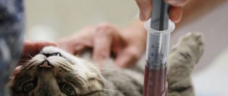
Types of blood tests, material studied
There are two main laboratory blood tests:
- general (or clinical);
- biochemical.
General (clinical) blood test for a cat
Shows the state of health of the body as a whole by the number and condition of formed blood elements. Also, this analysis can determine the presence of special parasites in the blood - hemobartenella and dirofilaria.
Basic indicators:
- hemoglobin;
- hematocrit;
- average content and concentration of hemoglobin in an erythrocyte;
- color indicator;
- ESR (erythrocyte sedimentation rate);
- leukocytes;
- red blood cells;
- neutrophils;
- lymphocytes;
- eosinophils;
- monocytes;
- platelets;
- basophils;
- myelocytes.
Material for analysis:
Venous blood of at least 2 ml, placed in a test tube with a special anticoagulant medium (heparin or sodium citrate), which prevents its coagulation and destruction of blood cells (blood cells).
Blood chemistry
Hidden pathologies in the cat’s body are revealed. The study provides information about damage to a particular organ or specific organ system, as well as an objective assessment of the extent of this damage. The result is determined by the work of the enzymatic system, reflected in the state of the blood. A biochemical blood test for a cat includes enzyme, electrolyte, fat and substrate indicators.
Basic indicators:
- glucose;
- protein and albumin;
- cholesterol;
- direct and total bilirubin;
- alanine aminotransferase (ALT)
- aspartate aminotransferase (AST);
- lactate dehydrogenase;
- gamma glutamyl transferase;
- alkaline phosphatase;
- ɑ-Amylase;
- urea;
- creatinine;
- calcium;
- magnesium;
- creatine phosphokinase;
- triglycerides;
- inorganic phosphorus;
- electrolytes (potassium, calcium, sodium, iron, chlorine, phosphorus).
Material for analysis:
Blood serum with a volume of about 1 ml (venous blood taken on an empty stomach and placed in a special tube that allows you to separate the blood serum from its formed elements).
Venous blood is drawn from the front or back paw by a veterinarian using topical anesthetic sprays. Usually it does not cause any discomfort to the pet if the doctor has certain skills.
Before a scheduled blood draw, the following should be excluded:
- excessive physical activity of the cat;
- administration of any medications the day before;
- any physiotherapeutic measures, ultrasound, x-rays and massages before the procedure;
- eating 8-12 hours before biochemical analysis.
How to understand a general blood test for your pet?
Contents hide
How to understand a general blood test for your pet?
It so happened that your pet needed the help of a veterinarian. Or maybe you yourself, without waiting for the disease, decided to get tested to make sure your pet is healthy. And here are the results in hand. What's next?
Photographer — Yulia Makurina
A blood test can tell a lot, but it is not always the main thing in diagnosing diseases. It is important in any situation to approach the examination comprehensively and carefully follow the specialist’s recommendations.
Today we will look at the meaning of general blood test indicators. In animals, as in humans, it includes determining its physical and chemical properties, studying the number and condition of blood cells.
ESR (ESR)
Norm in dogs and cats: 2-6 mm/hour.
Talks about how quickly red blood cells settle in one hour. If their rate is higher than normal, one can suspect inflammatory processes, poisoning, kidney and liver disease, injury, and pregnancy.
Hemoglobin (Hb)
Normal for dogs: 110-170; in cats: 100-140 g/l.
The blood protein responsible for the delivery of oxygen from the lungs to the tissues can increase physiologically - under heavy stress or excitement. In the absence of these, the indicator increases with dehydration, stress, hypertension, obesity or leukemia.
A decrease in hemoglobin more often indicates anemia (anemia) of various types, which, in turn, can occur due to blood loss, low content of vitamins or microelements in the feed, circulatory disorders, infections or parasitic diseases. A decrease is also possible in the second third of pregnancy.
Hematocrit (Ht)
Normal in dogs: 43-47.5; in cats: 42-47.5%.
The volume of red blood cells in the blood increases with dehydration and kidney disease. It is important to remember that in some breeds of dogs (for example, pointers) an elevated hematocrit is normal.
Accordingly, we have a decrease in this indicator with overhydration (increase in fluid in the body), anemia, and also in females during the period of milk feeding.
Red blood cells (RBC)
Norm in dogs: 5.2-7.4; in cats: 6.6-9.4 million/µl.
Along with the number of red blood cells, their shape and possible inclusions are considered. With dehydration, lack of oxygen, heart defects, kidney and liver damage, their number increases. But we remember that in some representatives of the same pointing dog breeds, relative erythrocytosis is the norm.
A decrease in red blood cells usually indicates their destruction under the influence of various toxins or microorganisms of infectious/invasive pathology. A low content of these cells is possible during bleeding, as well as in the last stages of pregnancy. And yet, with a sharp decrease in their number, it is recommended to make a differential diagnosis of anemia.
White blood cells (WBC)
Norm in dogs: 5.2-7.4; in cats: 10-20 thousand/µl.
An increase in the number of protective cells more often indicates infectious diseases, with which an intensified fight has already begun. Slight growth is possible during pregnancy or taking certain medications. But a significant increase in the number of cells may indicate inflammation, malignancy or allergies.
With a decrease in immunity, tumor processes, and some viral diseases (for example, feline panleukopenia), the number of leukocytes decreases.
Read more about some types of leukocytes below:
Increased eosinophils (normal in dogs: 3-8; normal in cats: 2-8)
often talks about parasitic diseases. But it can be observed with bronchial asthma, malignant neoplasms and allergies. And it usually decreases under stress.
Basophils may normally be absent.
Monocytes (normal in dogs and cats: 1-5) increase with various types of inflammation. They may decrease when treated with glucocorticoid drugs. Their complete disappearance - not the best sign - usually occurs with severe leukemia or sepsis.
It is worth noting that a general blood test often determines the relative number of cells, that is, their percentage. In the case of monocytes, their absolute number also matters (designated Monocytes abs. or Mon#).
Lymphocytes (normal in dogs: 21-40; in cats: 31-51) increase with leukemia, at the end of a viral disease, and also in the first time after vaccination. A decrease occurs during stress, inflammatory processes, lymph circulation disorders and at the initial stage of a viral disease.
Microscopic examination can also detect blood parasites: dirofilariasis, piroplasmosis.
We dare to note that this transcript is not complete and is for informational purposes only. That is, in any case, a diagnosis must be made by a veterinarian, based on a comprehensive examination and the personal characteristics of each animal.
Main indicators of blood tests and their characteristics
Each indicator is responsible for one or another degree of health/illness in the cat’s body, and also shows the functioning of individual organs or entire systems. Not only does each data matter individually, but also in relation to each other.
General (clinical) blood test
- Hematocrit is a conditional indicator showing the ratio of all formed elements of blood to its total volume. Another name is the hematocrit number and often the ratio of not all blood cells, but only red blood cells, is determined. In other words, this is the thickness of the blood. Shows how much blood can carry oxygen.
- Hemoglobin is the content of red blood cells responsible for transporting oxygen throughout the body and removing waste carbon dioxide. Deviation from the norm is always a sign of one or another pathology in the circulatory system.
- The average concentration of hemoglobin in a red blood cell shows in percentage terms how much the red blood cells are saturated with hemoglobin.
- The average hemoglobin content in an erythrocyte has approximately the same value as the previous indicator, only the result is indicated by the specific amount of it in each erythrocyte, and not by the general percentage.
- The color (color) blood index shows how much hemoglobin is contained in red blood cells in relation to the normal value.
- ESR is an indicator by which traces of the inflammatory process are determined. The erythrocyte sedimentation rate does not indicate a specific disease, but indicates the presence of disorders. In which specific organ or system can be determined in conjunction with other indicators.
- Erythrocytes are red blood cells that take part in tissue gas exchange and maintaining acid-base balance. It’s bad when test results go beyond the norm, not only in the direction of decrease, but also of increase.
- Leukocytes - or white blood cells that indicate the state of the animal's immune system. Includes lymphocytes, neutrophils, monocytes, basophils, basophils and eosinophils. The relationship between all these cells among themselves is of diagnostic importance: neutrophils - responsible for destroying bacterial infections in the blood;
- lymphocytes – a general indicator of immunity;
- monocytes - are engaged in the destruction of foreign substances that enter the blood and threaten health;
- eosinophils - stand guard in the fight against allergens;
- basophils - “work” in tandem with other leukocytes, helping to recognize and identify foreign particles in the blood.
- Platelets are blood cells responsible for blood clotting. They are also responsible for the integrity of blood vessels. Both the growth of this indicator and its decrease are important.
- Myelocytes are considered a type of leukocyte, but they are a somewhat separate indicator, because are located in the bone marrow and should not normally be detected in the blood.
Blood chemistry
- Glucose is considered a very informative indicator, because indicates the functioning of a complex enzymatic system in the body, including individual organs. The glucose cycle involves 8 different hormones and 4 complex enzymatic processes. Pathology is considered to be both an increase in a cat’s blood sugar level and a decrease in it.
- Total protein in the blood reflects the correct amino acid (protein) metabolism in the body. Shows the total amount of all protein components - globulins and albumins. All proteins take part in almost all vital processes of the body, so both their quantitative increase and decrease are important.
- Albumin is the most important blood protein produced by the liver. Performs a lot of vital functions in the cat’s body, therefore it is always determined by an indicator separate from the total protein (transfer of useful substances, preservation of reserve reserves of amino acids for the body, preservation of osmotic pressure of the blood, etc.).
- Cholesterol is one of the structural components of cells, ensuring their strength, and is also involved in the synthesis of many vital hormones. It can also be used to judge the nature of lipid metabolism in a cat’s body.
- Bilirubin is a bile component consisting of two forms - indirect and direct. Indirect is formed from erythrocyte breakdown, and bound (direct) is converted in the liver from indirect. Directly shows the functioning of the hepabiliary system (biliary and hepatic). Refers to “color” indicators, because when it is exceeded in the body, the tissues turn yellow (a sign of jaundice).
- Alanine aminotransferase (ALT, ALaT) and aspartate aminotransferase (AST, ACaT) are enzymes produced by liver cells, skeletal muscles, heart cells and red blood cells. It is a direct indicator of the functions of these organs or departments.
- Lactate dehydrogenase (LDH) is an enzyme that is involved in the final step in the breakdown of glucose. Determined to monitor the functioning of the liver and cardiac systems, as well as the risk of tumor formation.
- ɤ-glutamyltransferase (Gamma-GT) – in combination with other liver enzymes, provides insight into the functioning of the hepabilary system, pancreas and thyroid glands.
- Alkaline phosphatase is determined to monitor liver function.
- ɑ-Amylase – produced by the pancreas and parotid salivary gland. Their work is judged by its level, but always in conjunction with other indicators.
- Urea is the result of protein processing, which is excreted by the kidneys. Some remains circulating in the blood. Using this indicator, you can check your kidney function.
- Creatinine is a muscle byproduct excreted from the body by the renal system. The level fluctuates depending on the condition of the urinary excretory system.
- Potassium, calcium, phosphorus and magnesium are always assessed in complex and in relation to each other.
- Calcium is involved in the conduction of nerve impulses, especially through the heart muscle. By its level, you can determine problems in the functioning of the heart, muscle contractility and blood clotting.
- Creatine phosphokinase is an enzyme that is found in large quantities in the skeletal muscle group. By its presence in the blood, one can judge the work of the heart muscle, as well as internal muscle injuries.
- Triglycerides in the blood characterize the functioning of the cardiovascular system, as well as energy metabolism. Usually analyzed in conjunction with cholesterol levels.
- Electrolytes are responsible for membrane electrical properties. Thanks to the electrical potential difference, cells pick up and execute commands from the brain. In pathologies, cells are literally “thrown out” from the nerve impulse conduction system.
Norms of blood tests in cats
General (clinical) blood test
| The name of indicators | Units | Norm |
| % (l/l) | 26-48 (0,26-0,48) |
| g/l | 80-150 |
| % | 31-36 |
| pg | 14-19 |
| 0,65-0,9 | |
| mm/hour | 0-13 |
| million/µl | 5-10 |
| thousand/µl | 5,5-18,5 |
| % | 35-75 |
| % | 0-3 |
| % | 25-55 |
| % | 1-4 |
| % | 0-4 |
| million/l | 300-630 |
| % | — |
| % | — |
Blood chemistry
| The name of indicators | Units | Norm |
| mmol/l | 3,2-6,4 |
| g/l | 54-77 |
| g/l | 23-37 |
| mmol/l | 1,3-3,7 |
| µmol/l | 0-5,5 |
| µmol/l | 3-12 |
| U/l | 17(19)-79 |
| U/l | 9-29 |
| U/l | 55-155 |
| U/l | 5-50 |
| U/l | 39-55 |
| U/l | 780-1720 |
| mmol/l | 2-8 |
| mmol/l | 70-165 |
| mmol/l | 2-2,7 |
| mmol/l | 0,72-1,2 |
| U/l | 150-798 |
| mmol/l | 0,38-1,1 |
| mmol/l | 0,7-1,8 |
| Electrolytes | ||
| mmol/l | 3,8-5,4 |
| mmol/l | 2-2,7 |
| mmol/l | 143-165 |
| mmol/l | 20-30 |
| mmol/l | 107-123 |
| mmol/l | 1,1-2,3 |
Blood tests in cats (transcript)
All deviations in indicators are considered in complex and in relation to one data to another within the same results from the study of one blood sample. Only a specialist should decipher blood tests (results).
General (clinical) blood test
| The name of indicators | Promotion | Decline |
| 1. Hematocrit |
|
|
| 2. Hemoglobin |
|
|
| 3. ESR |
|
|
| 4. Red blood cells |
|
|
| 5. Leukocytes |
|
|
| 6. Segmented neutrophils (mature) |
|
|
| 7. Band neutrophils (immature) |
| |
| 8. Lymphocytes |
|
|
| 9. Monocytes |
|
|
| 10. Eosinophils |
| |
| 11. Platelets |
|
- incoagulability of blood. |
| 12. Basophils | hemoblastoses | Normally absent |
| 13. Myelocytes |
| Normally none. |
Blood chemistry
| The name of indicators | Promotion | Decline |
| 1. Glucose |
|
|
| 2. Protein |
|
|
| 3. Albumin | There is no true drop in albumin levels, most often due to dehydration and a decrease in total protein. | Likewise with the decrease in total protein. |
| 4. Cholesterol |
|
|
| 5. Direct bilirubin |
| |
| 6. Total bilirubin |
|
|
| 7. Alanine amino-transferase (ALT, ALaT) |
| |
| 8. Aspartate aminotransferase (AST, ASat) |
| infectious hepatitis (with a simultaneous increase in ALT). |
| 9. Lactate dehydrogenase |
| |
| 10. ɤ-glutamyl transferase |
| |
| 11. Alkaline phosphatase |
|
|
| 12. ɑ-Amylase |
|
|
| 13. Urea |
|
|
| 14. Creatinine |
|
|
| 15. Calcium |
|
|
| 16. Magnesium |
|
|
| 17. Creatine phosphokinase |
| |
| 18. Triglycerides |
|
|
| 19. Inorganic phosphorus |
|
|
| 20. Electrolytes | ||
|
|
|
|
|
|
|
|
|
|
|
|
|
|
|
Both clinical and biochemical blood tests are of great clinical importance for correct diagnosis and identification of hidden internal pathologies.
Author:
Grinchuk Ekaterina Andreevna veterinarian
Anemia, hemolysis, cachexia in a cat
Rosalia, Moscow
June 29, 2018
Good evening..We have the same problem. Cat, 8 years old, mixed breed, female. Vaccinated against rabies and vaccinated with Nobivak trio. The complaints began a year ago and gradually progressed: periodic vomiting and critical weight loss (cachexia, hypovolemia). Previously, we lived in another city, where veterinarians made incorrect diagnoses. We were admitted to one of the Moscow veterinary clinics on June 13, 2021, and had gastro and colonoscopy. A preliminary diagnosis was made: malabsorption, chronic gastroenterocolitis (histology is not ready yet). On the first day of admission, blood tests were taken, CBC, biochemistry and electrolytes. The CBC was almost perfect. The biochemistry was worse. The cat was in the hospital for 10 days, received treatment (amoxicalav, metrogil, B vitamins, omez, serenia - infusion), she felt much better, but on the day of discharge the potassium level dropped sharply (it was 3. a became 2.9) and symptoms of hypokalemia appeared. Magnesium was also significantly reduced. Magnesium was instilled - and its level reached the norm. Potassium was instilled and the symptoms of hypokalemia disappeared. Also, a CBC dated June 21, 2018 showed anemia and iron deficiency. Biochemistry from 21.06. on the contrary, it has become ideal - urea, creatinine, glucose, etc., ALT, AST - everything is ok. We were not diagnosed with chronic renal failure. Iron deficiency was present before (June 13), but it worsened significantly. In this regard, the doctor recommended not to waste time on using iron supplements, but to check reticulocytes and look for donor blood. There were zero reticulocytes. We found e.m. mass in the bank of cat donors, carried out compatibility (hemolysis reaction and agglutination, blood group). They said that everything was fine, we transfused the donor's e.m. mass to the cat on June 26th. The next day they took a CBC: hematocrit, hemoglobin and red blood cells increased significantly. But a day later (06/28) they took a CBC and biochemistry again. Doctors say that there is an active process of hemolysis. RBC, HGB HCT start to fall again. give unfavorable prognoses. Could the donor's blood still not fit and cause such a reaction? The leukocytes in the last CBC increased sharply (although before that I associated their gradual increase with lymphostasis and thrombosis of the hind leg - on June 22, the catheter ruptured the vein and the liquid dripped under the skin for some time). What could be causing such sudden onset of anemia? They checked for all infections (viral leukemia, panleukopenia, etc.) - everything is negative. At the same time, the cat is lively, shows interest in its surroundings, all reactions are present, there are no neurological reactions, the appetite is good () it’s just that she can be given Gorm Hills Health and Hills ID little by little and modular. Urination is also normal. Doctors say that, judging by the latest tests, multiple organ failure has begun (deterioration of biochemistry) and give us an unfavorable prognosis - a maximum of 3 days. My question is: what is indicated in the treatment of hemolysis? How can you stimulate your bone marrow? Is it possible to try to crush folic acid and instill hemobalance? Are hemodialysis and plasmapheresis indicated for us? Or re-transfer donor blood? Or it will be ineffective, since leukocytes have increased in this blood and biochemistry has worsened. Especially liver enzymes. But could all this be connected with the long-term use of amoxiclav and metrogyl intravenously? (some kind of toxic effect of antibiotics on the liver, for example)? I really hope for your comment. I can attach all the tests.. Thank you in advance for your answer.
The question is closed
anemia
hypovolemia
malabsorption
hemolysis


