4863Pavel
1
Ultrasound (ultrasound) is a widely used, accessible and painless method for diagnosing the internal organs of the abdominal cavity, both in humans and in cats and dogs. The technique is the use of ultrasonic waves to visualize the morphology, structure, and presence of pathological changes in the tissues of internal organs. The text will discuss how ultrasound is performed on cats and to whom the procedure is indicated.
An ultrasound machine is available in any modern veterinary clinic; it works on the principle of echolaction and emits high-frequency sound waves. These waves, hitting the organ, are deflected from it and returned back to the device, which interprets them as an image on the screen. However, these waves are difficult to pass through hollow organs and bones.
© shutterstock
Advantages of the method:
- Safe for the body.
- Completely painless.
- Non-invasive (no damage to tissues and organs).
- Has no contraindications.
- Informative and accurate.
- Shows the condition of internal organs using ultrasound waves: kidneys, spleen, liver, gall bladder, uterus and ovaries, stomach, pancreas, large vessels. Gives an image of the shape, size, structure, and the presence of pathological changes in organs.
- Shows pathological and functional changes in organs in the early stages of the disease, when clinical symptoms are not yet expressed.
- Allows you to monitor the results of treatment. You can compare images before and after treatment.
- Can be done repeatedly.
- Affordable price.
Ultrasound procedure in cats
To properly prepare for the procedure, the animal owner needs to find out how ultrasound is performed on cats.
The first thing to remember is that ultrasound examination does not cause any harm to the pet’s body. Therefore, it can be performed at any age and as often as necessary. Depending on which organ needs to be examined, additional preparation may be required.
Immediately before the procedure, the cat's fur is shaved from the examination area. This can be done by the owner himself at home, or by a doctor in a clinic. The cat is placed on the table on its back or side. The owner helps secure the animal. Then a special product is applied to the area of skin freed from fur. The gel is also applied to the ultrasound machine sensor. The examination usually lasts 15-20 minutes and is painless. However, being in an uncomfortable position, without the ability to escape, does not give the cat pleasure. She may be aggressive and try to bite or scratch her owner. Therefore, before the procedure, it is worth checking that your pet’s claws are trimmed and filed.
After the ultrasound is completed, the cat will have the gel removed from the skin and the owner will be informed of the results. The photographs or videos that the doctor took during the procedure are transferred to the owner. If desired, they can be used for consultation with another specialist.
Causes of intestinal obstruction in cats
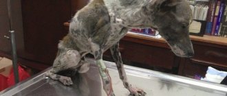
Intestinal obstruction occurs due to blockage of some part of the intestine, as a result of which gastric juice, feces and gases are not able to escape. This is a very dangerous condition that can lead to the death of the animal within a couple of days.
Juices in a cat's stomach are constantly produced. If there is a blockage in the intestines, they begin to accumulate in the stomach, which leads to vomiting. Many useful substances come out with the liquid, which leads to rapid dehydration of the body. The animal loses strength before our eyes.
Most often, the main cause of intestinal blockage is the ingestion of any large objects. However, obstruction can also develop against the background of other diseases.
The main causes of mechanical intestinal obstruction:
- Swallowing large objects . Cats are very curious, and they often try different objects to their teeth. However, not all of them are able to leave the body naturally. The most dangerous are: cellophane bags, rustling paper wrappers, New Year's tinsel, etc.
- Hairballs in the stomach . Cats lick themselves, and all the hair that falls out, especially during the molting period, ends up in the stomach. In most cases, the fur is easily removed on its own - this is what is associated with periodic vomiting in cats. There are times when a hairball begins to move further along the esophagus and stops the intestines.
- Infection with helminths . Although all stray cats suffer from worms to one degree or another, sometimes the situation can get significantly worse. Helminths multiply very quickly. They often stop various internal organs of the animal. The resulting lump of helminths can clog the intestines.
- Neoplasms in the intestines . A tumor does not have to be malignant to cause the death of an animal. Often the tumor grows slowly, and therefore the cat first begins to suffer from partial intestinal obstruction. The tumor will grow until it completely blocks the esophagus.
- Volvulus . One of the sections of the intestine is pinched. The situation is aggravated by the fact that necrosis of dying tissue is added to the obstruction.
- Hernia . In some cases, part of the intestine may prolapse into the abdominal cavity. It is compressed by tissues, which can lead to tissue necrosis and intestinal obstruction.
- Constipation . Poor nutrition leads to stool becoming increasingly dry. They have difficulty passing through the intestines. This can lead first to constipation, and then to the formation of fecal impaction.
In addition to mechanical, there is also dynamic intestinal obstruction. It is not associated with mechanical blockage of the intestines, but with a violation of intestinal contractions.
The main causes of dynamic intestinal obstruction:
- Abdominal surgery performed.
- Other serious diseases: ascites, peritonitis, lead to paralysis of the intestinal walls.
- Spinal injury.
- Poor circulation in the abdominal area.
- A strong spasm leads to compression of the intestinal muscle. In this case, the animal feels very severe pain. Spasm can be caused by inflammatory diseases (enteritis, enterocolitis), or severe mechanical trauma to the abdominal cavity.
You might be interested in: Why doesn't a cat step on its hind paws?
Examination of the abdominal cavity and genitourinary system
When examining the abdominal cavity and genitourinary system, the doctor looks at the condition of the stomach, intestines, liver, spleen, kidneys, bladder and uterus.
Ultrasound diagnostics of these organs is prescribed if the animal suffers from vomiting or diarrhea. If you have problems urinating or blood is detected in urine tests. Vaginal discharge appears, the animal has a decreased appetite, and weight loss is observed. After examining the animal and examining blood and urine tests, the doctor will prescribe a test.
If you need to do an ultrasound of the cat's abdominal cavity, then the last meal should be no later than 6 hours before the procedure. For several days before the examination, it is worth excluding some foods from your pet’s diet. Those that can cause increased gas formation (flour products, dairy products, beans, potatoes).
An ultrasound of the cat’s liver is performed to confirm diagnoses: cirrhosis, acute or chronic hepatitis, fibrosis. During the study, the animal's gastrointestinal tract should be free of food and feces. Before the procedure, your pet may need drugs that reduce gas formation and adsorbents.
Why do an ultrasound on a cat?
Ultrasound diagnostics is performed to confirm or exclude a diagnosis that the doctor may suggest during the examination. Using ultrasound, you can determine pathological changes in the structure of internal organs (enlargement or decrease, tissue proliferation, mineralization or other changes), assess the condition of the gastrointestinal tract, its peristalsis, possible inflammation; in some cases, ultrasound can help detect a foreign body. Ultrasound is very important for assessing the condition of the urinary system - this study helps determine the presence of suspension or uroliths (urinary stones) in the cat's bladder, ureters or kidneys, and inflammation can be seen. You can determine the presence of free fluid in the abdominal cavity.
An abdominal ultrasound may be performed for the following symptoms:
- decreased or lack of appetite in the cat;
- abdominal pain;
- diarrhea;
- vomit;
- constipation;
- urination disorders (absent or frequent);
- mucus/blood in stool/urine;
- discharge from the loop.
Cats, like people, can have an ultrasound of the abdominal cavity during pregnancy to check for its presence, estimate the approximate number of fetuses and their location.
How to carry out the procedure
Some doctors recommend examining the genitourinary system to check for a full bladder. However, this is not always necessary. Today, ultrasound diagnostic devices make it possible to conduct a high-quality examination of the uterus or kidneys for an empty bladder. Without the use of diuretics. If the veterinarian forces the cat to drink before the procedure or insists on using diuretics, his qualifications are questionable or the clinic has old equipment. In both cases, it is better to go elsewhere. However, if it is necessary to perform an ultrasound of the cat’s bladder, you will need to give the animal something to drink 2 hours before the procedure or give it a diuretic.
During the examination, the doctor may detect changes in the size and structure of internal organs. Tumors and fluid accumulation are also visible; assess the condition of the cat during pregnancy.
Ultrasound of the kidneys in cats makes it possible to diagnose urolithiasis, polycystic kidney disease, acute or chronic renal failure.
What diseases can be detected by ultrasound of the kidneys?
When performing an ultrasound of the kidneys, it is possible to determine the presence of a number of structural changes that may indicate various pathologies.
We can suspect signs of cystic kidney lesions, diffuse changes in the parenchyma, nephrolithiasis (kidney stones), we can see focal formations of the organ and suspect abscesses, hematomas, granulomas, neoplasia, signs of a decrease in the organ in size, suspected hypoplasia, aplasia, dysplasia of the kidney in a cat. We can see signs of acute kidney disease - nephritis (inflammation of the organ) or enlargement of the ureters, obstruction (blockage of the ureters due to a stone, clot or stricture) and so on.
To make a diagnosis, only the information obtained from ultrasound diagnostics is not enough. In some cases, a kidney biopsy may be necessary. Fine needle aspiration biopsy is a procedure that is performed under ultrasound guidance under general anesthesia. With a very thin needle, cells from the organ being examined are collected from the cat and sent for cytological examination to the laboratory.
As a rule, the kidneys are examined in conjunction with the entire genitourinary system, that is, the kidneys, ureters, bladder, urethra and genitals of cats and cats are examined. These organs are closely related to each other and the study of one of them is not very informative. An ultrasound scan of the kidneys is most often performed to monitor certain altered parameters (for example, in the presence of neoplasms).
When examining the bladder, you can detect suspension or uroliths (urinary stones), inflammation of the walls (cystitis), and neoplasms of the walls. The diagnosis of urolithiasis or cystitis in cats is usually made based on the results of an examination, medical history, ultrasound, and urinalysis.
Cats quite often have a problem with collecting urine for analysis - the easiest way to do this is to use an empty tray or a special kit with silica gel instead of litter, but not all cats agree to go into such a litter. Another disadvantage of such a collection will be the contamination of urine with bacteria from the external genital organs of the cat, the tray and the litter. It is also not always possible to immediately deliver the tests to the clinic, and for a correct study, no more than 4 hours should pass from the moment of urine collection (unless special tubes or refrigeration are used). Therefore, a routine procedure in veterinary medicine is cystocentesis (collection of urine by puncture directly from the bladder). It is the owners who are most afraid of this procedure, not the animals. The needle used is so thin that often the animals do not even feel the moment of puncture. This method is used in veterinary medicine due to the inability to collect urine from an animal in a sterile manner. And this is important for the doctor to understand whether the animal has a bacterial infection and whether it is necessary to use antibiotics, or inflammation of any other nature and a completely different therapy is needed.
Cats have very well developed compensatory mechanisms; this has developed evolutionarily and helped them survive in the wild. Therefore, unfortunately, our pets can hide diseases from us until the last stage. Very often, the only sign that something is hurting a cat is a decrease in activity, less active jumping, a change in sleeping position - this is often difficult to track, and such problems are often associated with age (the cat has matured, the cat has aged). To detect kidney disease at an early stage, it is recommended to undergo an annual medical examination after 4-5 years. Ultrasound of the kidneys is included in the standard set of studies during clinical examination in cats. Remember that cats are not people, and they cannot take revenge on us for anything when they go to the toilet in the wrong place. For a healthy cat, a clean, dry litter box is the optimal place to urinate; if she starts urinating on the sofa or folded things, in most cases it turns out that the cause is a bladder or kidney disease. If during this period you scold a cat that is already experiencing pain and discomfort, the problem can turn into a behavioral one and will be difficult to correct even after treatment.
Ultrasound of a cat during pregnancy
An ultrasound of a pregnant cat is performed in order to monitor the condition of the expectant mother and kittens. The first study is carried out 21 days after the mating took place. At this stage, you can see on the monitor whether pregnancy has occurred or not.
After some time, ultrasound can be used to assess the development of kittens in accordance with the period. Determine whether there are developmental pathologies, if any, count the heart rate. If necessary, based on the results of the study, the doctor will prescribe a cesarean section for the pet.
After pregnancy, complications may develop. One of the most common and dangerous is purulent inflammation of the uterus or pyometra. A timely ultrasound of the cat's uterus will make sure that everything went well. And if the disease develops, start treatment on time.
Treatment of intestinal obstruction in cats
The main mistake with intestinal obstruction in a cat is self-medication. It often causes even more harm, leading to a significant deterioration in the animal’s condition. If a cat stops eating and drinking, the owners try to force feed it. This leads to increased vomiting and dehydration.
If you have intestinal obstruction, you cannot feed or water your cat. Often, owners can give the animal anti-poisoning medications, which later makes it more difficult to make a correct diagnosis.
In this case, an enema is no less dangerous. Since intestinal blockage is often confused with constipation, owners can use this method to help their pet. An enema given for obstruction can lead to rupture of the intestinal walls. Because of this, the animal can die very quickly.
Vaseline oil, which is used for constipation, is also contraindicated for intestinal obstruction. It will only further complicate bowel function. In some cases, petroleum jelly can push a stuck foreign object further down the intestine, causing damage to its walls. Thus, you should not self-medicate. You need to listen to your doctor. After making an accurate diagnosis, strictly follow all its instructions.
In some cases, you can do without surgery and limit yourself to only conservative treatment. This method is used for partial intestinal obstruction. In this case, an enema or medications may be prescribed.
Often, surgery is still necessary. Often in its process it is necessary to remove the part of the intestine where necrosis began. Afterwards, stitches are applied, which can be removed after 14 days.
Sometimes the operation is complicated if the object that entered the body had sharp edges. Then there may be damage to the intestinal walls and internal bleeding, which must be stopped. There have been cases when cats swallowed threads with a needle, and stitching of the intestines occurred.
Caring for your cat after surgery
After surgery, the cat should not be fed for two days or given water for 24 hours. To avoid dehydration, saline is injected into the animal through a dropper. In some cases, your doctor may prescribe antibiotics to avoid complications caused by various infections.
The first time after surgery, the cat needs rest. A special bandage is put on the animal to prevent the cat from licking the postoperative wound.
The cat is put on a special diet. Doctors advise giving your cat easily digestible natural food during this period. Food portions should not be large, but they should be given more often. Most often, low-fat fermented milk products and lean meat are introduced into the diet. Vitamins are also added to the diet.
It is important to identify and eliminate the cause that led to intestinal obstruction. If the blockage is caused by helminth infection or disruption of the gastrointestinal tract, it is necessary to address this problem after surgery.
You might be interested in: Is it dangerous for a cat to have crusts on its ears?
To get rid of helminths, special drugs are used. A special diet and a complete review of the cat’s diet will help normalize metabolism.
Ultrasound of the heart
Some cat breeds have genetic heart disease. The risk group includes Sphynxes, Scots and Britons, Maine Coons and some others. They are most often diagnosed with the disease “hypertrophic cardiomyopathy.” It is deadly, and the sooner it is detected, the better. An ultrasound of the heart of a cat of these breeds is necessary on a regular basis. Especially for young cats, as problems may develop after the age of 1 year.
Cardiac echocardiography should be performed before surgery and other procedures that require anesthesia. Since any such intervention in the presence of heart disease can lead to death.
In the clinic or at home
You can perform an ultrasound on a cat either in a clinic or at home.
Home screening has its benefits. Any visit to the clinic for an animal is stressful. At home, the animal feels calm and tolerates the procedure more easily. The doctor brings with him the necessary equipment. Conducts research, issues a conclusion and all necessary documents.
It is worth considering that at home there are more factors that can influence the clarity of the picture during the procedure. Therefore, the results of such diagnostics are often unreliable. To avoid problems, the doctor performing the procedure must be highly qualified and experienced.
As for the procedure in a clinic, you should choose a clinic that has modern ultrasound equipment and qualified specialists.
The owner decides where to perform an ultrasound on a cat independently, based on his own preferences, the main thing is that the procedure is performed by a professional.
Prices for ultrasound
Depending on which organs require examination, the price for a cat ultrasound ranges from 400 rubles for one organ to 2000 rubles for a general examination of the abdominal cavity.
For a specialist to come to your home with equipment, you will have to pay a little more - from 2000 per visit. The price includes a doctor’s visit, ultrasound examination, ultrasound protocol and conclusion.
Unfortunately, pets can suffer from various diseases. Your veterinarian may order an ultrasound for your cat. In order for the correct diagnosis to be made and timely treatment to begin. The owner should take a responsible approach to the procedure and the choice of a specialist to carry out the procedure. In this case, there is every chance that the doctor will identify the problem. After which he will select the appropriate treatment that will allow the animal to maintain life and health.