Discopathy in dogs is considered a common disorder that affects all breeds. In the absence of treatment, the sick individual completely loses the ability to move, and the functions of other body systems are disrupted. Read more about the disease and its treatment in our article.
Discopathy in dogs: symptoms and treatment
General description of discopathy in dogs
This disorder refers to the pathological process of changes in the structure of the tissues of the intervertebral disc. Due to the resulting destruction, the spine loses sufficient mobility, becomes less elastic and cannot bear even minimal loads.
Discopathy is provoked in those individuals who suffer from insufficient regular walks or are constantly in an isolated room. In some dogs, the disease occurs in a form in which there is complete paralysis of the limbs with complete or partial loss of sensation.
A sick dog spends most of its time lying down
The main danger of the disease is its irreversibility. It is possible to stop an incipient problem only at the initial stage of the formation of discopathy. At the same time, diagnosis is not always made in a timely manner, due to the frequent asymptomatic course of the pathology in the first stages, and the intervertebral disc is already partially destroyed.
After the destruction begins, the metabolic process in the tissues deteriorates several times; they do not receive enough minerals and nutrients. Due to the constant degenerative process, pressure on the spinal cord increases, swelling of the spine and problems in the functioning of other body systems appear.
Video - Discopathy in dogs
Attention! Most often, discopathy in dogs, according to UK veterinarians, is diagnosed between the ages of three and eight years. The greatest peak of sick individuals occurs over a period of five years.
Causes of discopathy development and risk group
The term “discopathy” refers to pathologies of the intervertebral discs of an animal. This condition is caused by degenerative diseases, which result in irreversible changes in tissues. The disease can cause compression of the spinal cord. The reason for the development of this pathology may be a violation of the water-salt balance in the body.
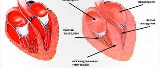
Intervertebral discs consist of a soft core and an outer dense ring. With discopathy, the internal contents begin to mineralize and thicken, which leads to a decrease in shock absorption. This leads to the fact that the pet begins to experience pain with any movement.
Gradually, the intervertebral disc stretches and changes its shape, and then begins to grow into the spinal canal. Over time, it becomes more and more deformed, and eventually the spinal cord is compressed, which is why the dog develops neurological symptoms (gait disturbance, decreased sensitivity in the paws, etc.).
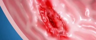
Development of the disorder in dogs
In normal condition, intervertebral discs are located between the cavities of all vertebrae. They are responsible for shock absorption and flexibility of the spine. When a dog develops discopathy, displacement of the intervertebral discs is observed, followed by its partial or complete destruction. Often, a hernia or complete rupture of intervertebral muscle tissue forms in the area of the affected area. This occurs due to the high load on the musculoskeletal system. A long-term degenerative process provokes protrusion of the fibrous ring located inside the intervertebral disc. Because of this, the functioning of the entire spinal cord is disrupted. This symptom develops abruptly and immediately leads to almost complete inability to move.
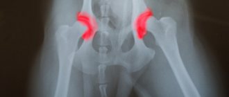
Damaged intervertebral disc on x-ray
Taking into account the severity of the negative changes that have occurred, the pet owner will observe different symptoms and a different clinical picture of discopathy. If the spinal cord is severely damaged, but its functions are not impaired, the animal suffers from a pain syndrome that is difficult to bear and the pet cannot stand on its paws.
Some individuals experience complete or partial paralysis. The clinical picture of discopathy in dogs can vary from several hours to several days, with the animal’s condition constantly worsening.
If your dog is paralyzed, you should immediately go to the veterinary clinic.
The disease can affect the cervical and thoracolumbar region. In the first case, the pet refuses or cannot turn its neck normally, and trembling of the neck and shoulder muscles is observed. Diagnosed in approximately 10-15% of cases, with this disorder the intervertebral disc between the 3rd and 4th vertebrae in the neck wears out. In the second type of disease, the pathology affects the area at the junction of the last thoracic and first lumbar vertebrae.
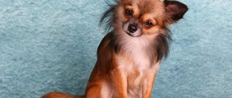
Attention! Dog breeds prone to discopathy are Dachshund and Pekingese. In dachshunds, a similar disorder develops in 65 out of 100 individuals. This feature is associated with unnatural body proportions, which causes a high load on the spine and the entire musculoskeletal system.
Four stages of intervertebral disc destruction
Reasons for the development of the disorder
Several factors can provoke discopathy in dogs:
- injuries, bruises and fractures of the spinal column;
- heavy load on the spine and the entire musculoskeletal system;
- congenital abnormalities of bone tissue structure;
- genetic predisposition of the breed;
- inflammatory processes in the tissues of the spine;
- other degenerative processes in joints, including arthrosis and osteochondrosis;
- increased body weight or lack thereof;
- low or excessive mobility.
Attention! If the animal is chained or confined most of the time, it is important not to become underweight or overweight. In this case, the likelihood of developing discopathy increases several times.
Special restraint device for paralysis of the hind legs
Symptoms of the formation of discopathy
In the initial stage of formation, it is quite difficult to notice any changes in the behavior or movement of the animal. Destruction of the intervertebral space can develop over several years. During this period, the intervertebral disc is almost completely replaced by pathological tissue, which cannot maintain the required degree of shock absorption and mobility of the spine.
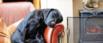
Large dog breeds are more likely to experience discopathy due to injury
Only after this does the dog develop typical manifestations of discopathy:
- the pet loses its activity and refuses to play;
- prolonged movement brings discomfort and pain;
- it is difficult for the dog to lift his limbs, which makes it almost impossible for him to walk up stairs;
- when palpating the abdominal wall, its increased tension is noted;
- with random movements, the individual may whine or squeal;
- the sitting position causes great discomfort, the sick dog tries to lie more in one position for a long time;
- the gait becomes uncertain, the dog often takes breaks;
- the back becomes arched to maintain the necessary balance and reduce pain;
- while standing, the pet begins to lift one of its limbs for no reason;
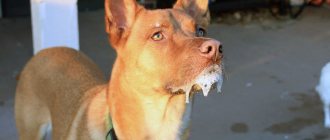
- the mood changes to aggressive, the dog does not allow himself to be touched, and may bite.
A closed space for a dog is a direct path to discopathy
When such symptoms appear, amateur owners often confuse the real problem with the individual’s fatigue and do not seek qualified help. Often, the owner can independently prescribe a painkiller to a sick dog, which only worsens the pet’s condition, since such a measure only temporarily relieves pain, but does not affect the underlying cause of the disease.
As the condition worsens, the dog finds it difficult to control its movements, its paws become uncontrollable, and problems with urinary function and bowel movements appear. Typically, such symptoms appear when the intervertebral disc is completely destroyed. In this situation, it is no longer possible to stop the disease; competent therapy can only improve the quality of life of the sick pet.
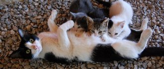
Dachshunds are most susceptible to abuse
Symptoms
In the first stages, the symptoms of the pathology are not immediately noticeable or begin and pass quickly, occurring periodically. The dog may suffer from cervical pain, so every movement of its head causes pain to it and it tries not to do it again. The animal can walk, slightly bending its paws at the knees, pain begins in the lumbar region. This can be seen by the way the dog refuses to sit down and does not allow himself to be stroked on the back. And while sitting or lying down, when suddenly standing up or jumping from a place, the pet squeals in pain. His peritoneum is tense, and thoracolumbar kyphosis develops with visually noticeable hunching.
When you try to pet the dog, he sometimes reacts with aggression. The dog tries to relieve the discomfort as much as possible by moving less. There is pain when defecating and urinating. It can drag one of the limbs when moving, as with osteochondrosis. The pet's gait becomes unnatural and very rigid, and the usual depreciation is not observed. He often trembles, tail spasms begin, and coordination of movements is impaired.
As the disease progresses, there may be paralysis of the limbs. This especially often develops after a walk or prolonged physical activity. For example, the owner ordered to bring a stick and the pet jumped and fell to the ground squealing and howling in pain.
If a dog was injured by an object while walking, hit, knocked down, or in other cases of mechanical impact, this does not apply to degenerative changes in the intervertebral disc. The pathology develops gradually.
Diagnosis of discopathy in dogs
Making a correct diagnosis requires an integrated approach, thanks to which it will be possible to refute or confirm the presence of the disease in the animal.
Table. Diagnostic methods for discopathy
| Diagnostics | Peculiarities |
| Visual inspection |
|
| X-rays in several projections |
|
| Myelography |
|
| MRI |
|
Attention! If the animal arrives at the hospital already in an immobilized state, the exact time of onset of paralysis is determined. If it happened more than 12 hours ago, it is almost impossible to restore the pet’s motor activity and most often a decision is made to euthanize. If the owner does not want this, the doctor tries to select possible options for supportive treatment until the dog dies a natural death.
Only a veterinarian can identify pathology
Diagnosis of the disease
To make a correct diagnosis, the veterinarian conducts a number of studies:
- Analysis of biochemical blood parameters.
- General analysis.
- Neurological tests:
- with a calm step, the doctor assesses the position of the animal’s head, back, limbs, and the tone of its muscles;
- the ability to run, jump, climb up and down from elevated positions is assessed;
- reflexes are checked;
- the pain threshold is examined by pinching the skin between the phalanges of the fingers, while the dog must withdraw its paw and bite; if it only whines, this means that sensitivity is reduced.
- An X-ray of the spine will show the location of the lesion and its extent. When a contrast agent is injected under the meninges, the X-ray result will be as reliable as possible.
- An MRI or CT scan is the most accurate diagnostic method, but is not available in all clinics and is quite expensive.
In addition to tests and instrumental studies, the veterinarian will ask the owner in detail about the symptoms that the dog has, and by combining all the results obtained, he will be able to make the correct diagnosis.
Drug treatment of the disorder
Treatment options for discopathy in dogs depend on the severity of the pathology. Therapy may involve the use of medications, surgery, and a number of additional techniques. When prescribing medications, it is necessary to use a whole range of drugs that relieve pain and other negative processes in bone, intervertebral and joint tissues.
"Dexamethasone"
A steroidal anti-inflammatory drug whose main effect is aimed at eliminating pain and inflammation. For treatment, a solution for intramuscular administration is used. It is recommended to administer no more than 0.3-0.5 ml of medication to small breed dogs, and no more than 0.7-1 ml to larger dogs. Dexamethasone is injected daily for an individually selected time.
"Dexamethasone" in ampoules
"Gabapentin"
It is used to relieve acute pain, including neuropathic pain. In addition to eliminating pain, it helps relieve convulsive and convulsive conditions. The dose of Gabapentin should be selected with your doctor, as it is affected by the age and weight of the pet. The tablets are taken daily, once a day with food. The duration of therapy is according to indications.
"Teraflex"
Belongs to the class of chondroprotectors. "Teraflex" normalizes the condition of joint and bone tissue, relieves swelling, inflammation, and starts regeneration processes. Medium and large breeds of dogs are treated with 1-2 tablets per day for a long course of up to six months. For small breeds, such as dachshunds and Pekingese, the dosage should be calculated by a doctor. The duration of therapy for them can also be six months. The minimum course of taking “Teraflex” is 12 weeks.
"Teraflex"
Vitamin B12
Necessary for improving cellular metabolism and normalizing tissue conductivity, improves cerebral circulation. For discopathy, an individually selected course is prescribed in the form of subcutaneous or intramuscular injections. The dosage depends on the dog's weight. Individuals up to 5 kg receive 250 mcg at a time, over five kilograms - 500 mcg, and those weighing more than 15 kg - 1000 mcg. Vitamin B12 does not show side effects and is well tolerated in the combined treatment of discopathy.
"Afobazol"
A sedative and relaxing drug that eliminates aggression and normalizes sleep in a sick dog. The dose should be carefully calculated based on the pet's weight. The largest breeds can take up to 30 mg of the active substance in three doses. Small and medium breeds receive no more than 5-20 mg of the daily dose of medication, which can be divided into several doses. The maximum course of therapy is 4 weeks for severe disorders. For dogs to normalize their condition, 2 weeks of active use of Afobazole is usually enough.
"Afobazol" tablets
"Diclofenac"
A non-steroidal anti-inflammatory drug that is better tolerated and safe compared to steroids. It is never used in combination with them, as such a combination can cause intestinal rupture and severe bleeding. Diclofenac relieves pain, inflammation, stiffness and swelling for discopathy. It is better to take it in the form of injections, the dose is selected by the doctor. Usually no more than three injections are given. Use Diclofenac once a day, regardless of food.
"Hondartron"
The main effect is aimed at restoring damaged tissues and stopping further degenerative disorders. For discopathy, it is recommended to administer the medication subcutaneously or intramuscularly to the dog; oral administration has less effect. Apply the solution morning and evening. The dose is classically selected taking into account the weight of the individual and is equal to 0.1 ml/kg. The duration of treatment is strictly according to the instructions of the veterinarian.
"Hondartron" for injection
"Chondrolon"
A very good chondroprotector, but can only be used on large breeds of dogs. It has a quick effect after just a few injections. When administered intramuscularly, it is better to hold the dog while administering it, as the injections are painful. The dose of the active substance is 100 mg every 48 hours. Used only on dogs over 30 kg in weight. The course consists of 25-30 injections.
Subcutaneous and intramuscular injections for dogs
"Diazepam"
It has become widespread in veterinary medicine due to its ability to relieve acute convulsions and the ability to relax muscles during discopathy. Diazepam can be administered intravenously or intramuscularly; in this case, the method of administration does not play a special role. For intravenous infusion, the dosage is 0.10-0.5 mg/kg, for intramuscular infusion - 0.3-0.5 mg/kg. Use once every 24 hours. If necessary, the dose is increased or decreased based on tolerability.
"Diazepam"
Acute intervertebral disc disease in dogs
Most dogs with intervertebral disc disease in the thoracolumbar spine present with back pain and paresis or paralysis of the hind legs. The back pain in these dogs is usually less severe than that seen with canine cervical disc disease, but affected dogs may stand hunched over and experience abdominal wall tenderness when pressed or palpated. The diameter of the spinal canal in the thoracolumbar spine is relatively small, so even a small volume of disc material extruded into the canal causes spinal cord compression and neurological deficits. In addition to the compressive effect of the disc material, there is often an impact injury to the spinal cord due to the explosive nature of the disc rupture. The majority (greater than 50%) of disc extrusions in this region are seen at the T12/T13 or T13/L1 level with 85% between T11/12 and L2/3. Disc extrusions in these disc spaces cause paresis or paralysis of the hind legs caused by upper motor impairment. neuron. Only 10-15% of dogs will have disc extrusion between the L3/4 and L6/7 disc spaces, damaging the spinal cord at the level of the lumbar enlargement and leading to lower motor neuron damage.
The severity of initial symptoms and the rate at which they progress depend not only on the volume of disc material extruded and the degree of resulting compression of the spinal cord, but also on the speed of extrusion. In some dogs, symptoms of pain and mild weakness due to partial disc rupture and mild compression of the spinal cord may occur for days or weeks before mild trauma or movement causes extrusion of a larger volume of disc material, causing paralysis. The neurological signs observed in dogs and cats with intervertebral disc disease are usually bilaterally symmetrical. Affected animals usually demonstrate pain when palpated in the spine directly above the site of the damaged disc, due to irritation of the spinal membranes and nerve roots at this location. When the spinal cord injury is severe and lies between T3 and L3, the cutaneous trunk reflex can be used to pinpoint the location of the injury.
Diagnostics
Trauma, fibrocartilaginous embolism, and spinal tumor are the main differential diagnoses considered in animals with acute disc extrusions in the thoracolumbar spine. The lesion should be localized as accurately as possible based on neurological examination data and identification of a specific area of pain in the spinal region. Plain radiography of the spine may be performed in an awake animal to look for evidence of intervertebral disc disease and to rule out other diagnoses. Careful placement of the suspected disc space into the center of the x-ray beam while the dog is anesthetized is necessary for radiographic identification of subtle lesions, but this examination is usually reserved for dogs who are potential candidates for surgery when preparation is being made for other diagnostic studies and decompressive surgery during that time. the same anesthetic episode.
The detection of calcified disc spaces confirms the presence of generalized canine intervertebral disc disease, but radiography is only 60-70% accurate in identifying the location of disc extrusion in the thoracolumbar region. Radiographic changes seen with a herniated disc in the thoracolumbar spine include a narrowed or wedge-shaped disc space, a small or cloud-shaped intervertebral foramen (“horse head”), narrowing of the articular facets, and calcified density within the spinal canal above the involved disc.
Myelography or advanced diagnostic imaging techniques (such as computed tomography and magnetic resonance imaging) should be performed to make an accurate diagnosis before surgery. Cerebrospinal fluid is usually collected from the cerebellar-medullary cistern prior to myelography. Cell counts can be performed quickly to rule out meningitis/myelitis, and the sample can be stored for further diagnostic testing if myelography does not show a compressive lesion. Lumbar contrast injection is preferred when performing myelography because the contrast agent must sometimes be injected under pressure to visualize the enlarged spinal cord in the area of the prolapsed disc.
Computed tomography is more accurate and faster than myelography; Because it is much more reliable in determining the location of disc material, it is useful for surgical planning. Magnetic resonance imaging is preferable to computed tomography when the extruded disc material is not mineralized and is the best method for assessing the spinal cord when the diagnosis of disc extrusion is unclear. The high sensitivity of computed tomography and magnetic resonance imaging may pose a challenge because clinically insignificant disc herniations that do not cause symptoms of spinal cord compression may also be detected.
Treatment
Treatment of acute intervertebral disc extrusion in the thoracolumbar spine can be nonsurgical or surgical. Non-surgical treatment is usually recommended when there are minimal or no obvious neurological deficits and the dog is still able to stand and ambulate without assistance. Strict confinement is the most important aspect of non-surgical treatment and should continue for at least 6 weeks to allow the annulus fibrosus to heal. Analgesics and anti-inflammatory drugs are often prescribed, as is the case for canine intervertebral disc disease in the cervical spine. Animals that are treated nonsurgically should be assessed frequently for neurological deterioration, as these dogs often deteriorate within 6 to 24 hours. If neurological symptoms do not improve within 5-7 days or even mild deterioration in neurological status is observed, surgery is indicated. Persistent or recurrent pain is also an indication for decompressive surgery.
Surgery is recommended for all patients who are unable to ambulate at the time of evaluation and for all dogs with symptoms suggestive of less severe spinal cord compression (eg, paresis, pain) unless neurologic signs improve rapidly with medical treatment. The rate of recovery is faster after decompressive surgery than after nonsurgical treatment, and the likelihood of residual neurological deficits is reduced. Decompression is usually performed using a hemilaminectomy and the disc material is removed from the spinal canal. Preoperative diagnostic imaging is necessary to identify diseased intervertebral spaces and determine where decompression should be performed to gain access to disc material.
Because clinical symptoms and myelography are not always reliable indicators of lateralized disc material, computed tomography or magnetic resonance imaging should be performed if possible. In addition to surgical decompression, many surgeons recommend simultaneous fenestration of adjacent high-risk disc spaces (T11–L3) to reduce the risk of herniation in generalized canine intervertebral disc disease in the thoracolumbar region.
After surgery, dogs should be kept in a clean and confined space. It is necessary to prevent bedsores in paralyzed animals by using soft bedding and frequent turning. In dogs that have lost voiding function, complete bladder emptying is required at least 4 times daily using manual pressure, placement of an indwelling urinary catheter, or intermittent aseptic catheterization. In dogs with bladder dysfunction due to upper motor neuron disease, drug treatment with phenoxybenzamine or diazepam can reduce sphincter tone, making manual pressure easier and the animal's attempt to urinate spontaneously.
Chronic intervertebral disc disease in dogs type 2
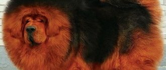
Some dogs may experience fibrous disc degeneration as part of the aging process, and this may result in a small amount of the disc core protruding into the annulus fibrosus. This results in a reaction of the anulus tissues causing a rounded, dome-shaped dorsal protrusion of the anulus that penetrates the spinal canal and causes slowly progressive compression of the spinal cord (Figure 1 C). This type of disc protrusion (i.e., Hansen's hernia type 2) is most often seen in older non-chondrodystrophic large breed dogs, especially German Shepherds, Labrador Retrievers, and Doberman Pinschers; however, it is also sometimes seen in small dog breeds.
Clinical features
Clinical signs arise primarily from slowly progressive compression of the spinal cord, although spinal discomfort is evident in a small number of dogs. Type 2 disc protrusion in the thoracolumbar region results in upper motor neuron symptoms in the hind limbs, with normal forelimbs. Type 2 disc disease in the cervical spine can be observed in Doberman Pinschers, especially in combination with malformation-malarticulation syndrome in the cervical spine (wobbler syndrome). In these dogs, the thoracic and pelvic limbs are affected, with upper motor neuron signs more commonly seen in the hind legs.
Diagnostics
Slowly progressive symptoms of spinal cord dysfunction in older dogs indicate the possibility of type 2 disc protrusion, degenerative myelopathy, and neoplastic disease. Neurological examination allows the lesion to be localized in the region of the spinal cord, but since the site of the lesion is usually not painful, palpation of the spine rarely allows the location to be clarified. Plain radiography of the spine is normal in most affected dogs. Some dogs may have narrowing of the disc space, osteophytes, and endplate sclerosis at the site of a type 2 disc protrusion, but these abnormalities are often seen in multiple locations in older large breed dogs and are therefore not very helpful in determining the location of the lesion. Myelography or computed tomography or magnetic resonance imaging is necessary to establish the extent and location of the lesion and to distinguish type 2 disc protrusion from a spinal tumor or degenerative myelopathy.
Treatment
Medical treatment with anti-inflammatory drugs (nonsteroidal anti-inflammatory drugs or low-dose prednisone) and muscle relaxants will be beneficial in dogs that experience discomfort when palpating or manipulating the site of the lesion. Neurological symptoms will, however, progress and surgery is recommended as definitive treatment. If the cervical spine is affected, ventral decompression is recommended, while if type 2 disc disease is present in the thoracolumbar spine, hemilaminectomy is recommended for decompression. Effective surgical decompression is often difficult to achieve due to the chronic nature of the lesion and the difficulty of removing the dorsal annulus. The goal of therapy is to stabilize the neurological status of the animal. The spinal cord is usually subject to significant chronic compression before clinical symptoms appear; therefore, complete recovery is rare. Some dogs exhibit temporary or permanent worsening of clinical signs after surgery.
From the book ” Fourth Edition, 2009 .
Articles on the topic;
- Castration status as a risk factor for the development of canine intervertebral disc herniation in dachshunds: a retrospective cohort study
- Acquisition of involuntary spinal locomotion (spinal gait) in dogs with irreversible spinal cord injury in the thoracolumbar region: 81 dogs
- Intervertebral disc degeneration in dogs: consequences, diagnosis, treatment, future developments
Additional treatments for discopathy
When using medications, veterinarians insist on the need for additional treatment, which will enhance recovery and reduce the likelihood of surgery. Several techniques are used for this.
- Acupuncture. Only carried out by a specialist. The doctor places the needles in a special order, which relaxes the muscles, relieves pain, reduces swelling and increases sensitivity.
- Massage. It is also recommended to conduct the first sessions with a good massage therapist, as it is important to learn how to perform the correct technique. Massage increases the flow of lymph and blood, prevents stagnant processes from developing, and relieves fatigue and pain. In addition, the nutrition of all tissues, including the brain, is normalized.
- Electrophoresis. It is carried out using small conductors that are applied to the diseased area. Using microcurrent, it is possible to improve tissue conductivity, relieve swelling, stiffness and pain.
- Magnetotherapy. It is also performed using small conductors, but already having a magnetic base. The main effect is aimed at suppressing the inflammatory process and accelerating tissue recovery.
- Physical activity. Swimming is usually prescribed for dogs with discopathy. Light and short exercises for flexion and extension of the limbs are also performed.
Video - Massage for spinal problems in dogs
Testing reflexes in dogs
Surgical treatment of discopathy
If there is no effect from medications and auxiliary non-invasive methods, as well as in the serious condition of the sick dog, the doctor decides on urgent surgical intervention. It consists of eliminating the deformed area.
- First, the surgeon completely or partially removes the cord on the diseased vertebra to gain an overview of the spinal cord.
- After opening, the core of the intervertebral space is completely removed.
- Only after this stage can the solid cavity in the spinal cord be opened.
- Taking into account the revealed state of the affected area, the doctor decides how much to really help a particular individual. To do this, the surgeon looks at the severity of the destruction of nerve fibers.
- To reduce the pressure resulting from the deformation of the intervertebral disc, the substance accumulated in the disc is removed using a special needle.
- Then, using a hemostatic sponge, the exposed solid cavity in the spinal cord is closed. Sometimes a medical omentum is used for this. Such a pad is necessary to prevent the formation of adhesions with the adjacent muscle corset.
- Once the incision is closed, it is completely sutured. A tight sterile bandage is applied.
Such interventions are performed only if more than two days have not passed since the moment of paralysis and loss of sensation in the limbs. Regardless of the success of the operation, the dog will still remain motionless or die.
Attention! After a successful operation, the dog is required to undergo a course of rehabilitation. It includes massage, taking anti-inflammatory drugs, chondroprotectors and physiotherapy.
Sometimes only surgery can save a dog
Possible complications of the disease
With the development of discopathy in dogs, in the absence of proper treatment, severe degenerative processes can develop in all body systems:
- voluntary urination and defecation;
- partial or complete paralysis of the front and hind limbs;
- disturbances in brain function, seizures;
- cerebrovascular accident;
- while maintaining the function of urination and defecation, these processes are characterized by severe pain;
- aggression, including attacks on owners trying to pet the animal;
- With strong compression of the spinal cord, respiratory function may stop.
Only comprehensive treatment can get a dog back on its feet.
Preventive measures
The occurrence of discopathy is often due to a hereditary predisposition, so affected individuals are excluded from breeding.
It is necessary to regularly monitor your pet's weight, since obesity significantly increases the risk of relapse of the pathology. Also, to prevent the development of the described pathology, owners are recommended to introduce chondroprotectors into the pet’s diet, which help maintain the working condition of the intervertebral discs for a longer period of time.
It is important to regularly monitor your pet’s behavior and begin treatment at the first manifestations of the disease - this gives a chance to completely restore motor functions and prevent the dog’s well-being from deteriorating.
In the video, the veterinarian tells everything about the disease discopathy in dogs.
Prevention of discopathy in dogs
Preventive methods must be used not only in healthy individuals, but also in those that have undergone a successful course of therapy. It is especially important to adhere to the recommendations of veterinarians to prevent discopathy for those owners who keep Pekingese and Dachshunds.
- Take your pet for walks regularly, but do not put too much stress on his spine.
- Teach your dog to swimming, as such physical activity significantly strengthens the muscle corset.
- Follow the principles of nutrition for the individual, taking into account its breed and age, avoiding a significant drop or increase in body weight.
- Periodically give your dog vitamin complexes and chondroprotector drugs to strengthen joint and bone tissue.
- Avoid injuring the spine and the entire musculoskeletal system. If your dog is injured, do not self-medicate and immediately consult a veterinarian.
A preventive examination by a doctor will reduce the risk of complications with discopathy
Prevention
Unfortunately, a set of preventive manipulations in the context of conservative treatment simply does not exist. Therefore, the likelihood of intervertebral disc pathologies can be reduced through careful selection work on a particular breed. Only healthy dogs with good pedigree can be allowed to breed. It is also worth monitoring your pet's body weight. Excess weight can create a traumatic load on the damaged spine. With increased weight, the duration of pain will also increase. Therefore, monitor your dog’s health: in 90% of cases, timely treatment and proper recovery after surgery bear fruit.
Life prognosis for dogs with discopathy
If specialists detect the violation in a timely manner, they give a good chance for the dog’s recovery. In the early stages, you can only get by with medication and non-invasive treatment, which brings about 65-70% of all sick dogs back on their feet. When undergoing surgery, prognosis for recovery is cautious and depends on many factors, including age, weight, and degree of advanced disease.
Attention! Dogs prone to discopathy should undergo preventive examinations by a veterinarian every few months to ensure early detection of possible problems.
Discopathy in dogs is a serious disorder that cannot be ignored. It is impossible to treat the pathology on your own, since it requires the use of a whole complex of medications and their proper combination with each other. If detected early, the disease can be stopped and the pet’s life can be extended as long as possible.