About dogs in human terms
01-10-2014 9:07 am
Some time ago, an article was published on the blog about the irrationality of docking dogs’ ears and tails. First of all, the communication of dogs with docked body parts is greatly affected, due to the fact that they cannot give intelligible and clear signals to their relatives. While studying the sources, I came across another equally interesting aspect.
In the book by Sotskaya M.N. “The skin and fur of a dog. Scientific, Veterinary and Cosmetological Aspects” describes the so-called violet or viola gland, which is probably responsible for communication between dogs.
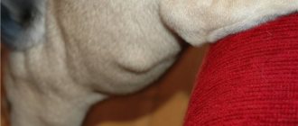
On the upper side of the dog's tail, near its root, is the violet gland. It is named so not because its secretion smells like violets, but because this flower resembles the contours of this gland in a cut. It is a glandular field where highly developed tree-branched sebaceous glands, ordinary sebaceous glands, and enlarged sweat glands are found. In dogs of different breeds, the violet gland is expressed to varying degrees, and it is not always easy to find. In huskies, German shepherds and other dogs with a “wild” coat type, a tuft of coarser hair grows at the location of the violet gland on the tail, and the surface of the skin at the base of the hair shafts is covered with a gray-yellow secretion.
The biological significance of violet glands has not yet been fully elucidated. A number of researchers believe that their secretion is associated with recognition of representatives of their species and for individual recognition. In many wild representatives of the canine family, it functions most actively during the rut. All dogs sleep curled up in a ball, with their nose buried in the root of their tail, where the violet gland is located. According to S.A. Korytin (1979), the secretion of this gland provides lubricant to the skin of the nose and enhances olfactory information by affecting the olfactory receptors. In a state of strong excitement in dogs, the tail is always raised high, the hair at the root of the tail bristles, which allows the smell to spread more intensely. At the same time, highly developed skin muscles contract and new portions of glandular secretion are released onto the surface of the skin (Sokolov et al., 1985). In smooth-haired dogs or those that are trimmed, in moments of excitement it is noticeable that the base of the tail often becomes noticeably wet to the touch.
Hormonal diseases that cause dog hair loss
For hormonal disorders, in particular:
• With hyperadrenocorticism (increased cortisol levels), dogs' hair falls out symmetrically, on both sides of the body.
• Lack of growth hormone (pituitary insufficiency), more often in males, causes bilateral symmetrical alopecia, except for the head and limbs during puberty. The disease is more common in breeds such as Chow Chows, Pomeranians, Toy Poodles, Airedale Terriers, and Boxers.
• Hyperestrogenism (excess estrogen) is observed in both females and males. With this pathology, bilateral symmetrical hair loss in dogs is observed in the perineum and around the genitals.
• Hypoestrogenism (lack of estrogen) causes baldness primarily around the vulva, and then throughout the body.
• With hypothyroidism, bilateral symmetrical hair loss in dogs without symptoms of itching is observed.
• Alopecia with acanthosis nigricans is also observed in dachshunds. Hair loss begins in the armpits and ears, and the smell from the skin is very unpleasant.
• Diminished alopecia can occur in any dog with diminished pigmentation. Hair loss in dogs occurs in areas with weakened pigmentation. Puppies are born with normal hair, and between the ages of six months and three years the hair begins to fall out.
Sweat glands
These glands look like a long tube, the end of which is often curled into a ball, and the rest is usually twisted in the shape of a corkscrew. The walls of the sweat gland consist of single-layer cuboidal epithelium, covered on the outside with a layer of smooth muscle fibers. The sweat gland develops in the form of a dense outgrowth of the main layer of the epidermis, growing deep into the dermis. Later, a channel appears in this outgrowth. Usually the sweat gland is formed along with the hair. Sweat glands most often open into the hair follicle. The secretion of the sweat glands is a product of the secretion of glandular cells that make up their walls. In addition, sweat glands filter intercellular fluid and are able to extract water with substances dissolved in it from the blood and lymph, participating in water-salt metabolism. Sweat contains sodium, potassium and chlorine ions, urea and other metabolic products. Thus, the sweat glands play the role of additional excretory organs.
There are two types of sweat glands, differing in their structure and functions. Human skin is dominated by small eccrine glands that secrete watery sweat, the evaporation of which from the surface of the skin causes cooling and plays an important role in thermoregulation. The intensity of sweating greatly depends on the ambient temperature, but can also occur under the influence of other factors, including emotional ones. Sweating is regulated by the endocrine system and nerve centers located in the brain and spinal cord. A dog has glands of this type on the soft parts of its paws. Since dogs do not produce liquid sweat, it is widely believed that they do not have sweat glands at all. However, this is fundamentally wrong.
Mammals covered with thick hair, including dogs, and also in small quantities in humans, have larger apocrine sweat glands, usually connected to hair follicles. Their secretion has a high content of organic substances, it is thicker and more fragrant. Mixing with the secretion of the sebaceous glands, it forms a natural fatty lubricant for the skin and hair. Unlike eccrine glands, apocrine glands are located in specific areas of the body and often begin to function only after puberty. A variety of apocrine glands are the glands of the eyelids and the glands that secrete earwax.
Sebaceous glands
Alveolar, i.e. pouch-shaped, holocrine-functioning sebaceous glands are found on the follicles of primary and secondary hair in all areas of the body. With the secretion of this type, the epithelial cells of the gland undergo fatty degeneration, and more and more fat droplets accumulate in their cytoplasm. Eventually, the cells are destroyed and form a sebaceous secretion, which accumulates in the cavity of the gland. The sebaceous glands appear during fetal development along with the formation of hair roots, but completely complete their development only with the onset of puberty. The size and shape of the sebaceous glands vary depending on the type of animal, breed, body area and type of hair follicle. Especially large and lobular glands are located on the dorsal side of the neck, on the back and on the tail, smaller and simpler ones are on the stomach. All sebaceous glands belonging to the same hair group are located approximately at the same level, in the reticular layer of the dermis.
The secretion of the sebaceous glands is discharged into the hair canal, where it is mixed with the secretion of the apocrine tubular glands. Glands of both types function, as a rule, simultaneously, having common external excretory ducts. This mixed secretion forms a sticky film on the surface of the skin. It protects against ultraviolet radiation, maintains elasticity of skin and hair and has a strong water repellent property, especially in animals with thick fur.
Inflammation of the sebaceous gland in a dog photo
Svetlana Belova, Estonian University of Life Sciences. The article uses the author's photo.
Introduction and etiology
Sebadenitis (SA) is a disease that is characterized by inflammation of the sebaceous glands of the skin and, as a consequence, deterioration in coat quality, peeling of the skin and alopecia. It occurs primarily in dogs, although rare cases of sebadenitis have been reported in cats, rabbits and horses.
The sebaceous glands produce sebum, which travels through the excretory duct to the mouth of the hair follicle, from where it is distributed over the stratum corneum of the skin and hair cuticle, forming a protective film that prevents moisture loss. In addition, sebum, due to the presence of fatty acids in its composition (linoleic, myristic, oleic and palmitic), also has antimicrobial properties.
Sebadenitis in dogs can be primary, when the sebaceous glands are the main and only target of the inflammatory process, and secondary, when they are involved in the inflammatory process “accidentally”, for example, in such primary pathologies as leishmaniasis, demodicosis and uveodermatological syndrome.
This article will talk about primary sebadenitis, which is characterized by sterile T-lymphocytic inflammation of the sebaceous glands, which can result in their complete destruction.
The first signs of sebadenitis usually appear at the age of 2–4 years. Predisposed breeds include Akita Inu, Poodle, Samoyed, English Springer Spaniel, Hovawart, Vizsla and Havanese Bichon. There is no gender predisposition.
The causes and pathogenesis of sebadenitis are not fully understood. The lymphocytic nature of inflammation and a good response to immunomodulators give reason to assume an immune-mediated etiology, and the breed predisposition is heredity.
Clinical picture
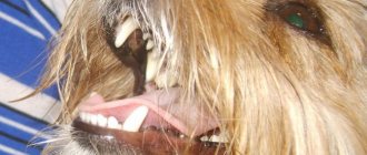
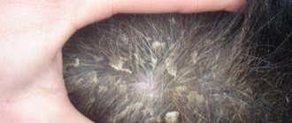
Due to the lack of natural “lubricant,” wool becomes dull and brittle, and normal keratinization processes are disrupted, incl. follicular. Thus, the characteristic signs of sebadenitis will be hypotrichosis and alopecia (photo 1), as well as peeling (photo 2) and the formation of voluminous follicular keratin casts, often “gluing” entire tufts of hair together (photo 3, 4). Skin lesions are symmetrical and most pronounced in the forehead, occiput, dorsal neck and back (photos 5, 6). The tail, due to severe alopecia, resembles a rat's (photo 7). In breeds with long and curly hair (for example, a poodle), the coat becomes shorter and loses its inherent curl (photo 8). In short-haired dogs, such as the Vizsla, sebadenitis manifests itself a little differently, namely as rounded, often merging areas of alopecia with peeling, involving the face, torso and even limbs.
| Photo 1. Lack of undercoat and alopecia in an Akita Inu with SA | Photo 2. Severe peeling with SA |
| Photo 3. Follicular casts made of keratin on wool | Photo 4. Follicular casts under a microscope - brown amorphous material |
| Photo 5. Skin lesions often start at the back of the head | Photo 6. Akita Inu with hypotrichosis and alopecia on the face and back |
| Photo 7. “Rat tail” | Photo 8. Short, straighter coat on a poodle with SA. |
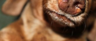
The inner surface of the auricle and the auditory canal itself are covered with dense scales, and earwax, due to the changed consistency and impaired self-cleaning mechanism of the outer ear, forms dry dark cerumen plugs, predisposing to external otitis (photo 9, 10).
Violet gland
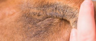
In all canids, not excluding dogs, on the upper surface of the tail in the area of the 9th caudal vertebra (the border of the upper third of the tail) there is the so-called violet gland (viola, violet gland, caudal gland). It is named so not because its secret smells like violets, but because this flower resembles its contours when cut. It is an oval-shaped glandular field 2.5-5 cm in length, where highly developed tree-branched sebaceous glands, ordinary sebaceous glands, and enlarged sweat glands are found. The hair in the area of the violet gland is larger in diameter, hard, coarse and differs in appearance from the surrounding hair. They grow each from their own hair follicle, while ordinary hair grows in a bunch of 6-11 hairs. The hair muscles in this area are more powerful; when they contract, they squeeze out the secretion of the gland. The sebaceous and apocrine glands in this area are large, extending deep into the dermis and subcutaneous tissue. The secretion of the gland consists of proteins and hydrophobic fats (molecules of fat, oil, wax, phospholipids and steroids).
In dogs of different breeds, the violet gland is expressed to varying degrees, and it is not always easy to find. In huskies, German shepherds and other dogs with a “wild” coat type, coarser hair grows at the location of the violet gland on the tail, and the surface of the skin at the base of the hair shafts is covered with a gray-yellow secretion. Some breeds of dogs (retrievers, ridgebacks, northern huskies) may have a darker colored area of fur in this area at an early age; with age, the color almost evens out. Some dogs develop bald spots over the gland (however, these should not be confused with tail hair loss as a result of disease, such as hormonal imbalances).
The biological significance of violet glands has not yet been fully elucidated.
Therapeutic measures
Treatment will depend on the stage of the disease, as well as the breed of the animal. It is important to take into account that the duration of therapy can and should be quite long, since “unfinished” pathology can easily turn into a complicated chronic form and appear within a few years, when it will be much more difficult to treat. So the final decision to discontinue therapeutic measures should only be made by an experienced veterinarian.
Some dogs are more sensitive to treatment than others. Veterinary practice shows that the most unpleasant in this regard are those same Japanese Akitu Inu. They respond poorly to therapy. All this suggests that these “Japanese women” have some kind of original genetic defect. However, let's not talk about sad things. How to cope with this pathology?
Typically, the following methods are used in practice:
- The dog's coat should be brushed more often to prevent the fur from permanently sticking together and forming tangles.
- Multivitamin complexes and corticosteroids are prescribed internally.
- The animal must be washed. It is better to ask your veterinarian about the type of shampoo, since conventional detergent compositions are contraindicated.
- If the affected skin has been contaminated with pathogenic microflora and there are signs of purulent inflammation, special antiseptic shampoos are used for washing. In most cases, antibiotic therapy cannot be avoided, since without antibiotics the risk of general sepsis is too great.
- If you are not squeamish, then, wearing surgical gloves, regularly pick out pieces of skin from your pet’s fur and cut out tangles of matted fur. By the way, it is generally advisable to cut dogs of long-haired breeds during the treatment period, since in this case it is possible to achieve better results.
Preputial glands
Large sebaceous and sweat glands are located in the skin of the prepuce. Their excretory ducts open into hair sheaths of large single guard hairs with a well-developed core. The size of the hair sheaths and excretory ducts increases with the age of the animal. The secretion of these glands lubricates the tuft of hair located at the end of the prepuce, through which urine flows during urination and protects them from its influence. In addition, during urination, this secretion joins the urine, which gives it an additional smell. Urine, as is well known, is the substance most actively used by mammals for marking territory.
Glands of the anal zone
A large number of different glands are concentrated in the anal area. The secretion of these glands is also associated with marking behavior and individual recognition. Around the anus there are relatively small and few circummunal glands. They consist of countless small and large lobules, separated from each other by connective tissue layers and muscles and together form an integral glandular layer surrounding the anus.
In the inner part of the anus, bordering the rectum, on the non-keratinized surface of the skin, proctodeal glands open through numerous ducts. These glands are located between the muscle bundles of the external sphincter, up to the internal sphincter, covering the rectum. According to their structure, they can be classified as complex polyalveolar-multitubular glands with branched ducts. The secretory part of these glands is much smaller in size than the system of their excretory ducts, which often expand in the form of cisterns and containers. A secretion accumulates in the reservoirs, the release of which probably facilitates the passage of feces and also gives them an individual smell. The emptying of the proctodeal glands is apparently associated with the work of the sphincter muscles, in the jejunum of which they are located.