What is eosinophilic granuloma in cats?
The disease, for which doctors still cannot find an explanation, can appear suddenly. Unfortunately, this disease is common among cats and is very common. Eosinophilic granuloma in cats appears at the age of 3 years.
An eosinophilic growth is a cluster of cells that were originally designed to protect the animal, but if they divide incorrectly and accumulate in large quantities, they form a malignant neoplasm that can kill a cat.
Eosinophilic growth
Important! Pets that roam freely outside are especially at risk of disease.
Description
Eosinophils are a subtype of leukocytes.
With the help of pseudopods, they move to the site of inflammation and absorb foreign cells, protecting the body. Eosinophils have cytotoxic properties, poisoning hostile cells. They bind and absorb a number of allergy and inflammatory mediators. Play a role in antiparasitic immunity. Granuloma in cats is a group of syndromes characterized by a persistent increase in the number of eosinophils. The mechanism is not fully understood, but it is known that such an increase causes a local reaction in the form of foci of inflammation on the skin. There are hundreds of times more eosinophils in the mucous membranes than in the blood, so granuloma in cats often affects the lips and oral cavity.
Although the disease is called eosinophilic granuloma complex (EGC), granulomas themselves do not always form in cats. In this regard, another name option was proposed - eosinophilic dermatosis. This is the same diagnosis, but the old name is used more often in the CIS countries.
It is believed that eosinophilic granuloma affects mainly young cats, but this is not entirely true. Simply, a hereditary predisposition to a malfunction of the immune system is detected at an early age. Cats get sick less often due to more stable hormonal levels.
This is a chronic condition that requires lifelong monitoring. Unfavorable factors provoke exacerbations and force a return to drug therapy. Cats and cats suffering from eosinophilic granuloma are removed from breeding.
Possible causes of the disease
There is still no single version about the causes of this disease. But all doctors agree that two factors have an influence:
- heredity;
- weak cat immunity;
- sensitivity due to breed.
Mycoplasmosis in cats: treatment, danger to humans
When two factors are combined, the risk of developing the disease increases. The development of the disease can be triggered by an insect bite or pollen falling on an area with an open wound.
The disease is not contagious and does not pose a danger to cats that are not infected.
Cats of artificially bred breeds that are highly sensitive to allergens and parasites are prone to the occurrence of eosinophilic granuloma.
Important! Owners of females are more likely to encounter granuloma than owners of males. The fact is that the cat’s body, prone to hormonal changes, is more vulnerable to disease.
The disease manifested in a kitten leads to death in 90% of cases, since the immune system is not strong enough to fight the disease.
Diagnostics
The veterinarian must distinguish this pathology from many other skin diseases that have similar symptoms. A similar clinical picture can be caused by fungal, bacterial or viral infections, tumors of benign or malignant etiology, abscesses, and idiopathic diseases. To confirm the diagnosis and exclude other causes, a professional histological examination of tissue is required. In this case, the specialist will identify characteristic signs of inflammation, as well as the presence of a huge number of eosinophils in the tissue. These are blood cells associated with the manifestation of the inflammatory mechanism. In addition, they are often detected in allergies.
Thus, microscopic phenomena with eosinophilic granuloma are relatively typical, and therefore there will be no particular difficulties in diagnosis. The difficulty lies perhaps in differentiating the various forms of this disease, but in practice the need for this is highly doubtful. Of course, an experienced veterinarian will be able to confidently make a diagnosis based on visual signs.
Once a diagnosis has been made, the specialist should focus on identifying the underlying cause (especially allergic etiology). Very often, this disease develops after something insignificant: a cat’s fleeting contact with household chemicals, an insect bite, or the introduction of a new type of food into the diet. So before prescribing treatment, an allergy test must be carried out.
Of course, in many cases, identifying a specific allergen can take a really long time (up to several weeks). But this is really important, since this approach allows you to correctly prescribe symptomatic treatment and avoid the use of drugs that may be dangerous. Unfortunately, some cases of eosinophilic granuloma may be associated with autoimmune diseases, which can be very difficult to identify and diagnose.
Be careful! The fact is that the same lichen that is transmitted to humans can in some cases cause similar clinical signs. So, if you have any problems with your cat’s skin, immediately contact a veterinary clinic, otherwise you may be in for an unpleasant surprise!
Types and symptoms of the disease
Piroplasmosis in cats: symptoms and treatment
An indolent ulcer develops in a cat in the form of a small wound on the upper or lower lip. Initially, the wound is small, may not be noticeable and may not cause discomfort to the pet. As the disease manifests itself, the wound begins to bother the animal. If you do not notice and start the process in time, an indolent ulcer in a cat will lead to tissue necrosis and spread to the entire pet’s face.
Important! The main sign of the appearance of eosinophilic granuloma in a cat is a neoplasm on the animal’s body that is recognizable by palpation.
Eosinophilic plaque
An eosinophilic plaque develops in the form of a small growth. Hair falls out at the site of the growth. The plaque appears either on the stomach or in the groin area of the cat. The growth immediately becomes a cause of concern for the pet. The main symptom is that the animal will pay a lot of attention to this area, bite, and lick the affected area.
Eosinophilic ulceration appears suddenly. The cat signals anxiety. Ulcers appear all over the cat’s body, secrete lymph and irritate the animal. The ulcerated side on the paw leads to lameness of the animal.
Eosinophilic plaque resembles ulceration in its symptoms, but is characterized by the rapid development and hardening of crusts on the surface of the skin. The cause of plaque is not only heredity, but also helminths in the cat’s body.
Ulcers in a cat
Painless ulcers are kind of a hoax. They don't hurt, but they grow at an incredible rate. If you don’t pay attention in time, the animal’s face will turn into one continuous ulcer. Area of development of painless ulcers: gums, oral mucosa, respiratory tract. High probability of developing a malignant tumor.
Important! Soreness is not the main problem with these ulcers.
If they are located on the gums, the root of the tongue, then this will be an obstacle to chewing and swallowing food. Because of this, there may be starvation, which will lead to weakened immunity and death of the pet.
Manifestations
In veterinary medicine, it is customary to distinguish several types of cat granuloma, which differ in localization and features of manifestation.:
- Ulcers . They are located on the tongue or upper lip of the cat, gradually increase in size and have a bright red color. The lip itself swells, the lesion covers the skin and mucous membrane. The initial size of such an ulcer is no more than 2 mm, but if treatment is not started in time, it will grow greatly and become more than 5 cm. The cat owner can easily visually detect such damage, since the ulcer is clearly visible, but the pet will not show any concern, the wound does not cause pain. Most often, the ulcer has slightly raised edges and does not bleed. More common in cats than in cats.
- Plaques . This variant of granuloma affects the pet's hips, groin or abdomen and is accompanied by severe itching. Inside each plaque is a liquid that leaks out when the cat scratches the lesion, corroding the skin and causing pain. This manifestation of cat eosinophilic granuloma can also be detected by visual examination: there is swelling at the site of the injury, it is red in color and approximately 3-5 cm in height. The surface of the lesion is shiny, hair does not grow on them. Can occur in animals of any sex.
- Raid . It is the formation of small bald patches, colored red, often ulcers form on their surface. The plaque affects the back, neck, and hips of the pet, but can also be found on other parts of the body. Often accompanied by itching.
Granuloma can also be located on cats' paws, on the chin, in the mouth, on the stomach or sides.
Diagnosis of the disease
Granuloma in cats develops quickly. Timely diagnosis is 50% of success. You should not self-medicate. As soon as anxiety or external signs of the disease become noticeable, you need to consult a specialist. The veterinarian will take blood tests from the cat and determine the cause of the concern. Additional analysis - biopsy and blood cytology will determine whether the ulcer is a malignant neoplasm.
Distemper in cats: symptoms and treatment at home
In addition, the doctor will test for allergens.
Important! If you decide to buy an animal from a cattery, you need to ask for cards of the kitten’s parents. It is necessary to check whether his parents have a predisposition to granuloma.
A person who does not have a veterinary education may mistake the disease for a fungus. For this reason, it is not recommended to self-medicate your cat.
Standard therapy: steroid hormones
Eosinophilic granuloma in cats: treatment
It is useless to treat eosinophilic granuloma in cats only with antibiotics and ointments. It is important to remember that granuloma only develops in adult cats. Kittens do not get sick from this because the hormonal system is weakly involved in their development, which means there is no reason for the disease. In order to cure the animal, the veterinarian prescribes hormone therapy.
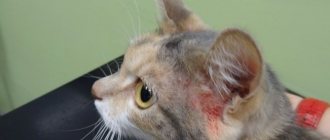
Ulcers near the ear
To reduce your pet's anxiety, the doctor prescribes hormonal glucocorticosteroids. They reduce the cat’s nervousness and help cope with inflammatory processes.
After taking hormonal medications, antibiotic therapy is prescribed, which eliminates the bacteria that served as a catalyst for the development of granuloma in the cat.
The duration of therapy is no more than 3 weeks. To be effective, it is worth supplementing the therapy with eye drops with an anti-inflammatory effect.
By the middle of the third week, you can notice your pet's lethargy. This is normal, as steroid therapy weakens the body. When the animal recovers, a vitamin complex can help the body recover.
To avoid an allergic reaction, a specialist prescribes antihistamines so as not to interfere with the body’s fight against the disease.
To reduce the cat's attention to the affected areas, Stop Itching spray is used. Using the spray will prevent new irritants from entering the affected area of the skin.
Important! The cat’s body cannot heal itself from this disease; the animal will need help.
Types of CEG
Three types of skin lesions have been described in eosinophilic granuloma complex:
- An eosinophilic plaque is a hairless, flat, raised lesion that is often red, moist, and shiny. Plaques are most often found in the groin and armpits or on the outer thigh. These lesions are often itchy and cats may lick them constantly.
- Eosinophilic granuloma is a hairless, raised, often whitish-yellowish lesion on the skin or mouth, often with ulcers on the surface. Most often, granulomas are found on the back of the thighs (you can find the name “ linear granuloma ”), they can also occur in the oral cavity and on the tongue. Less commonly, other parts of the body (chin, paw pads, etc.) may be affected.
- An indolent ulcer is a well-defined defect (erosion or ulcer) on one or both upper lips. First, the lip swells, then an ulcer forms. The ulcer is usually painless and does not bleed.
Anti-inflammatory and immunosuppressive therapy
If the disease was noticed late, then steroid therapy will only worsen the pet’s condition. If the treatment does not bring a positive effect, the doctor prescribes immunosuppressants. With the help of these drugs, the body activates and fights the disease on its own.
When prescribing immunosuppressants, the specialist must warn about possible complications, since treatment takes place after the use of hormonal therapy.
Treatment regimen with dosage calculation:
- Cyclosporine no more than 5 milligrams per day per kilogram of cat’s weight;
- Chlorambucil no more than 0.1 milligram per day per kilogram of cat’s weight;
- Solganol in injections. No more than 1 milligram per day per kilogram of cat’s weight.
Additional treatment includes adding vitamins and omega-3s to the cat's diet. The vitamin complex will help restore the integrity of the animal’s skin.
If the therapy does not produce the expected results, the doctor may prescribe a laser procedure. The affected area of the cat's skin is cauterized with a laser so that the affected area does not increase in the future.
There is no generally accepted treatment algorithm for cats. The doctor selects treatment based on age, gender, health and degree of disease.
Important! It is possible to cure eosinophilic syndrome in cats with strict adherence to the rules of care and drug treatment.
Indolent ulcer in cats treatment
The term feline eosinophilic granuloma complex (FEG) refers to a group of skin lesions. Traditionally, there are 3 groups: eosinophilic granuloma, eosinophilic plaque and indolent ulcer. These lesions are common and are more often recorded in cats that have hypersensitivity to aeroallergens, food allergies, and hypersensitivity to flea bites. There are publications that some eosinophilic granulomas located in the oral cavity of insect-hunting cats are the result of “immersed” particles of these same insects in the thickness of the lip tissue. However, many cases remain idiopathic.
Eosinophilic granuloma. Photo by Anastasia Sorochenko.
A combination of lesions may occur in the same cat. Quite often, these lesions are complicated by a secondary bacterial infection. There are no published studies on breed and sex predisposition. The term EEG itself is often used as a final diagnosis, but the root cause of the disease may be different.
Indolent ulcer. Photo by Nadezhda Neshko.
Eosinophilic granuloma can appear in the oral cavity in the form of whitish papules and nodules on the tongue, under the tongue, and on the palate. Lesions may be eroded or ulcerated and have areas of necrosis. Also, quite often the lesions are manifested by slight swelling of the chin with shiny skin on it. Dense, itchy lesions in the interdigital spaces, in the thickness of the paw pads and lesions in the form of intradermal thickening like a cord on the caudal surfaces of the thighs, sides, abdomen (linear granuloma) are also manifestations of eosinophilic granuloma.
Eosinophilic plaque - lesions are single or multiple, well-circumscribed, raised, with a moist, shiny surface. They can be eroded, ulcerated, or have necrotic foci. Most eosinophilic plaques are found on the abdomen, groin, and inner thighs. Often the lesions are complicated by a bacterial infection.
Eosinophilic plaque. Photo by Natalia Golomysova.
Indolent ulcer (or eosinophilic ulcer, or “flaccid” ulcer) – the lesions begin to appear as erosions on one or both upper lips. Then they increase in size and can develop into ulcers, in the center of which necrotic tissue appears. In some cases, purulent exudate may be expressed. The lips themselves are swollen due to the inflammatory infiltrate accumulated in the thickness of the skin. Itching and pain are very rare, but peripheral lymphadenopathy may be present. It is believed that the initial stage of development of an indolent ulcer is facilitated by the cat’s increased licking of other parts of the body due to its itching. One experimental study indicated that of 8 cats with flea allergy dermatitis, 5 cats had indolent ulcers. According to histological examination, “earlier” indolent ulcers had more pronounced neutrophilic inflammation, complicated by bacterial infection on its surface.
Indolent ulcer and eosinophilic granuloma. Before treatment. Photo by Olga Shamanova.
Differential diagnoses include deep bacterial and fungal infections, calicivirus and herpesvirus infections, mycobacterial infection, neoplasia, trauma (rodent and cat bites), dermatophytosis, immune-related diseases (drug reactions, pemphigus foliaceus).
Clinical symptoms are very characteristic, but nevertheless, cytological and histological studies are necessary for diagnosis. Data from the animal's life history and medical history will narrow the range of possible differential diagnoses. Skin scrapings, wool trichoscopy, wool culture to exclude dermatophytosis must be performed, “enhanced” anti-flea treatments for the animal must be carried out, and an elimination diet must be carried out.
Indolent ulcer and eosinophilic granuloma. After treatment 7 days. Photo by Olga Shamanova.
Cytological analysis (surface impression smear or fine-needle cytology) may reveal eosinophilia. However, the presence of intracellular bacteria, neutrophils and eosinophils indicates the presence of a secondary bacterial infection. Histological examination mainly helps to exclude other differential diagnoses. Eosinophilia in the blood can often be observed, but it may be absent. Eosinophilia in the blood is not a pathognomic sign of allergy in cats.
Eosinophilic plaque. Photo by Julia Mane.
Treatment of the disease depends on the underlying cause, but if it is not found, glucocorticosteroids are used as symptomatic treatment to relieve itching and reduce inflammation: prednisolone at a dose of 2 mg/kg once a day or methylprednisolone (0.8 * per dose of prednisolone). Some cats respond better to dexamethasone (0.15* per prednisone dose) or triamcinolone (0.25-0.8* per prednisone dose). Once remission is achieved, the dose can be reduced to a maintenance dose, given every other day, and then the dose can be reduced to the minimum effective for a given animal, which is given once every 3 days. Cyclosporine (7.5 mg/kg once daily) is also widely used and well tolerated. Polyunsaturated essential fatty acids and antihistamines have also been reported to be effective as adjunctive therapy.
To eliminate secondary bacterial infections, amoxicillin clavunate 15-20 mg/kg 2 times a day, cefovecin 8 mg/kg every 14 days, clindamycin 11-22 mg/kg once a day or marbofloxacin 2.75-5 mg/kg 1 are effective once a day.
Indolent ulcer. Photo by Daria Ivleva.
Immune therapy with recombinant feline omega interferon or recombinant human alpha interferon (30-60 IU/cat PO once daily for 30 days) is successful in some cats, although lesions recur after therapy ends. Surgical excision, laser surgery, cryosurgery, and radiation therapy are recommended for lesions refractory to medical treatment.
Linear granuloma. Photo by Ekaterina Budenkevich.
Eosinophilic plaque. Photo by Ekaterina Khudyakova.
Bibliography : _ _
1.Muller & Kirk`s. Small Animal Dermatology 7th Edition, 2013.
2.Tim Nuttall, Richard G.Harvey, Patrick J.McKeever. A color handbook of skin diseases of the dog and cat. Second edition, 2009.
3.Francesco Albanese. Atlas of dermatological citology of dogs and cats, 2010.
Golomysova Natalya Valerievna, veterinarian - dermatologist at the Center for Veterinary Dermatology, Moscow.
Rules for caring for a sick animal
If an animal is sick, it needs the following care:
- It is recommended to review your cat's diet. To minimize the risk of developing allergic reactions, the cat is switched to hypoallergenic food;
- The cat's sleeping area is disinfected daily;
- After each meal, the cat’s bowls are washed with a hypoallergenic detergent;
- The cat litter box is cleaned and washed daily with a hypoallergenic detergent;
- if there are other animals in the house, it is recommended to create a quarantine zone for the sick cat;
- if treatment was carried out with steroid drugs, the animal's immune system is weakened. You need to make sure that there are no drafts in the room in which the cat is located.
Forecast for the future and prevention of relapses
An allergy that led to the development of granuloma obliges the owner to transfer the cat to a lifelong hypoallergenic diet.
The sooner the owner turns to a specialist for help, the higher the likelihood that the cat’s illness will pass without a trace and will not in any way affect the future quality of life.
If it is observed that the animal has been injured somewhere, you need to treat the wound with a disinfecting solution and make sure that the cat does not disturb the treated area of skin for at least a day.
Preventative procedures against fleas and other parasitic insects are carried out regularly.
Throughout the life of the cat, daily hygiene is carried out. You need to get her used to brushing her teeth and ears. Once a month, the animal should be bathed with hypoallergenic cat shampoo.
Contact of the cat with detergents and washing powder is not allowed. The substances contained in detergents are dangerous to humans, and for a cat they can be fatal.
It is necessary to visit a veterinarian once every six months to prevent recurrence of the disease.
Throughout its life after the disease, the animal must take vitamin complexes to minimize the risk of decreased immunity, which will lead to the recurrence of the disease.
Important! Folk remedies for treating eosinophilic granuloma in cats do not help. Granuloma has the nature of an allergic reaction. It is very difficult to predict which substances you will be allergic to and which you will not. Treatment should be under the supervision of a physician.
Diagnosis and treatment
The veterinarian first of all carries out an external examination of the animal, but making an accurate diagnosis becomes possible only after conducting tests, in particular, histological examination of the tissue. A blood sample is taken to identify a specific allergen. Also, for the purpose of diagnosing an inflammatory disease, microscopy of skin scrapings (to exclude the effects of skin parasites) and cat skin biopsy (aimed at excluding malignant neoplasms) can be used. Microscopic phenomena with eosinophilic granuloma are quite typical, so for a veterinarian, as a rule, making a diagnosis will not cause any difficulties.
Treatment of eosinophilic granuloma in a cat most often begins with a course of antibiotics. Taking such drugs is not aimed at eliminating the root cause, but at improving the condition of the animal; the duration of the course is determined by the veterinarian individually, but most often it is 3-4 weeks. The following medications may be prescribed :
- Doxycycline (5-10 mg every 12 hours).
- Cyclosporine. Injections are given once a week for a month, the dose is calculated individually - 1 mg per kg of cat weight.
To relieve severe itching with granuloma, glucocorticosteroids are prescribed; they also quickly relieve inflammation. However, if the granuloma is caused by an allergic reaction, then such drugs can only intensify its manifestation and worsen the pet’s immunity. That is why taking this or that medicine on your own is unacceptable; only a specialist can determine treatment.
© shutterstock
Wound healing in cats with granuloma is stimulated by the drug methylprednisolone acetate or prednisolone, which are used until the skin is completely healed. Often, taking the medication may take several months, but relief will be noticeable after 30 days of use. The dosage for granuloma is determined by the veterinarian individually, but most often the following amounts of the product are used :
- Methylprednisolone acetate in the form of subcutaneous injections - 4 mg per kg of cat weight (every 2-3 weeks).
- Prednisolone - 2 mg per kg of weight (every 12 hours).
When the wounds go away, prednisolone is stopped, but not immediately, but gradually reducing the dose, this will avoid relapse of the granuloma. If contact with the allergen cannot be prevented or the allergen itself has not been identified, then methylprednisolone continues to be taken, but the dosage of the drug becomes the minimum possible - once every 2-3 months.
Antihistamines are prescribed to eliminate allergy symptoms. It is very important to identify the allergen and prevent its exposure to your pet, so your veterinarian may suggest a special diet.
There are situations when eosinophilic plaques are resistant to prednisolone. In this case, the veterinarian may prescribe other medications :
- Dexamethasone – 0.4 mg orally every 24 hours for each kg of cat’s weight.
- Triamcinolone - 0.8 mg orally every 24 hours for each kg of cat's weight.
When the eosinophilic plaques have healed, the drug intake is reduced, the goal is to achieve the minimum effective dose (taking the drug every 2-3 days, no more often).
Danger to people and other domestic animals
Feline granuloma is not dangerous for humans. But when caring for a cat, a person can be a carrier of allergens. So you need to pay attention to hand hygiene and cleanliness of clothes.
You can treat a sick animal at home.
Granuloma is not dangerous for neighboring animals; it is not transmitted by airborne droplets or saliva. But for a sick pet, other animals are a potential danger, because animals can carry allergens and parasites, which will lead to a relapse.
Thus, eosinophilic syndrome in cats is not a dangerous disease for humans, which also does not spread to other animals. Harm to your pet can be minimized if you provide timely medication and change the conditions of detention as prescribed by the veterinarian.