Breathing disorders are one of the most dangerous complications. A prolonged lack of oxygen can be fatal, so it is very important not to miss the first signs of illness. Similar pathologies include collapse of the trachea in dogs, accompanied by deformation of the rings of the respiratory tube.
What is tracheal collapse
To understand the real danger, it is necessary to understand the structure of the trachea. It is located at the intersection of the larynx and bronchi and performs 2 functions: conductive and protective.
Anatomical features
The breathing tube is hollow inside, and outside it is surrounded by rigid half rings that form its frame. Their open side faces the spine, where it is covered by a special membrane. It is very mobile and consists of muscle fibers and connective tissue. It makes the rings look complete.
Such a well-thought-out structure does not restrict the mobility of the neck and facilitates the passage of food and air currents. The flexible organ constantly contracts and expands, regulating inhalation and exhalation during the breathing process.
No less interesting is what is inside. The walls of the hollow tube are covered with ciliated epithelium, containing a huge number of special cilia. It is responsible for the production of mucus, which traps harmful microorganisms. Bacteria stuck in it are coughed up along with sputum or spat out with saliva.
Collapse and what it can be confused with
As a result of tissue softening, the tracheal half-rings are flattened and deformed. The membrane covering them sags into the tube, narrowing the existing lumen. The walls of the deformed organ begin to come into contact with each other, irritating the nerve endings.
In response to these changes, the body launches an inflammatory reaction, accompanied by a debilitating cough. In this way, he tries to clear the clogged passage, as the epithelial cells cease to cope with their work. Gradually they are replaced by fibrous tissue.
Due to lack of oxygen, internal pressure also changes. It increases in the pulmonary artery, causing right ventricular failure. Without timely medical care, the animal may die not only due to hypoxia, but also due to cardiac arrest.
The symptoms of the pathology are similar to most respiratory diseases. For this reason, it is necessary to be confident in the diagnosis and not self-medicate before contacting a veterinarian.
Symptoms
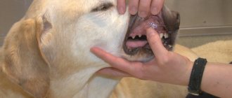
This pathology is often diagnosed by performing a chest x-ray. But there are symptoms characteristic of this disease. The most important thing is the cough. It appears when pressing on the trachea, with excitement.
With this disease, the animal experiences shortness of breath, and due to lack of oxygen, the mucous membranes become blue. In advanced cases, dogs become very anxious and worry, which increases the frequency of breathing movements, which results in an attack.
The disease also affects other dog systems:
- Respiratory. Due to the collapse of the trachea, paralysis or inflammation of the larynx may develop. The latter is observed due to a violation of the procedure for cleaning the respiratory tract from pathogenic microorganisms.
- Cardiovascular. This usually occurs with a severe form of the pathology, when pulmonary hypertension (increased pressure in the lungs) is added to the underlying disease.
- Nervous. It is observed as a result of insufficient oxygen entering the body, which often results in the pet fainting.
Symptoms get worse with age. The pet’s condition is affected by additional factors, which include obesity, heart failure, respiratory diseases, and pressure on the trachea, which is observed when wearing a tight collar.
Main causes of attacks
The wear and tear of cartilage tissue that causes tracheal collapse in dogs occurs due to a genetic predisposition or as a complication of a number of diseases. In the first case, the disorder is often diagnosed 6 months after birth.
Adverse factors and health problems
Tissue deformation develops against the background of acute glucosamine deficiency. This substance is part of the fluid that lubricates joints. Its deficiency results in faster wear of the articular surfaces and loss of shock-absorbing function.
Unfavorable factors that accelerate the process of compression of the tracheal tube include:
- excess weight;
- frequent colds;
- allergic reactions and regular inhalation of aggressive substances that irritate the nasopharyngeal mucosa (household chemicals, tobacco smoke, perfume);
- indoor air is too dry;
- prolonged stress or excessive emotionality;
- mechanical damage to the neck;
- reverse sneezing syndrome;
- an incorrectly selected collar that compresses the trachea.
The secondary type of pathology is diagnosed in the presence of cardiovascular diseases, extensive neoplasms, respiratory infections and inflammation of the respiratory system. In these cases, the main treatment is aimed at eliminating the underlying cause.
Breed predisposition
The risk group includes decorative dog breeds that suffer from a lack of physical activity. Miniature Poodles, Chihuahuas, Pomeranians, Pekingese and Yorkies spend most of their time on the couch. If you don’t monitor their weight, it will go up very quickly.
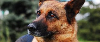
But the mentioned factor is not fundamental. It only exacerbates a problem that has been present since birth. The fact is that all of the listed breeds have much softer tracheal rings than those of shepherd dogs, Labradors and other large dogs. Signs of a congenital anomaly worsen with age, reaching their peak by 6-7 years.
Treatment
Medicines are selected only by a doctor - it depends on the degree of pathology and the characteristics of each pet.
With a mild form of tracheal collapse in dogs, some veterinarians first advise fighting excess weight, using harnesses instead of a collar, and treating secondary inflammatory foci located in the respiratory tract.
If this does not help, drug therapy is prescribed:
- Hormonal drugs. They will help relieve swelling and relieve symptoms for a certain period. Prescribed only after taking a smear, if there are indications. The most commonly prescribed drug is Prednisolone.
- Bronchodilators. The drugs relieve spasms and tension, help reduce pressure in the trachea, and increase the lumen of blood vessels. Veterinarians often prescribe Terbutaline or Theophylline.
Additionally, antitussive drugs are needed; if necessary, antibiotics or antiviral agents may be prescribed.
If you consult a doctor when the first symptoms appear in the first or second stage of the disease, then in 70% of cases drug treatment has a positive effect, resulting in long-term remission. But if there is no effect within 2-3 weeks after the start of therapy, the pet owner will be offered surgery.
Thanks to surgical intervention, the size of the lumen can be adjusted. The positive effect of the procedure is more than 85% even at the third stage of tracheal collapse.
Methods:
- Implant installation. With this method, doctors will replace the deformed area.
- Stenting. Using this method, a stent is installed in the place where the trachea narrows, which completely preserves air flow. The only drawback is that after the operation the dog will still have a cough, so therapy with antitussive drugs can be continued.
The main goal is to make life easier and slow down the progression of the disease. After surgery, it is important to take the necessary preventive measures to avoid secondary infection.
To avoid postoperative complications, the owner needs to regularly bring the pet for examination to the veterinary clinic for x-rays.
Symptoms and manifestations
When the trachea collapses, the dog develops a chronic cough. This is the main symptom, the severity of which can be used to judge the advanced stage of the disease.
Other symptoms include the following:
- cyanosis of the mucous membranes, paw pads and ears on the inside;
- gurgling wheezing and shortness of breath;
- fatigue and lethargy;
- attacks of suffocation and short-term loss of consciousness.
Breathing problems can occur when you inhale and exhale simultaneously, or when you inhale air exclusively. The first option is noted when the thoracic vertebrae are affected, and the second option when the cervical vertebrae are affected.
The latent period of the disease, accompanied by rare coughing, often drags on, preventing timely diagnosis. Despite this, distinguishing pathology from a common cold is not so difficult. When the tracheal rings are damaged, most attacks occur during physical exertion, accidental pulling on the leash, and excessive expression of emotions at the sight of their beloved owner.
Causes of tracheal collapse in dogs
Tracheal collapse differs from other diseases in its chronic form. It consists of severe deformation of the respiratory lining. During such an illness, there is a strong narrowing of the space of the trachea tube itself. The C-shaped rings of cartilage and tubes become severely flattened while the tissue becomes flabby. Wear and tear in cartilage can occur for a variety of reasons.
The prerequisites for the development of tracheal collapse in dogs have not been fully established. Experts say that this pathology can develop both independently and against the background of other diseases.
The primary form of the disease in most cases is based on genetics. Predisposition can only manifest itself in a pet of a certain breed; it can often be detected already in puppyhood. The congenital defect may manifest as initially soft cartilage of the tracheal rings.
As a secondary disease, collapse can occur as a special complication of damage to the pet’s respiratory system, heart failure, as well as disturbances in the circulatory process. Often the background for this is inflammation of the upper and lower sections of both the larynx and lungs.
There are other factors that influence the development of collapse in a dog. In most cases, there is a connection with allergic reactions and frequent inhalation of dirty air (smoke, smog, dust, various chemical components). In addition to all this, the pet’s excess weight negatively affects the overall muscle structure and the cartilaginous tissue of the trachea.
All of the above factors can develop an acute deficiency of glycosamines and glycoproteins in tissues. During low levels of such substances, the rings lose their correct shape and also become several times shorter. The respiratory lumen narrows significantly, and its resistance to body weight increases. The tracheal membrane begins to constantly move: when the cartilage moves in the chest cavity, it protrudes, and when the cartilage moves in the cervical region, it retracts when inhaling. Fluctuations provoke complete obstruction, irritate the mucous membranes of the body, and also further develop inflammation.
Severity of the problem
The method of treatment depends not only on the cause, but also on the severity of the disease. Depending on the nature and scale of the deformation, there are 4 degrees of severity:
- The tracheal lumen is narrowed within 25%
. The sagging of the membrane at this stage is insignificant, so the rings operate as usual without losing their functionality.
- The lumen narrows by half
. The membrane membrane sags heavily, and the cartilage becomes denser. There is a partial loss of function.
- The percentage of narrowing increases to 75%
. The sagging membrane almost touches the rings, which take the shape of an ellipse.
- The lumen narrows to 95-100%
completely blocking breathing. The membrane shell fits closely to the rings. At this stage, suffocation occurs even with complete rest and can result in the death of the animal.
It is important to note that the recommended rate of full tube dilatation is not typical for Yorkies. The tracheal cartilages of these dogs have an ellipsoidal shape from birth.
Is a polyp in a dog really dangerous?
Despite the fact that the growths are benign formations, nevertheless, polyps can pose a danger to the health and life of the pet. First of all, constant mechanical trauma to tissue leads to the development of an inflammatory process.
If the tumor reaches a significant size, it can mechanically block, for example, the nasal passage, trachea, urethra and lead not only to health problems, but also cause the death of the animal.
Among experienced breeders there is an opinion that a benign formation can develop into cancer over time. According to veterinary experts, there is a risk of transformation, for example, of hyperplastic polyps into oncological structures.
How is diagnostics carried out?
It is possible to understand how to treat tracheal collapse in a dog only after a complete diagnosis. To do this, you will have to make an appointment at the veterinary clinic.
The condition of the affected organ is examined using:
- palpation, revealing noticeable deformation of the walls of the tracheal tube;
- X-ray, which determines the size of the lumen and the presence of foreign bodies;
- fluoroscopy, which allows you to observe a specific area in dynamics;
- endoscopy, necessary to take mucus samples and assess the actual extent of the lesion;
- Ultrasound to check for tumors and associated diseases;
- clinical and biochemical blood test confirming pathological deviations in basic parameters.
X-rays and endoscopy are performed under sedation, since the dog’s movement may interfere with taking a picture or taking a piece of biomaterial. For other studies, the administration of anesthetic is not practiced.
Fluoroscopy is considered the most effective diagnostic method. Despite the high radiation dose to everyone present, the data obtained turns out to be 100% correct. This avoids the false positive and false negative results that are common with x-rays.
Tracheal collapse in dogs: treatment
Treatment for trachea in dogs will depend on the stage of the disease. At the second and first stages, you can fight the disease in a conservative way. At a later stage, an operation is already underway. Traditional recipes during such an illness will not be effective, but, on the contrary, will cause significant harm.
Even the most minimal symptoms of the disease require timely assistance from a veterinarian who will help you choose quality therapy. During any treatment method, the animal will be prescribed a special diet, replacing the collar with a harness, regular ventilation and installing a special air humidifier in the room where the pet spends most of its time.
The process of drug treatment for a dog
The main conservative treatment is the use of drugs of a certain group. If the main causes lie in another disease, then it is necessary to prescribe medications to eliminate them (antiviral, cardiac, antibiotics, anti-inflammatory and others).
The intensity of respiration and tissue condition will be regulated mainly by glucocorticoid hormones. It is also worth using antitussives and bronchodilators, since there is simply no way to relieve attacks of tracheal collapse with suffocation without them.
Surgical treatment of an animal
During surgery, the doctor corrects the lumen in the trachea, followed by general restoration. The operation should be performed when the membrane is very sagging, the cartilage at this time almost does not retain its shape, and the threat to the pet’s life exceeds the risks of developing complications after the operation.
The operation can occur in two ways:
- Implantation - the damaged area is replaced, while its anatomical shape is repeated. The implant maintains normal pressure, but may lead to the process of peeling off the base material.
- Stenting is the installation of a mesh stent with lumens in the area of severe narrowing of the trachea. With their help, air permeability is maintained. This technique is considered the safest for the body.
How to help your pet
Emergency assistance from the owner is permissible only in case of a severe attack. In all other cases, the animal must be treated by a veterinarian.
Conservative therapy is effective for disease of 1st and 2nd severity. Four-legged patients with a more severe diagnosis are required to undergo mandatory surgery.
First aid for an attack
First aid is aimed only at temporarily improving the condition, so the victim will still have to be shown to a veterinarian. If you experience an attack of suffocation, follow these steps:
- Calm your pet with an approving voice and strokes. Try to remain calm, as your excitement will certainly be transmitted to the patient.
- Try to eliminate the provoking factor. To do this, you need to remove the collar and ventilate the room if you are not outside.
- Wet your dog's head with cool water and apply a cold compress wrapped in a thick cloth to his chest.
- Contact your veterinary clinic by phone to receive personalized advice from your veterinarian.
An oxygen cylinder, which can be purchased at a regular pharmacy, will help normalize breathing. Codeine, which blocks the cough center, also copes well with the problem. If you don’t have any of these remedies on hand, don’t waste precious time looking for them and go to the veterinary clinic.
Drug therapy
To stimulate respiratory function, glucocorticosteroids (Prednisolone) are used, and to suppress spasm and cough - bronchodilators (Albuterol, Terbutaline, Theophylline), antitussives (Butamirate) and expectorants (Mukaltin, Bronchipret) drugs. The list of other medications depends on the cause of the disease. It may include:
- cardiotonics and vasopressors that lower blood pressure and normalize the force of heart contraction;
- antibiotics (Baytril, Amoxicillin, Tsiprovet) and antiviral (Anandin, Neotime, Fosprenil), eliminating the causative agent of infection;
- anti-inflammatory, dulling the body's response to irritation;
- immunomodulators that restore defenses;
- sedatives that have a calming effect.
Thanks to drug therapy, stabilization of the condition is observed in 71-93% of four-legged patients, depending on the severity of their disease. In other cases, surgical intervention is resorted to.
If a large amount of mucus is detected, the respiratory tract is sanitized. The frequency of procedures depends on the rate of sputum formation and the characteristics of the cough, since when it is dry, the natural discharge of pathological secretions is difficult.
Surgical intervention
The operation carries many risks, so it is used only when the threat to life outweighs the possible complications. Its main task is to correct the narrowed lumen using prosthetics. To do this, use one of two methods:
- Installation of external implants
, replacing damaged areas. Suitable exclusively for pathologies of the cervical spine and is often accompanied by rejection of the material.
- Installation of a special metal stent
, performing a frame function, from the inside. It is used for dysfunction of both the cervical and thoracic vertebrae, but does not solve the problem of constant coughing and copious sputum discharge.
In addition to possible rejection, the disadvantages of implants include the high likelihood of damage to the laryngeal nerves during surgery, which can lead to paralysis of the larynx. For this reason, many owners agree to stenting, which is attractive not only due to lower risks, but also due to more gentle anesthesia.
The persistence of chronic cough during such an operation is explained by the atrophy of the cilia on the mucous membrane, which are forced to constantly contact the tube inserted into the lumen. Despite this, stenting does make breathing easier, so with stable use of symptomatic medications, the patient’s condition is successfully stabilized.
Causes of polyps in dogs
In veterinary medicine, polyps in pets include benign tissue formations that rise above one or another organ. A tumor-like substance is most often found on the mucous membrane. Polyp in dogs is a common pathology, characteristic of the nasal cavity, trachea, stomach, small and large intestines, and uterus. Often neoplasms affect the bladder and rectum.
According to veterinarians and experienced dog breeders, there are several reasons that provoke the formation of polyps in pets:
- Genetic predisposition. Heredity plays a primary role in the etiology of benign tumors. As a rule, if a polyp is diagnosed in a parent, there is a high risk of detecting a tumor in the same organ in the offspring. In this regard, breeders, if possible, do not allow such animals to be bred.
- Inflammatory process. At the site where the local inflammatory reaction occurs, tissue often grows in the form of a polyp as a result of atrophic processes. Most often, against the background of inflammation, growths appear in the mucous membrane of the nose, intestines, and uterus. The cause of a polyp in the trachea in a dog, according to veterinary specialists, is chronic bronchitis due to damage to the mucous membrane of the respiratory tube organs.
- The inflammatory etiology of the occurrence of tumors in tissues can also be traced if they are localized in the stomach and intestines. Gastritis, chronic duodenitis, enterocolitis are the main causes of growths in the thickness of the mucous membrane of the digestive system.
- Allergic reaction . According to experienced breeders, the cause of the growth of benign tumors in the body is food allergies. Dyes, stabilizers and flavor enhancers, preservatives have a constant irritating effect on the mucous membrane of the intestinal tube, which leads to polyps in pets.
- Chronic diseases of the digestive system . A burdened gastroenterological history associated with disruption of epithelial cells is the most common cause of the development of polyps in the stomach and intestines. Chronic constipation, impaired secretory function and motility lead to hyperplastic processes in the mucous membrane, which is accompanied by its pathological condition.
The so-called embryonic theory contributes to the etiology of the development of polyps. According to scientists, already at the stage of fetal formation in the womb, conditions are created for the further growth of benign tumors in the tissues of the body.
We recommend reading about why your dog has pus coming out of its nose. From the article you will learn about the causes of purulent nasal discharge, symptoms of sinusitis in dogs, diagnosis and treatment.
And here is more information about the dangers of a runny nose in dogs.
Are there any complications?
As a result of deformation of the tracheal tube, not only the organs of the respiratory system, but also the heart and brain are affected. Without timely treatment, the disease provokes the development of the following disorders:
- pulmonary hypertension, characterized by an increase in blood pressure due to narrowing of blood vessels;
- right ventricular heart failure, which can lead to death due to gradual heart failure;
- death of brain neurons suffering from prolonged lack of oxygen.
Long-term use of antibiotics can also cause complications. These drugs destroy not only pathogenic microorganisms, but also their own immune cells. As a result of such therapy, the existing disease is often complicated by a secondary infection.